




 Servicios personalizados
 Inglés (pdf)
 Articulo en XML
 Referencias del artículo
 Traducción automática
 Enviar articulo por email
 Citado por SciELO
  Citado por Google
  Similares en
SciELO
Similares en
SciELO  Similares en Google
 Permalink
INTRODUCTION
Hypophosphatasia (HPP) is a rare genetic disorder caused by loss-of-function mutations in the ALPL gene, which encodes the tissue-nonspecific isoenzyme of alkaline phosphatase (TNSALP) (1). It was first described by J.C. Rathbun in 1948 (2). This enzyme plays a crucial role in bone and dental mineralization as it hydrolyzes substrates such as inorganic pyrophosphate (PPi), a potent inhibitor of hydroxyapatite crystal formation. Deficiency of TNSALP leads to extracellular accumulation of PPi and other substrates, such as pyridoxal-5´-phosphate (PLP) -circulating form of vitamin B6- and phosphoethanolamine (PEA), impairing normal mineralization (3). The accumulation of PLP should also be considered, as their effects may extend beyond bone metabolism, potentially influencing neurological function. E.g.: impaired hydrolysis of PLP can cause vitamin B6 deficiency in the central nervous system and reduced neurotransmitter synthesis, potentially leading to pyridoxine-responsive seizures in severely affected infants (4).
The first mutation in the ALPL gene was identified in 1988 in a Canadian infant with the lethal form of HPP (5). Currently, over 480 mutations linked to HPP have been reported, the majority of which are missense variants (6). HPP can be inherited in either an autosomal recessive or autosomal dominant pattern. The more severe clinical forms, are typically inherited in an autosomal recessive manner. By contrast, milder forms of the disease may result from either autosomal recessive or dominant inheritance (7). The wide variability in genotype-phenotype correlations raises challenges for clinical prediction and management, especially among heterozygous carriers (8). Importantly, heterozygous individuals can also manifest clinical symptoms, and recent data from the Global HPP Registry indicate that most patients with non-life-threatening forms of HPP carry a single ALPL variant (9,10). It has been demonstrated that 3 categories of alleles, classified according to their severity and dominant effect (s: severe recessive, Sd: severe dominant, and m: moderate), occur with a similar prevalence across different regions of Europe, suggesting that the prevalence of HPP is likely consistent (11). Even though, the current estimate of potential undiagnosed HPP cases in countries such as Spain could rise to 27,177 cases (12). This would make the prevalence of HPP higher than suspected.
There is traditionally considered to be an inverse relationship between age at onset and clinical severity in HPP, with the most severe phenotypes typically occurring in the perinatal and infantile forms. However, this concept has evolved as growing evidence shows that adults can also present with substantial disease burden, and in some cases with symptoms that rival or exceed those seen in children. Many adults experience chronic musculoskeletal pain, impaired mobility, recurrent fractures, dental abnormalities, fatigue, and even require assistive devices such as wheelchairs or surgical procedures such as intramedullary rodding to stabilize femoral fractures (13,14). The high variability in symptomatology, particularly in adults, contributes to frequent diagnostic delays, often by several years or even decades, as historical classifications have tended to exclude adults with pediatric-onset disease (15). Furthermore, recent data show that HPP is a dynamic and evolving disorder: symptoms can change over time and multiple clinical complications accumulate progressively throughout life, rather than being static or confined to a single body system (13,16). While early-onset forms are associated with high mortality, many patients live into adulthood yet endure a considerable and persistent disease burden, affecting physical function, daily activities, and overall quality of life (14,17). Consequently, HPP is increasingly understood as a lifelong, multisystemic condition rather than solely a pediatric or bone-related disease, and diagnosis remains based on persistently low serum alkaline phosphatase levels, alongside clinical, radiological, and often genetic findings (18).
Regarding pediatric forms, perinatal HPP is the most severe and typically lethal form of the disease, presenting before or at birth (19,20). It is characterized by profound skeletal hypomineralization, resulting in short, deformed limbs and caput membraneceum. Additional features may include a high-pitched cry, pyridoxine-responsive seizures, episodes of apnea with cyanosis and bradycardia, fever of unknown origin, irritability, bone marrow failure (myelophthisic anemia), intracranial hemorrhage, and occasionally underdeveloped lungs (21,22). Survival is very rare.
Infantile HPP presents before 6 months of age (23). Symptoms such as poor feeding, failure to thrive, and delayed milestones are usually found in these patients (24). It is marked by skeletal abnormalities, metabolic imbalances (such as hypercalcemia and hypercalciuria), and complications such as fractures, respiratory issues, and pyridoxine-dependent seizures (25). Thoracic deformities can lead to pneumonia, a major cause of death. Despite initial normal development, the condition often progresses rapidly, with a historically high infant mortality rate (11).
Childhood HPP presents after 6 months of age, and it can be considered mild or severe depending on the clinical presentation (7,26). Premature loss primary teeth is a common feature of childhood HPP, and in severe cases, all deciduous teeth may be lost early (7). Skeletal deformities resembling rickets are often present, including skull abnormalities, bowed legs, knock-knees, and enlarged joints due to metaphyseal flaring. Children may experience significant bone pain, short stature, muscle weakness, delayed walking, and a waddling gait. In rare cases, joint swelling and radiographic changes may resemble chronic recurrent multifocal osteomyelitis (7,25,27,28). Importantly, this traditional classification based on age of presentation does not fully capture the complexity of the disease, as many adults may have pediatric-onset HPP that persisted undiagnosed for years, while others develop symptoms later in life (adult-onset HPP). Thus, adult patients may belong to either group, underscoring the need to consider disease onset, progression, and cumulative burden rather than age alone for accurate characterization.
Based on this, increased awareness and the implementation of screening protocols are essential for early recognition and management, particularly as enzyme replacement therapy is now available and approved by major regulatory agencies such as the FDA and the EMA (24,29).
METHODS
STUDY POPULATION
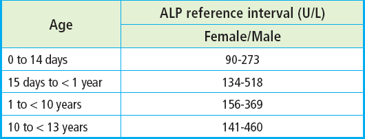
We conducted a retrospective study in the province of Granada (Spain) on the pediatric population with suspected HPP. Patients were identified through the laboratory database by selecting those who had at least one recorded test of low serum ALP levels, defined according to the reference values for the age and sex of the patient (Table I) according to the CALIPER study (30). The electronic health records of these patients were subsequently review to collect clinical information.
Data on age, sex, medical history and usual medication were collected for each patient. All the information was incorporated into an anonymized database for analysis, guaranteeing confidentiality in accordance with current data protection regulations. From the total number of identified patients, exclusion criteria were applied to rule out secondary causes of HPP, as low alkaline phosphatase (ALP) levels are not pathognomonic for this disease and may also be observed in a variety of other conditions (8). Patients were excluded if they presented with any of the non–HPP causes of hypophosphatasemia listed in table II.
Table II Secondary causes of hypophosphatasemia
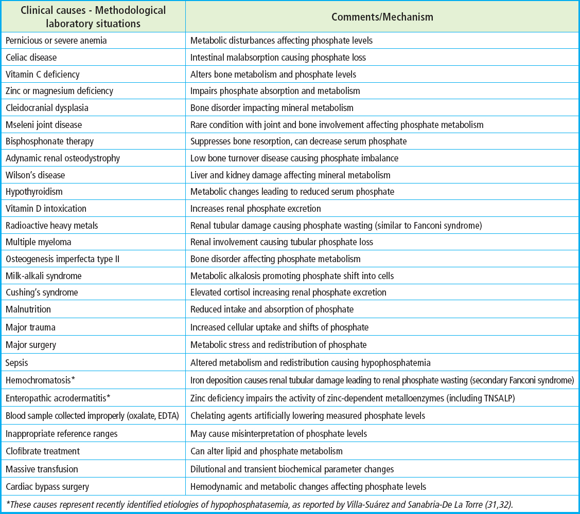
*These causes represent recently identified etiologies of hypophosphatasemia, as reported by Villa-Suárez and Sanabria-De La Torre (31,32).
Potential HPP cases were contacted by their referring specialists or primary care physicians to undergo screening within the framework of the Phase 3 Study of ALXN1850 in Treatment-Naïve Pediatric Participants with HPP. As part of the study protocol, these patients were referred to Hospital Universitario La Paz (Madrid, Spain) for the necessary diagnostic procedures, including genetic sequencing of the ALPL gene and determination of serum PLP levels. These patients signed the corresponding informed consent.
The present study was approved by the ethics committee of Granada in full compliance with the principles outlined in the World Medical Association Declaration of Helsinki (project ID: 0768-N-17 on 28 June 2017).
RESULTS
A total of 271 pediatric cases aged between 0 and 12 years having at least one recorded blood test indicating low serum ALP levels were identified. The flowchart applied during the screening process is shown in figure 1.
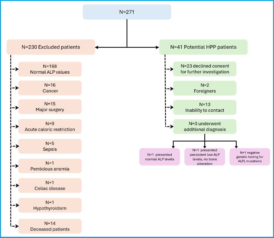
Figure 1 Flowchart illustrating patient selection and outcomes. Patients with low serum ALP were first divided into 2 groups: excluded cases and potential HPP cases. Excluded patients were categorized according to the identified secondary causes of HPP. For the potential HPP group, the chart details subsequent follow-up actions and outcomes, including additional diagnostic testing, lack of consent and inability to contact.
Of the 271 patients initially considered, 168 (62.0 %) were excluded because at least one analytical determination during follow-up showed normal ALP levels, suggesting that the initial low value was transient or nonspecific. An additional 16 patients (5.9 %) were excluded because of a diagnosis of cancer, either under active treatment or within follow-up periods that could affect bone metabolism and serum ALP levels. Fifteen patients (5.5 %) had undergone major surgical procedures in the days preceding the detection of low ALP levels, representing another potential confounding factor, and were therefore excluded.
Additionally, 9 cases (3.3 %) were found to have experienced acute caloric restriction, either due to medical conditions such as severe GI disorders or restrictive eating behaviors including anorexia, all of which are known to affect ALP levels and bone turnover. These were also excluded. Five patients (1.8 %) exhibited sepsis at the time of laboratory evaluation, which could influence liver and bone enzyme levels. These cases were also discarded from further consideration. Furthermore, isolated cases of pernicious anemia (n = 1), celiac disease (n = 1), and hypothyroidism (n = 1) were identified, each of which constitutes a potential secondary cause of low ALP, and they were therefore removed from the candidate group.
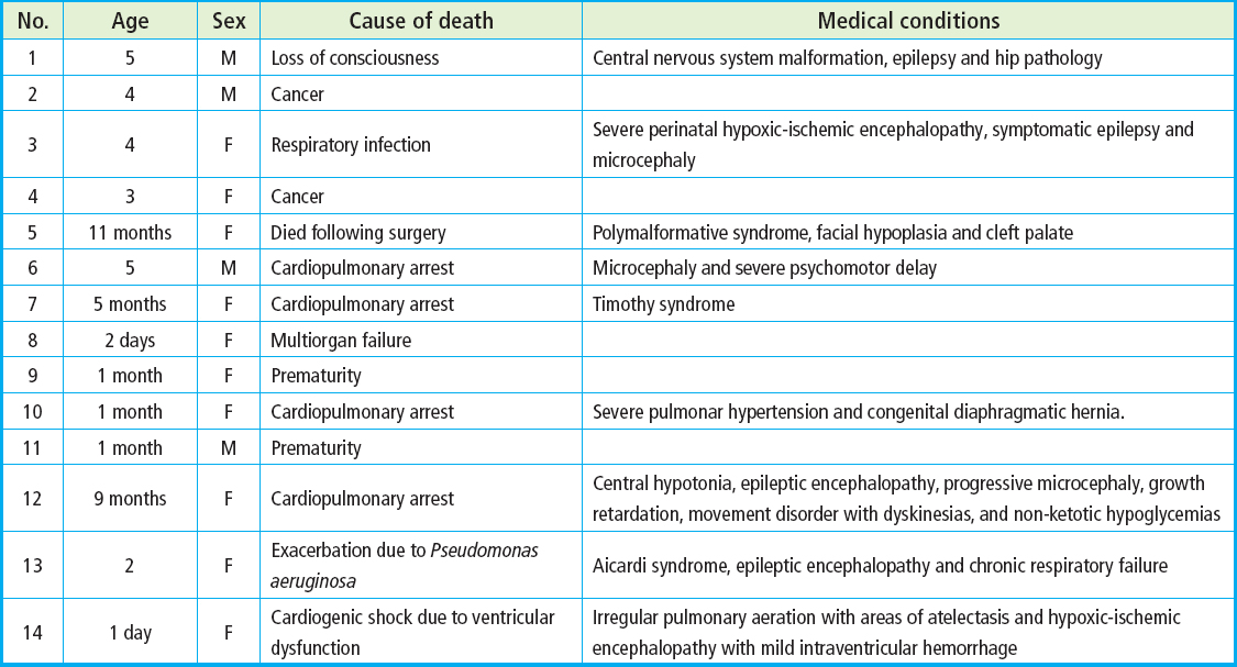
A total of 14 patients (5.2 %) were found to be deceased at the time of data collection, and sufficient follow-up data to evaluate the clinical course or confirmatory diagnostics were unavailable, precluding their inclusion in the final analysis. Their clinical profiles were heterogeneous, and in most cases, complex comorbidities were present (Table III). First, 2 patients (ages 4 and 3) died of cancer, without evidence suggestive of HPP. Another 4 patients died shortly after being born, associated with perinatal complications such as prematurity, hypoxic-ischemic encephalopathy or congenital diaphragmatic hernia. In these cases, the underlying cause of HPP remained unexplored due to early death. Eight of the deceased patients exhibited congenital malformations and neurological impairments or their main cause of death was shock, which may be consistent with HPP and therefore could not be definitively excluded as HPP patients.
After applying all exclusion criteria, 41 cases (15.1 %) with no identifiable secondary cause of hypophosphatasemia constituted the group of potential HPP cases, pending further diagnostic workup.
Of the 41 potential HPP cases identified, the primary care physicians of 28 patients (68.2 %) were successfully contacted. Among these, 3 patients underwent additional diagnostic testing and were evaluated with expanded clinical, biochemical, and radiological assessments, including confirmatory enzymatic studies and, when appropriate, ALPL gene sequencing. Two patients (4.9 %) were identified as foreign nationals who had received isolated medical care at centers in Andalusia and could not be followed up. Twenty-three families (56.0 %) declined consent for further investigations. The primary care physicians of the remaining 13 patients (31.7 %) could not be contacted because of missing or outdated contact information.
Among the 3 patients (7.3 %) who underwent further diagnostic evaluation, none were ultimately diagnosed with HPP (Table IV). In one case, a repeat blood test showed normal ALP levels, leading to exclusion. The remaining two patients continued to exhibit persistently low ALP levels and underwent bilateral wrist and knee radiographs. One of them showed radiographic signs consistent with rickets, while the other had only mild, nonspecific changes, which were not considered sufficient for HPP diagnosis. The patient with radiographic signs of rickets also exhibited short stature and a pectus excavatum deformity; however, subsequent genetic testing for ALPL mutations returned negative, thereby excluding the HPP diagnosis.

DISCUSSION
This retrospective study aimed to identify potential pediatric cases of HPP based on persistently low serum ALP levels and to assess the clinical consequences of under recognition in this population. Out of 271 children aged 0-12 years with at least 1 recorded episode of hypophosphatasemia, 41 patients (15.1 %) were identified as having persistently low ALP levels without identifiable secondary causes of HPP. These individuals constitute a group of high clinical interest, underscoring the potential diagnostic value of unexplained hypophosphatasemia in routine pediatric care and highlighting a window of opportunity for earlier identification of HPP.
Among the patients who underwent extended evaluation, 2 maintained persistently low ALP and one presented with short stature, chest deformity (pectus excavatum), and radiological signs compatible with rickets. Although the clinical and biochemical picture raised strong suspicion of HPP, sequencing of the ALPL gene did not identify any pathogenic variants. However, this result must be interpreted with caution, as conventional sequencing methods typically assess only coding regions of the gene and may fail to detect pathogenic variants located in intronic, promoter, or other regulatory non-coding regions (33,34). In such cases, functional ALPL gene expression may be impaired despite the absence of detectable coding mutations. Therefore, a diagnosis of HPP cannot be definitively excluded in this patient, reflecting the diagnostic limitations in clinical practice, particularly in patients with borderline or atypical phenotypes. According to the article by Rush et al., patients may be diagnosed with HPP not only when pathogenic or likely pathogenic variants in the ALPL gene are identified, but also when variants of uncertain significance (VUS) are present, or even in the absence of detectable variants, provided that the clinical and biochemical features are consistent with the disease (35). In these cases, additional assessments are recommended to be performed such as whole exome sequencing to identify potential mutations in non-coding regions of ALPL gene or in other ALP-regulatory genes.
Despite the rigorous inclusion criteria and systematic screening approach, no confirmed cases of HPP were diagnosed in this cohort. This finding is consistent considering the reported prevalence of HPP in its more severe forms. Estimates suggest that the combined prevalence of perinatal and infantile HPP (typically inherited in an autosomal recessive pattern), ranges from 1 in 100,000 to 1 in 300,000 live births (11,12). Therefore, given the limited sample size and geographic focus of this study, the absence of confirmed diagnoses aligns with statistical expectations for severe forms. Additionally, a significant proportion of potentially eligible patients (31.7 %) could not be evaluated due to loss of contact with primary care physicians or refusal of consent by the families. This represents a substantial diagnostic gap that may have masked the identification of true HPP cases, especially among patients with milder or atypical presentations. These logistical limitations, which are common in retrospective studies involving rare diseases, highlight the challenges of case detection in real-world clinical settings.
One of the most concerning findings of this study was the identification of 14 pediatric patients who died before further evaluation could be performed. Only two of these (patients 2 and 4) had a clearly documented oncologic cause of death, with no clinical suspicion of HPP. In the remaining 12 patients, HPP could not be ruled out as a contributing or primary cause of death. Notably, several of these individuals presented with clinical features that may be consistent with severe forms of HPP, including early-onset neurological impairment, epileptic encephalopathy, congenital malformations, growth delay, and severe respiratory dysfunction. Patients #1, #3, #12, and #13 exhibited epilepsy or epileptic encephalopathy, signs potentially related to PLP accumulation in the setting of TNSALP deficiency. TNSALP plays a critical role in the dephosphorylation of PLP to pyridoxal, thereby enabling its cellular uptake and transport into the central nervous system (36). Accumulation of PLP may interfere with neurotransmitter synthesis, contributing to seizure activity (37).
Additionally, patients #3, #6, #7, #10, #12, #13, and #14 died from cardiorespiratory arrest or respiratory complications. Patient #14 had documented pulmonary atelectasis, a finding that may be mechanistically linked to impaired phospholipid biosynthesis in the setting of TNSALP deficiency, although rib cage deformities and hypoplastic lungs are more commonly recognized as contributing factors in HPP-related respiratory failure (38,39). Furthermore, multiple patients presented with syndromic phenotypes involving facial or skeletal malformations (eg, patients #5, #12, and #13), which could be consistent with the perinatal or infantile forms of HPP that are often lethal if not recognized and treated early (21).
The mortality rate observed in this cohort (4.4 %), excluding oncologic causes, should be interpreted with caution, as the retrospective design and substantial loss to follow-up limit the strength of the conclusions. Nonetheless, this finding underscores a potentially important diagnostic gap in pediatric HPP. Several unexplored deaths occurred in children with persistent hypophosphatasemia, whose reported symptoms closely resembled the classical presentation of HPP. While a direct causal relationship cannot be established, these observations raise concern that some fatal cases might have been related to undiagnosed HPP. This highlights the critical importance of early recognition and diagnostic evaluation in pediatric patients with persistently low alkaline phosphatase levels, as timely identification could prevent severe disease progression and potentially avoid fatal outcomes.
These findings underscore the potential value of developing structured diagnostic algorithm that incorporates ALP as a frontline screening biomarker, particularly when levels are markedly low and not attributable to secondary conditions. In any child with persistently low ALP and signs of poor mineralization, delayed milestones, or unexplained seizures, HPP should be included in the differential diagnosis. Repeat testing to confirm ALP persistence and exclusion of confounding factors are essential steps, followed by targeted biochemical and genetic testing.
Moreover, our results support the implementation of neonatal biochemical screening strategies for early detection of HPP. Although widespread genetic screening via the heel-prick test remains financially and logistically unfeasible, a more accessible and cost-effective approach could involve measuring ALP activity in capillary blood samples collected shortly after birth. Such a biochemical screening tool, if incorporated into routine newborn testing, could help identify neonates with severe TNSALP deficiency before the onset of life-threatening symptoms. Although further studies are required to assess its feasibility, cost-effectiveness, and clinical utility, infants flagged by this approach could subsequently undergo confirmatory testing and early intervention, including consideration of enzyme replacement therapy with asfotase alfa, which has demonstrated significant survival benefits in perinatal and infantile HPP (20,40).
Despite its limitations, including its retrospective nature, reliance on existing laboratory records, and the inability to follow up with a significant proportion of potential cases, this study provides valuable insights into the magnitude of underdiagnosis in pediatric HPP. A major limitation was the difficulty in contacting primary care providers, compounded by time constraints and the high workload within primary care settings, which likely contributed to missed follow-up opportunities. Furthermore, the lack of access to advanced genetic or enzymatic diagnostic tools across the healthcare system limits clinicians’ ability to fully characterize or confirm suspected cases (41).
Nevertheless, this study has notable strengths. To our knowledge, this is one of the first studies to apply a systematic screening approach based on real-world ALP data to a pediatric population, offering a practical framework for identifying children who may benefit from further diagnostic workup. The clinical data gathered from deceased patients highlights the potentially severe consequences of undiagnosed HPP and demonstrates how routine biochemical markers, if correctly interpreted, can uncover life-threatening conditions that might otherwise remain hidden. By focusing on the pediatric population, this study addresses the group at highest risk for the severe, life-limiting forms of the disease and helps to define the profile of patients who would benefit most from early detection and intervention.
In conclusion, this study highlights the importance of persistent hypophosphatasemia as an early warning sign of HPP in children and underscores the substantial mortality burden that may go unrecognized in the absence of structured diagnostic protocols. The development of standardized clinical algorithms and the implementation of biochemical neonatal screening strategies are critical next steps to improve diagnostic yield and reduce avoidable morbidity and mortality in pediatric HPP. In the era of effective enzyme replacement therapy, early identification is no longer optional, it is essential.

