




 Servicios personalizados
 Inglés (pdf)
 Articulo en XML
 Referencias del artículo
 Traducción automática
 Enviar articulo por email
 Citado por SciELO
  Citado por Google
  Similares en
SciELO
Similares en
SciELO  Similares en Google
 Permalink
INTRODUCTION
Postmenopausal women face an increased risk of osteoporosis and fractures. Hormone replacement therapy (HRT) can reduce the fracture incidence rate—as evidenced by the Women’s Health Initiative (WHI) trial, which demonstrated fracture prevention benefits of estrogen-progestin therapy. However, the WHI also revealed that HRT’s serious side effects (eg, cardiovascular risks) often outweigh its benefits (1). Furthermore, estrogen plays a crucial role in protecting vs muscle damage. Therefore, its deprivation in menopause leads to a dual problem: decreased bone density and fracture risk, along with muscle weakness and fatigue (2,3). Hence, menopause-related muscle and skeletal pain complaints increases, and many postmenopausal women seek a non-HRT alternative symptomatic treatment to relieve their pain and discomfort.
There are many agents from different chemical groups fall into “centrally acting muscle relaxants” such as tizanidine (TZ), thiocolchicoside (TCC), cyclobenzaprine (CBZ) and are used in clinical situations such as muscle pain and muscle spasm. They work by affecting the central nervous system, specifically at the brainstem level, inhibiting motor neuron activity and reducing muscle spasms. Typically, they are used for short-term relief in acute musculoskeletal conditions, and some of them are available in many countries as over-the-counter medication (4-6).
It has been reported that skeletal muscle contractions, whether from purposeful exercise or regular daily movements, contribute to more than 70 % of the mechanical load on bones bear (7). Through the administration of centrally acting muscle relaxants, this stress response is mitigated. This biomechanical effect might be deleterious on highly vulnerable bone tissue on postmenopausal period when the muscle mass is already decreased (8).
RANKL (receptor activator of NF-κB ligand) is responsible for osteoclasts maturation which leads to bone resorption. OPG (osteoprotegerin) prevents excessive bone breakdown by blocking RANKL (9). Even a special human monoclonal antibody, denosumab, has been recently developed to inhibit human RANKL and is indicated for the treatment of postmenopausal women with osteoporosis (2). Although these drugs have been used for decades, their long-term biomechanical effects (eg, bone and muscle integrity) and biochemical effects (eg, metabolic processes) require further investigation.
This research could help to identify potential bone metabolism risks and benefits associated with the use of muscle relaxant drugs in postmenopausal women and fill a knowledge gap and provide valuable information about their safety. For this purpose, a menopause model has been established in rats via ovariectomy, and the effects of the drugs on bone has been evaluated.
MATERIALS AND METHODS
All animal procedures complied with the ARRIVE guidelines and the Guide for the Care and Use of Laboratory Animals and were approved by the Aydin Adnan Menderes University for Animal Experiments Ethics Committee (HADYEK 64583101/2023/25); anesthesia, analgesia, and humane endpoints were implemented to minimize pain and distress.
In this study, 50 adult female Wistar Albino rats were obtained from Aydin Adnan Menderes University Experimental Animals Production Laboratory. At 3 to 4 months of age, the rats were divided into 5 groups, with 10 rats in each group. Ovariectomy was performed in 40 rats under general anesthesia with ketamine and xylazine (10).
The experimental groups were as follows:
Sham group: group in which the abdominal incision was made and then closed without removing the ovaries.
– OVX: group subjected to ovariectomy without any other procedure for 10 weeks.
– OVX + TZ: following ovariectomy, at the end of eight weeks, daily oral 2 mg/kg tizanidine (Sirdalud tablet®, Novartis, Istanbul, Turkey) was administered.
– OVX + TCC: following ovariectomy, at the end of eight weeks, daily intramuscular 2 mg/kg thiocolchicoside (Muscoril ampoule®, Bayer, Istanbul, Turkey) was administered.
– OVX + CBZ: following ovariectomy, at the end of eight weeks, daily oral 2 mg/kg cyclobenzaprine (Flessi tablet®, Menarini, Istanbul, Turkey) was administered.
Drug doses were adjusted according to weekly changes in body weight. All drugs were administered in two divided doses daily for 2 weeks.
At the end of the study, under anesthesia with ketamine (50 mg/kg) and xylazine (5 mg/kg), blood samples were obtained by cardiac puncture, centrifuged (1000 × g for 10 minutes), and the separated serum was stored at −20 °C for biochemical analyses. Muscle and soft tissues attached to the femur were removed and cleaned from surrounding tissues, and the bones were wrapped in saline-moistened paper towels and stored frozen at −20 °C for subsequent 3-point bending testing.
3-POINT BENDING TEST STUDIES
The bones were thawed at room temperature before mechanical testing. Then, the lengths of the bones were measured, and the midpoint of the bone was marked. The midpoint was determined as the loading point for the 3-point bending test. After the mechanical test, cranio-caudal, medio-lateral endosteal and periosteal diameters (External Diameter) were measured. In the 3-point bending test, Zwick Roell Z0.5 (Ulm, Germany) mechanical testing machine was used in the TARBIYOMER laboratories of Aydin Adnan Menderes University. For the mechanical test, the support points (L) (Span Length) were determined as 15 mm, preload as 2 N, and strain rate as 1 mm/min. A cranio-caudal load was applied from the midpoint of the bone. After the test, medio-lateral and cranio-caudal endosteal diameters (Internal Diameter) were measured from the fractured bones. Using the endosteal and periosteal diameters, the cross-sectional moment of inertia of the bones was calculated. The stiffness was calculated from linear regression of the Force-Displacement graph using the software testXpert (Zwick/Roell, Ulm, Germany) obtained after mechanical testing. Using the stiffness, moment of inertia, bone diameter and distance between the support points, ultimate strength and elastic modulus were calculated by using the formulas specified in the references (11-14).
BIOCHEMICAL STUDY
Commercially available kits for Total Oxidant Status (TOS, Elabscience Biotechnology Inc, USA; sensitivity: 2.5 µmol H2O2 Equiv./L), Total Antioxidant Status (TAS, Elabscience Biotechnology Inc, USA; sensitivity: 23 mmol Trolox Equiv./L), Osteoprotegerin (OPG, BT Lab China; sensitivity: 5.45 ng/L) and Receptor Activated Nuclear KappaB Ligand (RANKL, ELK Biotechnology CO. Ltd. USA; sensitivity: 5.7 pg/mL) were used according to the instructions of the manufacturer and plates were read at suggested nm wavelength by a spectrophotometer (MultiscanGo, Thermo Fisher Scientific Inc, USA).
STATISTICAL ANALYSIS
Body weights were evaluated by a percentage change amount in the groups. All the data obtained was averaged for the left and right bones. The data of normal distribution was checked with the Shapiro-Wilk test. One-Way ANOVA was performed for normally distributed values and Kruskal-Wallis intergroup comparison was performed for non-normally distributed values. Data were expressed as mean ± standard deviation, values of p < 0.05 were accepted as significant. Analyses were performed using IBM SPSS Statistics for Windows, Version 25.0 (IBM Corp., Armonk, NY, USA).
RESULTS
CLINICAL FOLLOW-UP
No rats were lost during the study. The percentage change in body weight over the 10-week menopausal period was 30.70 ± 3.18 % in ovariectomized (OVX) rats, 22.37 ± 1.60 % in the TZ-treated group, 25.93 ± 3.37 % in the TCC-treated group, and 24.56 ± 3.79 % in the CBZ-treated group, whereas the control group showed a change of 8.18 ± 2.00 %. Only TZ treatment resulted in a statistically significant reduction in body weight gain compared with the OVX group (p < 0.05).
3-POINT BENDING TEST RESULTS
The data obtained after the mechanical test was shown in table I. The study data showed no significant differences between the groups in terms of bone lengths, fracture force, deformation values, and stiffness values derived from the force-deformation graph.
Table I Changes in biomechanical parameters in all groups
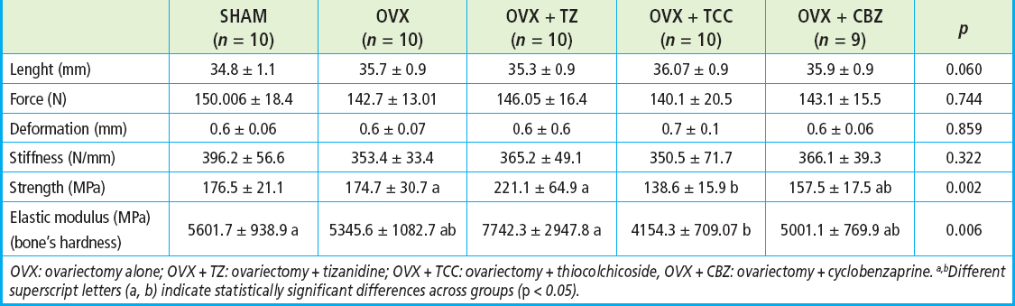
OVX: ovariectomy alone; OVX + TZ: ovariectomy + tizanidine; OVX + TCC: ovariectomy + thiocolchicoside, OVX + CBZ: ovariectomy + cyclobenzaprine. a,bDifferent superscript letters (a, b) indicate statistically significant differences across groups (p < 0.05).
A statistical difference was noted between the groups in the strength value representing bone strength. The OVX + TZ group had a value that was 25 % higher than the sham group, whereas the OVX + TCC group had a value that was 21 % lower than the sham group (p = 0.002).
The groups differed in the elastic modulus value, indicating the bone’s hardness. The OVX + TZ group had a value 38 % higher than the sham group, while the OVX + TCC group had a value 25 % lower than the Sham group (p = 0.006).
BIOCHEMICAL ANALYSES
The data obtained from biochemical tests were shown in table II.
Table II Biochemical features of all experimental drugs. Values are mean ± SD from 9-10 rats
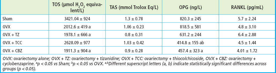
OVX: ovariectomy alone; OVX + TZ: ovariectomy + tizanidine; OVX + TCC: ovariectomy + thiocolchicoside, OVX + CBZ: ovariectomy + cyclobenzaprine. ap < 0.05 vs Sham; bp < 0.05 vs OVX. a,bDifferent superscript letters (a, b) indicate statistically significant differences across groups (p < 0.05).
In TOS analyses, there were significant decreases in OVX (p < 0.05), TZ (p < 0.05) and CBZ groups (p < 0.05) vs the Sham group (Sham 3421.04 ± 924, OVX 2012.66 ± 419, TZ 1978.12 ± 666, TCC 2628.09 ± 977, CBZ 1911.38 ± 904 µmol H2O2 equivalent/L).
There were no significant differences among groups in TAS study (Sham 1.39 ± 0.78, OVX 1.06 ± 0.23, TZ 0.86 ± 0.31, TCC 1.03 ± 0.42, CBZ 0.92 ± 0.28 mmol Trolox Eq/L.).
When looking at the OPG value, there was no difference between the control and OVX groups. TZ tended to decrease, TCC significantly reduced OPG levels (p < 0.05) vs both the Sham and OVX groups. (Sham 820.37 ± 245, OVX 818.52 ± 581, TZ 631.24 ± 244, TCC 414.81 ± 155, CBZ 457.41 ± 323 ng/L).
Regarding serum RANKL levels, TZ tended to increase RANKL, whereas TCC and CBZ tended to decrease it. However, no statistically significant differences were observed among the groups (sham, 5.71 ± 2.24 pg/mL; OVX, 4.83 ± 3.10 pg/mL; TZ, 6.45 ± 2.88 pg/mL; TCC, 4.51 ± 1.44 pg/mL; CBZ, 4.01 ± 1.72 pg/mL).
DISCUSSION
Postmenopausal women frequently experience non-osteoporosis-related generalized musculoskeletal achiness (15), which often results in them intermittently taking over-the-counter muscle relaxants. Therefore, we designed our study to mimic this intermittent usage pattern by restricting the drug exposure period in the rats to two weeks. Since muscle relaxants might further weaken muscles already compromised in postmenopausal women, this study investigates their safety regarding their potential impact on bone health with biomechanical and biochemical parameters. Bones need significant mechanical loading stress to stay strong (7).
It has been reported that Masson trichrome-stained sections in microscopy and micro-CT images revealed significant osteolytic changes and a decrease in trabecular bone in the femur from the OVX group at eight weeks after surgery (16). Based on this research, we designed our study; we waited eight weeks to establish an osteoporosis model before applying treatments for fifteen days. Therefore, the non-treated group underwent 10 weeks of OVX. Another research by Khera, Kanta (2) has also reported that post-menopausal osteoporosis developed four weeks later of bilateral ovariectomy. It has been stated that the duration of the remodeling cycles (includes four sequential phases of activation, resorption, reversal, and formation) in normal trabecular bones is approximately 6 days in rats (17). These researchers also emphasized that trabecular bone mass in femoral neck decreases 30 days after OVX.
Compared with previous researches by Khera (2), Lee (16), Yousefzadeh (17), we did not observe prominent changes in bone biomechanical values. This suggests that a 10-week post-ovariectomy period may be too early to establish well-developed osteoporosis in this animal model. However, we did observe a trend toward decreased flexibility and a significant reduction in the elastic modulus in the OVX group. This suggests that the cortical bone was partially affected, though the effect was less pronounced than typically observed in trabecular bone at ten weeks post-ovariectomy. Additionally, our choice to use 3- to 4 months-old Wistar rats for the surgery was made to eliminate the confounding effect of age-related findings. This younger age may contribute to the differences observed in comparison to existing literature, resulting in our rats being more resistant to the induction of osteoporosis.
The relationship between load applied to a structure and deformation response to the load is called a load-deformation curve. The curve can be divided into two regions: elastic deformation and plastic deformation. Stiffness is calculated in the region where the deformation in the bone increases linearly with increasing load. Stiffness is the extrinsic rigidity of the structure. There are viscous effects during deformation, due to fluids in the bone matrix, which cause some of the elastic energy to be lost. Bones show viscoelastic properties. Due to this bone feature, elastic modulus and strength values, which are intrinsic properties of bone, are calculated by engineering formulae (14). Focusing on intrinsic properties in bone research provides reliable results. This study found no significant difference in the extrinsic properties of bone tissue, including bone length, fracture force, deformation, and stiffness values. However, there was a distinction between the groups in the intrinsic properties, such as elastic modulus and strength values of TZ and TCC.
Additionally, we did not detect oxidative stress in the 10-week post-menopausal rats. Our results show that TAS levels tended to decrease, it bears in mind that antioxidants might have been consumed at this term of the model. These antioxidant enzymes are essential for removing reactive oxygen species, which can cause cellular damage. By maintaining a balanced redox state, these enzymes help protect cells from oxidative stress. Our previous study with 16-month post-menopausal rats showed a trend of increasing TOS and decreasing TAS (10). However, our analysis only looked at the total amount of oxidants and antioxidants in the serum. Considering that oxidant and antioxidant are in a delicate balance in the body, it seems TAS could compensate for the TOS increase at some point during this early period of animal model. Measuring specific types of oxidants and antioxidants could provide more detailed information about the menopausal progression of experimental model.
RANKL, a signaling molecule belonging to the Tumor Necrosis Factor (TNF) family, plays a critical role in transforming monocytes into bone-resorbing osteoclasts. This essential cytokine is produced by osteoblastic/stromal cells and directly leads monocytes to become osteoclasts (6). We expected an increase in RANKL levels especially in OVX group due to the high osteoclast activity in menopausal rats, but there was no significant change. Meanwhile, we expected a decrease in OPG levels of the OVX group, but it was the same between our control and OVX groups. Therefore, we assume that osteoporosis development was in its early stages in our current study. The role of OPG, RANK, and RANKL in the pathogenesis of postmenopausal or age-related osteoporosis have been stated as a controversial issue. Estrogen exerts a stimulatory effect on OPG production in osteoblasts and bone marrow stromal cells. Nevertheless, despite a decline in OPG production by bone marrow stromal cells with advancing age, serum OPG levels exhibit an age-related increase in both genders. Furthermore, postmenopausal women with osteoporosis have been reported to have significantly higher serum OPG levels vs age-matched non-osteoporotic women (18).
When examining the bone effect of abovementioned drugs on this model, our study found a remarkable increase in bone strength and elastic modulus (25 % and 38 % respectively) treated with TZ vs the sham group. Interestingly, while TZ has demonstrated cytotoxicity vs various cancer cells (including U2 osteosarcoma, MCF-7, HOP92, and A549), it has also been found to be toxic to normal human osteoblasts (4, 19, 20). Therefore, when considering its therapeutic use, it must be kept in mind that the dosage of TZ may lead to deleterious effects on healthy bone tissue. On the other hand, prevention of body weight gain by TZ suggests a potential benefit in menopause. Further evaluation is needed regarding its effects on energy expenditure, glucose metabolism, insulin sensitivity, and food intake.
Our study found that TCC treatment, unlike TZ, resulted in a decrease in bone strength (21 %) and elastic modulus (25 %). Elastic modulus, which indicates bone hardness, was negatively affected by TCC. Contrary to our findings about the negative aspect of the drug on bone mineralization, previous research by Reuter, Gupta (6), showed that TCC suppresses RANKL-induced osteoclastogenesis and inhibits cancer cells through the inflammatory nuclear factor-κB (NF-κB) pathway.
Toll-like receptor (TLR)-4 activation in RAW264.7 macrophages signals through NF-κB and MAPK (mitogen-activated protein kinase) for inflammation (5); consequently, this study proposes that CBZ may reduce inflammation by inhibiting the TLR-4/NF-κB pathway (5). Notably, the anti-nociceptive effect of TZ is also attributed to the inhibition of the TLR4/NF-κB pathway in a nerve injury model (21).
Literature reveals very limited published data on the inhibitory effects of TZ, TCC, and CBZ on the RANKL, NF-κB, TNF-α, and other inflammatory cytokine pathways. Upon reviewing existing anti-cytokine therapies, we found few available drugs; studies have shown that non-steroidal anti-inflammatory drugs (NSAIDs) like Aspirin (increasing bone mineral density and NF-κB inhibition (22,23), Indomethacin (suppressing IL-1-induced osteoclast formation (16), and Celecoxib [inhibiting osteoclast formation in culture and mice (9, 24)], exhibit beneficial bone effects. The specific cytokines, notably IL-17, IL-6, TNF-α, and IFN-γ, are known to stimulate osteoclast formation and inhibit osteoblast differentiation, leading to substantial bone loss (2). Therefore, muscle relaxant has effects on the other cytokines and pathways should be explored on the different time points of osteoporosis model. A lower dose of drugs used in our study and/or the short treatment period to mimic the intermittent usage of muscle relaxation for pain clinics.
Despite being widely used as a muscle relaxant in the treatments, there is still a need for more information regarding their effects on bones. Former studies have reported a similar risk of fracture associated with baclofen, tizanidine, and cyclobenzaprine (25). Our findings further support the need to carefully evaluate the potential skeletal side effects of these drugs, especially in postmenopausal osteoporotic women. This study suggests a direction for future research, highlighting the necessity of advanced techniques such as micro-CT, DEXA, immunohistochemical analysis, and inflammatory cytokine assays, which represent the limitations of our current methodology. The effects of high doses and longer-term usage—such as that indicated for some spastic diseases—should also be further investigated when making rational drug therapy decisions.
CONCLUSIONS
Safe daily drugs are critical for managing osteoporosis in post-menopausal women. Our study suggests that TZ may be a safer option vs TCC when considering its effects on bone metabolism. Additionally, it significantly decreased body weight gain, which represents a highly favorable effect during menopause. However, further research is needed to fully understand the higher doses and long-term impact of these drugs on bone health with other cytokines contribution.
AUTHORS´ CONTRIBUTION
Ugur Berkay Ince performed the research, performed the statistical analysis, wrote the manuscript, and had primary responsibility for the final content of the manuscript. Figen Sevil Kilimci performed the bone strength research, performed the statistical analysis, wrote the draft. Tolga Esmerligil performed the research, helped for the statistical analysis. Sumeyye Nur Gursoy performed the research, helped for the statistical analysis. Buket Demirci contributed to conceptualization, methodology, supervision, validation, visualization, review and editing of the draft. Turhan Dost contributed to conceptualization, designed the research, formal analysis, methodology, project administration, supervision, validation, visualization, writing the original draft, and review and editing of the draft.

