




 Servicios personalizados
 Inglés (pdf)
 Articulo en XML
 Referencias del artículo
 Traducción automática
 Enviar articulo por email
 Citado por SciELO
  Citado por Google
  Similares en
SciELO
Similares en
SciELO  Similares en Google
 Permalink
INTRODUCTION
Peak bone mass, corresponding to the maximum accumulation of bone mineral density (BMD) achieved primarily during childhood and adolescence, represents a crucial stage of human development and determines bone strength throughout life. Evidence indicates that higher BMD levels attained during youth are strongly associated with reduced fracture risk and delayed onset of conditions such as osteoporosis in later decades (1). This association is partly explained by the high sensitivity of the skeleton to mechanical stimuli during prepuberty and early puberty. During this period, impact activities such as jumping and running not only promote greater structural gains in bone mass but also increase bone formation markers and reduce bone resorption markers (2).
School-based interventions involving impact exercises performed over several months have demonstrated significant increases in BMD, especially in vulnerable regions such as the lumbar spine and femur (3). In addition to increasing BMD, exercise stimuli promote modifications in bone geometry and cortical thickening. An 8-year longitudinal study indicated that vigorous physical activity during childhood is associated with continuous gains in cortical thickness maintained into adulthood (4).
Participation in moderate- to vigorous-intensity exercise during adolescence has also been associated with greater hip bone strength in adulthood, even among individuals who are currently inactive (5). The combination of physical activity and an adequate diet, including balanced calcium, protein, and vitamin D intake, enhances bone development. However, further studies are needed to investigate the isolated effects of each factor (6). In this context, the present study is noteworthy for specifically examining the association between physical activity during adolescence and bone health in adulthood, highlighting the originality of the investigation.
Considering the importance of BMD, skeletal sensitivity during growth, and the long-term effects of early physical activity, further understanding of this association is needed. Therefore, the primary objective of the present study was to investigate the association between sports participation during childhood and adolescence and BMD in older women. As a secondary objective, the study aimed to estimate the 10-year fracture risk according to sports participation during childhood and adolescence. This study seeks to contribute to the understanding of how early physical experiences may influence bone health decades later, assisting healthcare professionals in developing preventive strategies from childhood focused on promoting bone health and reducing the risk of osteoporotic fractures in older age.
METHODS
STUDY TYPE AND SAMPLE SELECTION
We conducted this cross-sectional, retrospective, quantitative study (7) at a university in Recife, Pernambuco, Brazil. The protocol was approved by the Research Ethics Committee (HUOC/UPE; opinion No. 6.855.902), and data collection was performed between January and June 2025 in accordance with the Declaration of Helsinki.
Eligible participants were postmenopausal women aged ≥ 60 years who were sedentary or insufficiently active (categories A and B) (8), were not receiving pharmacological treatment or vitamin supplementation for osteoporosis and had no diagnosis of dementia or cognitive impairment. Women classified as active or very active according to established criteria (8), those with difficulties understanding the interview, or those with physical limitations preventing BMD assessment were excluded.
Participants were recruited through a public invitation disseminated on social media. Interested individuals underwent an initial prescreening to verify eligibility according to the inclusion and exclusion criteria, followed by scheduling of the assessments.
Sample size was calculated according to World Health Organization guidelines for cross-sectional studies (9), adopting a 95 % confidence level, an absolute margin of error of 11 %, and an expected prevalence of 20 % for organized sports participation during childhood or adolescence, based on previous Brazilian data (10). The minimum estimated sample size was 50 participants. As the sample size calculation was based on exposure prevalence, analyses involving BMD outcomes should be interpreted as exploratory.
STUDY DESIGN
After the initial selection and prescreening, 67 older women were recruited and had their evaluations scheduled at a university laboratory. On the day of evaluation, participants underwent a sociodemographic interview that included assessment of current and past health conditions, as well as administration of the International Physical Activity Questionnaire (IPAQ) (8), both aimed at identifying participant characteristics, health status, and verifying compliance with the previously established inclusion and exclusion criteria.
Of the 67 older women recruited, 15 were excluded because they were classified as active or very active, resulting in a final sample of 52 participants. The exclusion of these individuals aimed to ensure the study focus on women with low or insufficient levels of physical activity. This measure was intended to avoid bias resulting from physiological adaptations associated with regular and intense sports practice, which could influence the evaluated parameters, such as BMD.
Participants who met the eligibility criteria completed an interview regarding early sports participation during childhood and adolescence, as proposed by Fernandes and Zanesco (11). Based on their responses, participants were allocated into the following groups:
- G0: group without sports participation during childhood or adolescence (n = 29).
-
- G1: group with sports participation during childhood and/or adolescence (n = 23):
Finally, participants underwent BMD assessment using dual-energy x-ray absorptiometry (DXA) at the following sites: whole body, lumbar spine (L1-L4), forearm, and femoral neck (12). Each evaluation session was conducted by the same examiner to ensure procedural standardization. BMD assessment was performed by a trained examiner blinded to participants’ group allocation.
INSTRUMENTS
Current physical activity level was assessed using the short version of the IPAQ, validated for the Brazilian population (8). The instrument evaluates the frequency and duration of physical activities performed during the previous 7 days and classifies individuals as sedentary, insufficiently active, active, or very active.
Sports participation during childhood (7-10 years) and adolescence (11-17 years) was retrospectively assessed using 2 dichotomous (yes/no) questions regarding engagement in organized and supervised sports activities outside school for at least 1 year (11). This instrument was selected because of its ease of understanding and high reproducibility (κ = 1.00; p = 0.001) and has been used in epidemiological studies (10,12,13). To facilitate recall, examples of common sports and school-stage temporal references were provided during the interview.
BMD was assessed by DXA using a Hologic device (Discovery CI/WI; software QDR4500W, version 11.2). Total body BMD and regional BMD at the lumbar spine (L1-L4), femoral neck, and forearm were evaluated according to standardized positioning protocols (14). All DXA scans were performed by a single trained examiner blinded to group allocation.
Fracture risk was estimated using FRAX adapted for the Brazilian population (15), incorporating femoral neck BMD and clinical risk factors, including age, sex, history of fractures, family history of fractures, corticosteroid use, smoking, alcohol consumption, rheumatoid arthritis, and secondary osteoporosis. The model estimated the 10-year probability of major osteoporotic and hip fractures (16).
STATISTICAL ANALYSIS
Numerical variables were described as mean ± SD. Data normality was assessed using the Shapiro-Wilk test, and homogeneity of variances between groups was evaluated using Levene test, both considered prerequisites for the application of parametric tests. Data entry was performed blindly, without prior knowledge of the group to which each participant belonged, and was subsequently verified by a second investigator to ensure data accuracy.
For comparisons between groups G0 (no sports participation during childhood and/or adolescence) and G1 (sports participation during this period), the independent-samples Student t test was applied to variables with normal distribution and homogeneity of variances. For variables that did not meet these assumptions (p < 0.05 in the Shapiro-Wilk or Levene tests), the Mann-Whitney U test was used. In both cases, a significance level of p < 0.05 was adopted. Effect size was calculated according to Cohen d formula and interpreted as follows: negligible (≥ -0.15 and < 0.15), small (≥ 0.15 and < 0.40), medium (≥ 0.40 and < 0.75), large (≥ 0.75 and < 1.10), and very large (≥ 1.10 and < 1.45).
For comparisons among groups G0 (no sports participation), G1a (participation only during childhood), G1b (participation only during adolescence), and G1c (participation during both childhood and adolescence), 1-way ANOVA was used for variables that met assumptions of normality and homogeneity of variance. When these assumptions were not met, the nonparametric Kruskal-Wallis test was applied as an alternative. All statistical analyses were performed using IBM SPSS Statistics software, version 28.0.
RESULTS
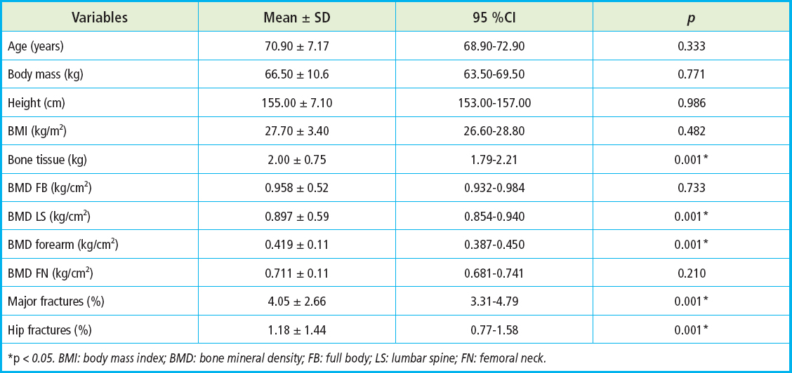
The sample consisted of 52 older women, with a mean age of 70.9 ± 7.2 years. Participants were allocated into groups according to early sports participation. Detailed anthropometric and clinical characteristics of the total sample and each group are presented in table I.
Table I Sample characterization (n = 52)
*p < 0.05. BMI: body mass index; BMD: bone mineral density; FB: full body; LS: lumbar spine; FN: femoral neck.
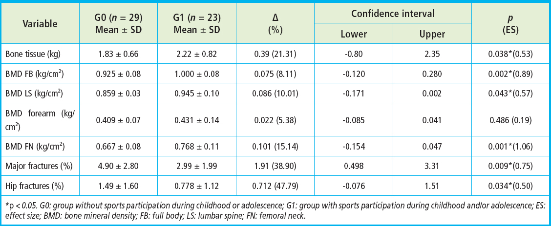
When comparing bone parameters between groups G0 and G1, group G1 showed significantly higher values. Total bone tissue was 21.31 % greater in G1 (p = 0.038), with a medium effect size (ES = 0.53); total body BMD was 8.11 % higher (p = 0.002), with a large effect size (ES = 0.89); lumbar spine BMD showed a 10.10 % increase (p = 0.043), with a medium effect size (ES = 0.57); and femoral neck BMD was 15.14 % higher (p = 0.001), with a large effect size (ES = 1.06). Furthermore, the estimated 10-year fracture risk was lower in group G1: major osteoporotic fractures showed a 38.90 % lower probability compared with G0 (p = 0.009), with a large effect size (ES = 0.75); and hip fractures were 47.79 % less likely (p = 0.034), with a medium effect size (ES = 0.50) (Table II).
Table II Differences between the group without sports participation during childhood or adolescence (G0) and the group with sports participation during childhood and/or adolescence (G1) in bone tissue, bone mineral density (full body, lumbar spine, forearm, and femoral neck), and fracture risk (major osteoporotic fractures and hip fractures)
*p < 0.05. G0: group without sports participation during childhood or adolescence; G1: group with sports participation during childhood and/or adolescence; ES: effect size; BMD: bone mineral density; FB: full body; LS: lumbar spine; FN: femoral neck.
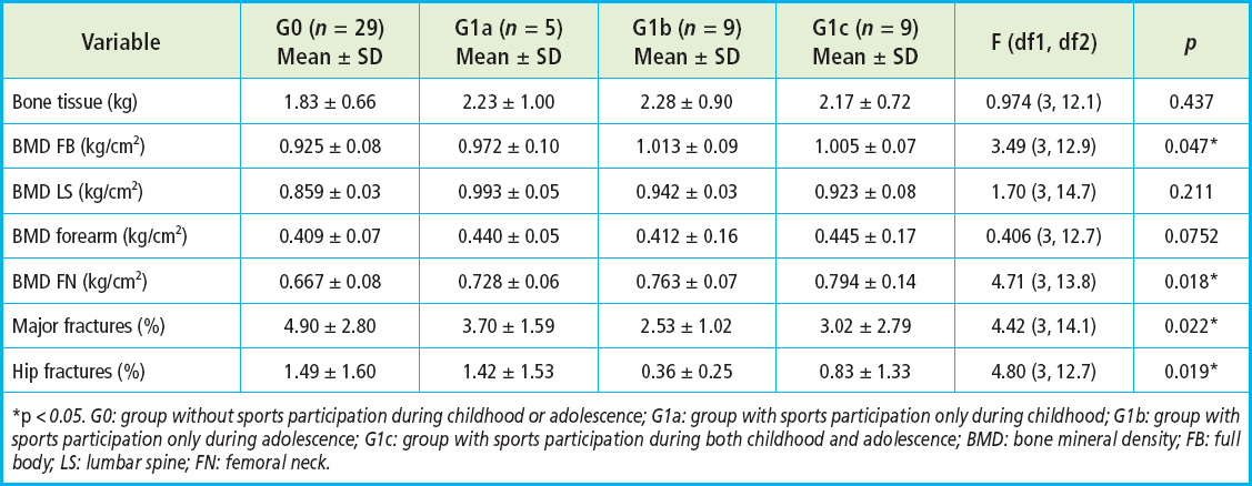
When comparing bone parameters between group G0 and the subgroups that practiced sports during childhood and/or adolescence (G1a, G1b, and G1c), significant differences were observed in total body BMD (p = 0.047), with group G1b showing the highest values (1.013 ± 0.09 kg/cm²), corresponding to values 17.93 % higher than those of G0. Regarding femoral neck BMD (p = 0.018), the best results were found in group G1c (0.794 ± 0.14 kg/cm²), with values 19.04 % higher than those of G0.
Regarding the 10-year fracture risk estimates, the lowest values were observed in group G1b. For major osteoporotic fractures (p = 0.022), this group had a risk of 2.53 ± 1.02 %, representing a 48.73 % lower risk compared with G0. For hip fractures (p = 0.019), the risk in G1b was 0.36 ± 0.25 %, corresponding to a 74.10 % lower risk of hip fracture compared with G0 (Table III).
Table III Differences among the group without sports participation during childhood or adolescence (G0), the group with sports participation only during childhood (G1a), the group with sports participation only during adolescence (G1b), and the group with sports participation during both childhood and adolescence (G1c) in bone tissue, bone mineral density (full body, lumbar spine, forearm, and femoral neck), and fracture risk (major osteoporotic fractures and hip fractures)
*p < 0.05. G0: group without sports participation during childhood or adolescence; G1a: group with sports participation only during childhood; G1b: group with sports participation only during adolescence; G1c: group with sports participation during both childhood and adolescence; BMD: bone mineral density; FB: full body; LS: lumbar spine; FN: femoral neck.
DISCUSSION
The results of this study show that older women with a history of sports participation during childhood and/or adolescence have better bone health parameters and a lower estimated fracture risk compared with those who did not practice sports during these periods (p < 0.05). The group that reported early sports participation (G1) showed significantly higher BMD values at the total body, lumbar spine, and femoral neck compared with the group without this history (G0), in addition to a lower estimated risk of major osteoporotic fractures and hip fractures over the subsequent 10 years. These findings indicate that early exposure to structured physical activity may contribute to long-term skeletal benefits, even in the absence of regular physical activity during older age.
The association between sports participation during youth and better bone health in later life reinforces the role of physical activity during growth as a key determinant of achieving higher peak bone mass (1,2), which may exert a protective effect even in the presence of low levels of physical activity during senescence. Previous studies suggest that combined interventions involving adequate diet and physical activity during childhood and adolescence promote BMD accumulation, although evidence regarding the long-term persistence of these effects remains limited (6). Although the present study focused on childhood and adolescence, it is important to emphasize that regular physical exercise during older age—particularly resistance training—may also positively influence BMD in postmenopausal women. High-intensity training (≥ 70 % 1RM), performed 3 times per week for at least 40 minutes per session, appears to be optimal (17).
These findings reinforce the importance of understanding the impact of different life stages on bone health and suggest that the benefits of early physical activity may persist over time, even in the absence of continuous stimuli throughout adulthood.
The magnitude of the differences observed between the groups analyzed in this study reinforces the clinical relevance of the findings. Femoral neck BMD, a skeletal site highly vulnerable to osteoporotic fractures, was 15.14 % higher in group G1, with a large effect size (ES = 1.06). Furthermore, the 47.79 % difference in FRAX-estimated hip fracture risk suggests a potential protective effect of early physical activity, even among sedentary or insufficiently active older women. Reduction in fracture risk is a particularly relevant finding, considering that falls—a common precursor to fractures—represent the leading cause of death from unintentional injuries among adults aged ≥ 65 years (18). In this context, a recent systematic review demonstrated that physical exercise interventions are associated with reduced falls across several high-quality trials and provide significant benefits for multiple health outcomes (19).
Thus, the findings presented here may have important implications for public health policies aimed at preventing falls and fractures by highlighting the importance of building a robust skeletal foundation during youth.
Subgroup analysis revealed that the benefits varied according to the developmental period during which sports participation occurred. Group G1c, which practiced sports during both childhood and adolescence, showed the highest femoral neck BMD values, whereas group G1b, with sports participation exclusively during adolescence, exhibited the lowest fracture risks, especially for major osteoporotic fractures. These findings suggest that adolescence may represent a critical period for consolidating bone adaptations induced by mechanical stimuli, consistent with the literature indicating puberty as the stage of greatest velocity of bone mass acquisition, influenced by hormonal and biological maturation changes (3). Considering that most Brazilian adolescents currently do not participate in sports activities and fail to meet minimum physical activity recommendations (20), it is necessary to develop and implement strategies that facilitate physical exercise across multiple settings, including schools and leisure-time activities.
Prioritizing physical activity during adolescence should therefore be considered a long-term investment in musculoskeletal health, with the potential to reduce the burden of osteometabolic diseases in the aging population.
Despite its cross-sectional and retrospective design, the present study used a validated self-report instrument to assess sports participation during childhood and adolescence, which has been widely applied in adult populations and has demonstrated high reproducibility in previous studies (11,12). In addition, the exclusion of participants who were currently active or very active helped isolate the potential long-term effects of early sports participation, thereby reducing confounding related to current physical activity levels.
Nevertheless, some limitations should be acknowledged. Owing to the cross-sectional and retrospective design, causal relationships cannot be established, and the findings should be interpreted as associations. The assessment of early sports participation relied on a dichotomous self-report measure (yes/no) encompassing broad developmental periods, which limited exposure precision because information regarding intensity, duration, type of sport, and mechanical loading characteristics was not collected and may have been subject to recall bias despite the high reproducibility of the instrument.
The sample size was relatively small, and subgroup analyses resulted in small numbers of participants, which may have reduced statistical power and contributed to instability in some estimates. In addition, recruitment through social media may have introduced selection bias, with possible overrepresentation of individuals with greater digital literacy and healthier profiles, thereby limiting the generalizability of the findings. Important factors potentially influencing BMD throughout life—such as nutritional intake (eg, calcium and vitamin D), sun exposure, medication use, hormonal status, reproductive history, and menstrual characteristics—were not controlled for and should be addressed in future investigations.
Despite these limitations, the findings support the hypothesis that regular sports participation during growth is associated with more favorable bone health indicators during older age, reinforcing the relevance of early-life physical activity as a potential contributor to long-term skeletal integrity.
CONCLUSIONS
This study aimed to investigate whether participation in organized sports during childhood and adolescence is associated with bone health and fracture risk in older women. The findings indicate that women who reported early engagement in sports activities presented higher BMD values at the total body, lumbar spine, and femoral neck, as well as a lower estimated 10-year risk of major osteoporotic and hip fractures compared with those without such history. These findings suggest a positive association between early exposure to mechanical loading and skeletal parameters later in life.
Although the cross-sectional and retrospective design does not permit causal inference and potential recall bias should be considered, the findings suggest that participation in organized sports during youth may be associated with more favorable bone health outcomes in later life. Promoting structured physical activity during childhood and adolescence may therefore represent an important strategy within broader public health approaches aimed at supporting skeletal health throughout the lifespan. Future longitudinal and controlled studies including larger and more diverse populations are necessary to confirm these associations and to better elucidate the long-term relationship between early sports participation and bone health.

