




 Servicios personalizados
 Articulo en XML
 Referencias del artículo
 Traducción automática
 Enviar articulo por email
 Citado por SciELO
  Citado por Google
  Similares en
SciELO
Similares en
SciELO  Similares en Google
 Permalink
INTRODUCTION
Osteoporosis (OP) is a condition characterized by reduced bone mass and deterioration of bone microarchitecture. This process leads to bone fragility and a consequent high probability of fractures. Dual-energy x-ray absorptiometry (DEXA), which measures bone mineral density (BMD), is considered the gold standard for the diagnosis of osteoporosis and osteopenia. DEXA expresses results in terms of T-score, a statistical value indicating the number of standard deviations below the average value of young White adults. BMD is commonly measured at the lumbar spine, left femur, and left femoral neck. Wrist BMD can also be measured when both femurs are unavailable (eg, because of bilateral prostheses), whereas DEXA total-body scanning is currently limited to the study of sarcopenia through the measurement of muscle mass and body fat mass (1,2).
According to WHO criteria, 4 different degrees of bone mineral density can be defined: a) normal, when the BMD value is within 1 standard deviation of the T-score reference range (T-score ≥ −1); b) osteopenia, when the BMD value is between 1 and 2.5 standard deviations below the young adult mean; c) osteoporosis, when the BMD value is 2.5 standard deviations below the young adult mean; and d) severe osteoporosis (or established osteoporosis), when the BMD value is 2.5 standard deviations below the young adult mean and 1 or more fragility fractures have occurred. Fragility fractures are the consequence of low-energy trauma, defined according to WHO criteria as a fall from standing height (1,3,4). The correct densitometric diagnosis is based on the lowest T-score found in the spine, femur, or femoral neck (5,6). However, it is clear that BMD is only 1 among several fracture risk factors. For this reason, DEXA should be associated with fracture risk assessment tools.
FRAX (Fracture Risk Assessment Tool, developed in 2008 by the Centre for Metabolic Bone Diseases of the University of Sheffield; the Italian version was revised in 2013) (7), DeFRA (software developed by the Italian Society for Osteoporosis, Mineral Metabolism and Bone Diseases in 2012 and recently revised), and FRA-HS (developed by the Italian Society for General Practitioners in 2017) (8,9) are currently used alongside DEXA to estimate the risk of major fractures. These algorithms are designed to predict the 10-year fracture risk based on the most common and severe risk factors for fragility fractures: spine and femoral BMD, smoking, daily alcohol intake (> 3 units), previous fragility fractures, corticosteroid use (daily dose), arthritis or chronic inflammatory connective tissue diseases, age at menopause, low body weight (BMI < 18.5), and family history of major osteoporotic fractures. Clearly, DEXA can evaluate only BMD, but not bone quality; therefore, alterations in bone microarchitecture are probably responsible for the high number of fragility fractures occurring in patients with osteopenia (10,11).
Given that DEXA alone cannot be considered a reliable predictive tool for preventing fragility fractures, the development of additional tests capable of measuring trabecular structure properties (quality), rather than only bone density, is needed.
TBS (Trabecular Bone Score) is a textural index of bone microarchitecture derived from DEXA images. It analyzes gray-level variations in the lumbar spine, providing information regarding trabecular bone microarchitecture (12).
However, TBS remains under discussion because of several limitations. It can only be applied to lumbar spine DEXA images, which are themselves limited by the frequent presence of osteophytes and aortic calcifications (13,14).
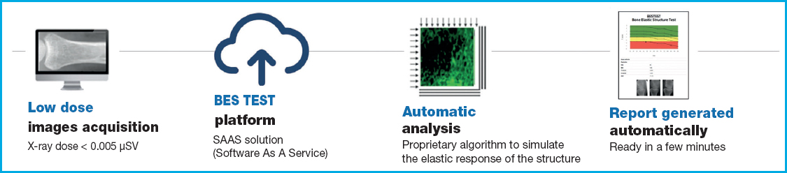
Conversely, the Bone Elastic Structure Test (BES TEST) measures the elastic response of bone to loading. Based on a direct discrete numerical approach (15), the BES TEST analyzes low-dose (< 0.005 mSv) planar radiographic projections of the proximal epiphysis of the first phalanx of the hand to perform a noninvasive biomechanical evaluation of trabecular bone microarchitecture through engineering simulations of load application, thereby quantifying pathological alterations in bone microarchitecture (16). BES TEST is a CE-marked, registered software medical device based on an internationally patented method. The BES TEST software processes radiographic images by transforming them into numerical structural models that are used to simulate compression loading through the Cell Method approach, which is highly effective in terms of robustness, computation time, memory requirements, and accuracy of results (17,18).
The simulation outcomes are combined into an index, the Bone Structure Index (BSI), which reflects the ability of trabecular bone structure to absorb loads (19). The BES TEST outputs are independent of bone mineral density as measured by DEXA (19).
Because of the clear visualization of trabecular structure on standard radiographs, the proximal phalanges of the second, third, and fourth fingers are used as the most suitable regions for evaluation (19). The outcomes obtained with the BES TEST are expressed as BSI T-scores. As with DEXA, these data are derived by comparing the patient’s BSI with the average BSI of young White women aged 22 to 45 years and calculating the difference in number of SDs (20).
Previous studies suggest that the BSI T-score may predict fracture risk over the subsequent 3 years and that a value below −1.4 could represent the cutoff point (19). Moreover, no correlation has been observed between BSI and DEXA T-score (20).
For these reasons, we performed a randomized controlled trial to assess the reliability of the BES TEST in patients with fragility fractures and normal or osteopenic DEXA T-scores.
PARTICIPANTS, STUDY DESIGN, MATERIAL, AND METHODS
We conducted a clinical trial approved by the Ethics Committee of Istituti Clinici Scientifici Maugeri - IRCCS, Pavia (EC2387; February 4, 2020). The study was conducted in full compliance with the principles outlined the Declaration of Helsinki.
Fifty consecutive female outpatients were enrolled and divided into 2 groups. Group A consisted of 25 female patients with no history of recent fragility fractures, whereas group B consisted of 25 female patients with a recent fragility fracture. Sample size calculation was also approved by the same ethics committee on the basis of previous data.
All patients underwent baseline lumbar spine, femoral, and femoral neck DEXA (Hologic QDR 4500 densitometer) together with a baseline BES TEST. Both examinations were repeated after 18 to 24 months, with the addition of spine morphometry.
BES TEST performance characteristics were as follows: intraoperator CV, 0.06; 95 %CI, ± 8 BSI; interoperator CV, 0.11; 95 %CI, ±10.8 BSI (21), in line with current OP diagnostic standards.
Inclusion criteria were female sex, age between 40 and 75 years, and lumbar and femoral DEXA T-scores > −2.5 SD.
Exclusion criteria included treatment with glucocorticoids or antiosteoporotic drugs.
The trial began in 2020 but was interrupted because of the COVID-19 pandemic, resulting in the dropout of 3 patients. At present, 44 patients have completed the study and follow-up.
STATISTICAL ANALYSIS
Statistical comparisons between groups were performed using the unpaired Student t test. Statistical significance was established at p < 0.05.
Currently, we are collecting data and analyzing the final patient cohort. Furthermore, all patients are being contacted to update their clinical status. We are particularly interested in determining whether: a) they experienced a new fragility fracture during the year following the second BES TEST and DEXA evaluation; b) they are currently receiving treatment for osteoporosis; c) new fracture risk factors have been identified; d) they have experienced falls; and e) they have undergone a new DEXA scan, morphometric evaluation, or other relevant blood or urine tests.
RESULTS
Preliminary data showed that, at baseline and after 18 to 24 months, no significant differences were observed in DEXA T-scores between the fractured (F) and nonfractured (NF) groups; p = 0.09. In contrast, the BSI T-score differed significantly between the F and NF groups; p = 0.0001.
No significant differences were observed in femoral neck DEXA T-scores at baseline vs 18- to 24-month follow-up. Similarly, no significant differences were found in femoral DEXA T-scores or spine DEXA T-scores between baseline and follow-up evaluations.
In group B (fractured patients), no significant differences were observed in BSI T-score between baseline and 18- to 24-month follow-up. Likewise, no significant differences were detected in femoral neck DEXA T-scores, femoral DEXA T-scores, or spine DEXA T-scores between baseline and follow-up assessments (Fig. 1).
DISCUSSION
The BES TEST does not measure bone quantity but rather evaluates the elastic structural integrity and biomechanical competence of bone, thereby providing insight into functional bone strength. By focusing on trabecular bone elasticity, bone alterations can be monitored within weeks. BES TEST complements densitometry as a low-dose monitoring tool for bone follow-up (20) in rheumatology (22), oncology (23), nephrology (24), and rare diseases (25).
Evidence from a small study applying the test to both the hand and foot demonstrated similar trends across anatomical sites, indicating sensitivity to systemic physiological changes and supporting the concept that skeletal fragility reflects a generalized condition rather than a site-specific phenomenon (“La valutazione della Bone Elastic Structure (BESTEST™) in segmenti scheletrici sottoposti a diverso carico,” oral presentation at the SIOMMMS National Conference 2022; abstract unpublished).
This concept is further supported by studies showing strong correlations between hand bone measurements and fracture-relevant skeletal sites. Bone mineral density assessed at peripheral hand bones correlates with femoral neck DEXA values, and quantitative ultrasound measurements at the finger phalanges can effectively assess fracture risk and detect age-related bone changes, with diagnostic sensitivity comparable to lumbar densitometry (26-28).
In a previous study (20), 351 consecutive White women were enrolled in a population study and contacted after a 3-year follow-up period to evaluate the incidence of fragility fractures; 166 of 351 responded to follow-up contact.
A total of 91 out of 351 patients experienced fractures, whereas 75 remained fracture-free. Fractured patients were slightly older than nonfractured patients; p = 0.00485. Body mass index (BMI) was similar between groups; p = 0.1243, and no correlation was found between BMI and BSI T-score in either group.
The mean BSI T-score was −1.5 (range, −3.4 to 0.8) in fractured patients and −0.4 (range, −3.2 to 2.4) in nonfractured patients. These findings suggested that the BSI T-score may predict fracture risk during the following 3 years and that patients with values below −1.4 should undergo careful evaluation (20).
In 2020, we published the first pilot study comparing BES TEST with DEXA (22). We enrolled 9 patients younger than 74 years with recent fragility fractures despite normal or osteopenic femoral neck T-scores on DEXA evaluation (group A). Conversely, group B consisted of volunteer female patients who had undergone DEXA and BES TEST evaluations in 2015 during the Trieste NEXT 2015 event (Ethics Committee of the University of Trieste approval No. 66, November 11, 2015).
All patients in both groups were analyzed according to the previously described BES TEST method, and results were expressed as mean ± SD. Comparisons between groups were performed using the unpaired Student t test. Statistical significance was established at p < 0.05.
In that second study (22), we confirmed that no correlation exists between BSI and DEXA T-score. Moreover, fractured patients showed significantly lower BSI T-scores compared with femoral neck DEXA T-scores. In contrast, nonfractured patients had normal BSI T-scores despite osteopenic femoral neck DEXA T-scores (22). Indeed, fracture risk in these patients may be more accurately assessed by combining DEXA with FRAX or DeFRA algorithms (28).
CONCLUSIONS
Current clinical recommendations suggest treatment of nonfractured osteoporotic patients only when FRAX or DeFRA indicate a substantial fracture risk, in order to avoid overtreatment and the consequent increase in adverse effects (29,30).
Because a high proportion of fragility fractures occur in patients with normal or osteopenic DEXA T-scores, this phenomenon is probably related to the limited ability of DEXA to distinguish and quantify bone quality. This limitation is especially evident in patients with secondary osteoporosis, such as those receiving chronic glucocorticoid therapy. Preliminary data from our trial confirm the reliability and reproducibility of both DEXA T-score and BSI T-score measurements. The expected outcome is confirmation that BSI, when combined with DEXA and FRAX or DeFRA scores, may improve the assessment of fragility fracture risk.
BES TEST should be further investigated to determine its ability to detect rapid skeletal changes during pharmacologic treatment, particularly in chronic inflammatory diseases such as rheumatoid arthritis. In these conditions, inflammation combined with glucocorticoid therapy may rapidly induce bone fragility that DEXA alone is unable to identify. Therefore, BES TEST could represent a valuable complementary tool, potentially helping to tailor pharmacologic therapy and, when necessary, rehabilitation programs aimed at reducing fall risk.

