




![Evaluación de la distribución y captación de [18F]FDG mediante microPET/CT en un modelo murino de infarto de miocardio](/img/es/next.gif)


 Servicios personalizados
 Inglés (pdf)
 Articulo en XML
 Referencias del artículo
 Traducción automática
 Enviar articulo por email
 Citado por SciELO
  Citado por Google
  Similares en
SciELO
Similares en
SciELO  Similares en Google
 Permalink
Highlights
This study provides evidence of the hepatoprotective potential of Cynara scolymus extract against thioacetamide-induced liver injury through the modulation of antioxidant defence mechanisms.
The study provides a strong foundation for the development of Cynara Scolymus Extract-based hepatoprotective nutraceuticals in clinical applications.
Introduction
Liver disease is a serious, worldwide problem with high morbidity and mortality. The main causes of liver diseases are excessive alcohol consumption, viruses, toxins, parasitic diseases, hepatitis, hepatotoxins, antibiotics, and chemotherapeutic agents including paracetamol, carbon tetrachloride (CCI4), and thioacetamide (TAA). TAA is a hepatotoxin frequently used to generate experimental liver damage.
The liver has a great capacity to detoxify toxic substances and produce useful end products. Acute liver injury can be caused by excess alcohol consumption or exposure to hepatotoxins and may progress to severe hepatic diseases such as hepatitis, cirrhosis, and cancer. Acute liver injury is linked to oxidative stress that produces harmful intermediates such as free radicals and redox-active reactants. It can then progress to acute liver failure, which results into hepatic encephalopathy and multiple organ failure, with a high mortality rate 1.
Drugs can cause liver disease in several ways. Some drugs are directly injurious to the liver; others are transformed by the liver into chemicals which subsequently may cause injury to this organ. There are three types of liver toxicity: dose-dependent toxicity, idiosyncratic toxicity, and/or drug allergy 2.
The incidence of hepatic adverse drug reactions (ADRs) remains unknown in the general population. Sgro et al (2002) reported that the main drugs implicated in ADRs were anti-infectious, psychotropic, hypolipidemic agents, and nonsteroidal anti-inflammatory drugs (NSAIDs) 3.
Jabbar et al (2023) investigated the protective effects of silymarin (20 and 40 mg/kg daily for 2 months) against TAA fibrosis in rats (200 mg/kg TAA three times weekly for two months) by histopathological and immunohistochemical assays. The results showed that silymarin is a hepatoprotective compound due to its inhibitory effects on fibrosis, hepatotoxicity, liver cell proliferation, up-regulation of HSP 70, and downregulation of α-smooth muscle actin (α-SMA) expression, inhibiting lipid peroxidation by decreasing malondialdehyde (MDA) production, while retaining the liver index (serum bilirubin, total protein, albumin, and liver enzymes) and antioxidant enzymes to normal 4.
The role of silymarin on the inflammatory response induced by carbon tetrachloride (CCl4) as an example of xenobiotics on liver tissues in male rats has been assessed 1. Silymarin reduced CCl4 induced-liver inflammation by overcoming the oxidative stress process and inflammatory cytokines production.
TAA-induced hepatotoxicity has been reviewed 5. TAA induces acute and chronic liver injury due to its effects on the synthesis of proteins, RNA, DNA and γ-glutamyl transpeptidase activity.
TAA administration is an established technique for generating rat models of liver fibrosis and cirrhosis. Oxidative stress is believed to be involved as TAA-induced liver fibrosis is initiated by thioacetamide S-oxide, which is derived from the biotransformation of TAA by the microsomal flavine-adenine dinucleotide (FAD)-containing monooxygense (FMO) and cytochrome P450 systems 6.
Treatment of mice and rats with TAA induced liver cell damage, fibrosis and/or cirrhosis, associated with increased oxidative stress and activation of hepatic stellate cells 7.
The protective effects of silymarin in TAA-induced liver damage have been investigated 8. This study determined gene expression changes in the liver at the level of microRNA (miRNA), and found that treatment with silymarin before exposure to the toxin successfully altered its effects on the animals in the study.
Jimenez-Escrig et al. (2003) reported that artichoke (Cynara scolymus L.), an edible vegetable from the Mediterranean area, is a good source of natural antioxidants such as vitamin C, hydroxycinnamic acids, and flavones 9. Extracts from Cynara scolymus leaves have long been used in folk medicine for their choleretic and hepatoprotective activities, which are often related to the cynarin content 10.
The hepato-curative effects of Cynara Scolymus extract (CSE) on CCl4-induced oxidative stress and liver injury has been investigated in rats 11. CCl4 was administered at a dose of 0.2 ml/kg twice daily. CSE was given orally for 2 weeks at a dose of 1.5 g/kg after CCl4 application on the curative group. Significant decreases in serum alanine-aminotransferase (ALT) and aspartate-aminotransferase (AST) and MDA levels were detected. Significant increase in superoxide dismutase (SOD) and catalase (CAT) activities were obtained. C. scolymus leaf extract application normalized the DNA % fragmentation, p53 and caspase 3 levels in the liver.
CSE has antioxidant and anti-inflammatory effects, and countless medicinal properties 12. Artichokes inhibit the production of reactive oxygen species (ROS) and free radicals due to the phenolic acids and flavonoid compounds they contain, and also suppress the activation pathway of NF-κB. Therefore, CSE reduces oxidative stress and inflammatory factors. On the other hand, CSE has been shown to be effective on the in vitro inhibition of LDL oxidation.
In a previous study, we showed the hepatoprotective role of CSE on a TAA-induced liver injury model 13. The objective of the present study was to delve deeper into the mechanism of action of CSE, focusing on the gene expression of antioxidant markers (CAT, GPX, SOD).
Methods
Cynara scolymus (Artichoke)
The Cynara scolymus plant is a traditional vegetable crop of the Mediterranean basin. Cynara cardunculus L. variety C. scolymus L. (artichoke) is cultivated for its fleshy heads. It belongs to the Asteraceae family 14. The Cynara scolymus leaves were purchased from a medicinal plant farm at the Faculty of Agriculture of the University of Banha, Egypt. The leaves were collected in March 2023. The plant was identified by Dr. Mostafa Hamza Mohamed, Assistant Professor of Vegetable Crops, Horticulture Department, Faculty of agriculture, Benha University, (Voucher # 96647).
Drugs and chemicals
Thioacetamide (TAA)
TAA is used for experimental induction of liver injury in animal studies 15. It was obtained from Alamia company, Benha, Qalioubia Governorate, Egypt. TAA powder was dissolved in saline at a concentration of 40 mg/ml and administered at the standard dose of 100 mg/kg body weight 17.
Silymarin
Silymarin was produced by Medical Union Pharmaceuticals (MUP), Abu Sultan, Ismailia, Egypt, under the commercial name Hepaticum®. It is presented as 50 mg / 5 ml suspension.
Reagents used for the assessment of antioxidant markers
Phosphate buffered saline (PBS) solution, pH 7.4 containing 0.16 mg/ml heparin. Cold buffer (100 ml potassium phosphate, pH 7.0, containing 2 ml EDTA per gram tissue). Other standard laboratory chemicals and solutions were also used: 70 % hydroethanolic alcohol, distilled water, normal saline solution (sodium chloride 0.9 %), among others.
Laboratory animals
Thirty male Wister albino rats weighing 150-170 g were obtained from the Faculty of Veterinary Medicine, Benha University, Egypt. The animals were kept in a standard environment with controlled temperature (25 °C), humidity (45-75 %), and photoperiod (12-hr/12-hr light/dark cycle). All animals had free access to food and water. The animals were housed in stainless steel wire mesh cages with wood chip bedding. They were fed a standard diet (vegetable feed with 19 % protein). The animals were given a 15-day acclimatization period. Research was conducted in accordance with the Committee of Experimental Animal Care and Procedure (Faculty of Veterinary Medicine, Benha University, Benha, Egypt).
Preparation of C. scolymus leaves extract
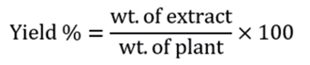
Leaves around the stems ofC. scolymuswere obtained and cut into smaller pieces and then dried at room temperature. The extraction was done by maceration of chopped leaves with 70 % v/v (ethanol/distilled H2O). The mixture was left for 72 hr. in a refrigerator with intermittent shaking. The extract was filtered through muslin mesh, concentrated at 70 °C for 3 days to determine the weight of crude extract, and then stored at 4 °C until needed. The high dose extract was prepared by dissolving 4 g in 200 ml saline solution (0.9 %). The low dose extract was prepared by dissolving 2 g in 200 ml saline solution (0.9 %). Concentrations were 40 mg/ml and 20 mg/ml, respectively. Both extracts were used to evaluate the hepatoprotective effect of artichoke extract in high (200 mg/kg) and low (100 mg/kg) doses in rats 16. This procedure was repeated weekly to obtain fresh extracts. Yield was determined using the formula:
Study design
The experiment was conducted according to the Guide for Care of Laboratory Animals and approved by the Ethical Animal Committee, Faculty of Veterinary Medicine, Benha University (Approval # 96647).
The rats were divided into 5 groups of 6 rats each, as follows:
Group I: Negative control group. The rats received no drugs, only vehicle (saline solution orally and intraperitoneally).
Group II: Positive control group (or diseased group). The rats received TAA at a dose of 100 mg/kg, interperitoneally, twice weekly for 8 weeks to develop liver injury.
Group III: Standard group. This group was made up of rats with liver injury as in group II and they were treated with silymarin (100 mg/kg, orally, daily for 8 weeks).
Group IV: Small artichoke dose. This group was made up of rats with liver injury as in group II and they were treated with CSE (100 mg/kg BW, orally, daily for 8 weeks).
Group V: Large artichoke dose. This group was made up of rats with liver injury as in group II and they were treated with CSE (200 mg/kg BW, orally, daily for 8 weeks).
Sampling
The animals were sacrificed by cutting the carotid arteries with a scalpel and forceps under light ether anesthesia. The livers were examined macroscopically and then removed from the animals. Pieces (100 mg) were placed in Eppendorf tubes and submerged in Trizol for RNA isolation and PCR of hepcidin and interleukin-1 and -2.
Other liver pieces were placed in sterile tubes for homogenate preparation. For this purpose, liver pieces were washed in ice-cooled saline, dried, weighed and kept in buffer saline, and then homogenized with an electric homogenizer. Then the homogenate was centrifuged at 4 ºC for excluding debris. The transparent supernatant was used for the evaluation of antioxidant markers 18.
The remaining parts of the liver lobes were preserved in 10 % formalin for histopathological examination.
Measurements
Spectrophotometric análisis
SOD was determined spectrophotometrically using a kit from Biodiagnostic (29 Tahreer St., Dokki, Giza, Egypt), based on the methodology described by Nishikimi et al (1972) 19. GPx was determined spectrophotometrically with a kit from Biodiagnostic, based on the methodology described by Paglia and Valentine (1967)20. CAT was determined spectrophotometrically using a kit from Biodiagnostic, based on the work by Aebi (1984) 21.
RT-PCR of catalase, GPX, SOD in hepatocytes
Total cellular RNA was extracted from the hepatic specimens with Trizol reagent following the methodology described by Chomczynski & Sacchi (2006) 22. RT was performed with RNA random primers using a RT-PCR kit (Takara Bio INC., Shiga, Japan) following the manufacturer’s instructions.
PCR was performed using the following primers (Rikaken, Nagoya, Japan). Catalase, forward: 5′- GCAGATACCTGTGAACTGTCCCT-3′; catalase, reverse: 5′-GTAGAATGTCCGCACCTGAGTGA-3′; GPX, forward: 5′- CGGTTTCCCGTGCAATCAGT-3′; GPX, reverse: 5’-ACACCGGGGACCAAATGATG -3′ for GPX; SOD, forward: 5′-GGTCCACGTTTCTTGTTCTGC-3′; SOD, reverse: 5′- CAATCACACCACAAGCCAAGC-3′.
The PCR products were separated in 1.5 % agarose gel electrophoresis starting with 70 V for 20 min and continuing with 120 V until the fragments reached the desired separation levels. The DNA bands were visualized with an ultraviolet trans-illuminator (UVP, CA, USA). Quantification of amplified DNAs was carried out by densitometric analysis using Scion Image 4.02 software (Scion Corporation, Maryland, USA).
Histopathological examination
After euthanizing the animals, a small portion of each liver was fixed in 10 % formalin. Each piece was dehydrated in an increasing gradient of ethanol and finally cleared into toluene. The pieces of liver were then embedded in molten paraffin wax. Sections were cut with a microtome at 5 mm thickness and stained with hematoxylin and eosin (H&E) and examined under a light microscope at magnification powers of 200x and 400x.
Data Presentation and Analysis
Data of spectrophotometric analysis are expressed as mean ± standard error of the mean of 6 separate observations. Observations were compared with an ANOVA followed by a posteriori Tukey post-hoc test (P < 0.05 was considered statistically significant). Gene expression was done in triplicates and calculated as fold-change compared to the control. All statistics and graphs were generated using GraphPad Prism® version 8 software (GraphPad Inc., CA, USA).
Results
Thioacetamide-induced liver injury and effects of CSE
Figure 1 shows macroscopic and microscopic images of livers from all study groups. Those from TAA-intoxicated rats show diffuse degenerative lesions, including vacuolation, fibrotic and hyperplastic changes at both macroscopic (a) and microscopic (a/) levels. Adjunct administration of artichoke extracts significantly protected the liver tissue from the toxic effect of thioacetamide in a dose-dependent manner (d & d/; e & e/, respectively) reaching results similar to those of the Silymarin group (c & c/).
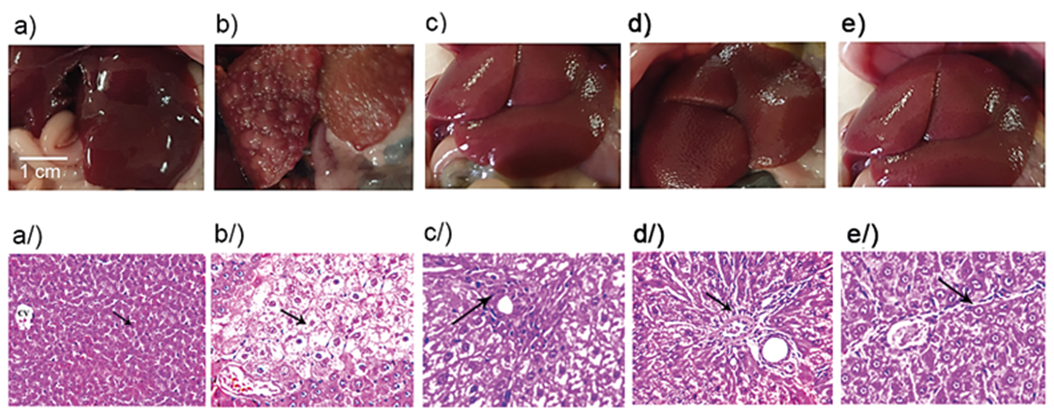
Figure 1 Normal liver tissue (a & a/) and the effects of thioacetamide without (b & b/) or with Silymarin (c & c/), or artichoke extract at small or high dose (d & d/; e & e/). (a) shows normal histology (x 200). (b) shows extensive lesions, namely, hydropic degeneration (arrow) of hepatocytes characterized by swollen, pale, vacuolated cytoplasm (arrow, x400) with mild hyperplasia. (c) shows mild peri-portal fibrous connective tissue proliferation (arrow, x400), with mild leukocyte infiltration. (d & e) show fine strands of fibrous connective tissue (arrow) in the hepatic parenchyma (arrow, x400). CV, central vein. Staining, H&E.
Effect of CSE on liver CAT, GPx and SOD
As shown in table 1, control liver samples showed normal levels of antioxidant markers CAT, SOD and GPx, while those of group 2 revealed a significant decrease in these enzymes compared with control samples. Administration of CSE along with thioacetamide induced significant increases (improvement) in these parameters in comparison with the TAA group. This improvement was dose-dependent and parallel to that in the silymarin group. The potential of protection produced by CSE were 49.26 % after small dose and 87.024 % after large dose (CAT); 49.18 % and 95.44 % (GPx); and 57.93 % and 81.84 % (SOD), as effective as silymarin, respectively.
Table 1: Effects of CSE on liver CAT, GPX and SOD
| Group | CAT (IU/g wet wt) | GPx (IU/g wet wt) | SOD (IU/g wet wt) |
|---|---|---|---|
| Control (saline) | 5.24 ± 0.32a | 72.46 ± 1.4a | 126 ± 2.36a |
| TAA 100 mg/kg | 1.97 ± 0.11d | 41.34 ± 1.28c | 56.39 ± 4.67d |
| TAA 100 mg/kg + Sil 100 mg/kg | 4.19 ± 0.19b | 70.35 ± 1.56a | 107.9 ± 1.95b |
| ISD (TAA 100 mg/kg + CSE 100 mg/kg) | 3.07 ± 0.07c | 55.61 ± 1.17b | 86.23 ± 1.08c |
| ILD (TAA 100 mg/kg + CSE 200 mg/kg) | 3.91 ± 0.09b | 69.03 ± 1.07a | 98.55 ± 2.39b |
Data are expressed as mean ± SEM of 6 observations/group. The values in the same column with different letters are significantly different from each other (P < 0.05). SOD, superoxidase dismutase; GPx, glutathione peroxidase; TAA, thioacetamide; CSE, artichoke extract; ISD, Intoxicated rats treated with small dose of CSE; ILD, Intoxicated rats treated with large dose of CSE.
The group treated with TAA revealed significant decreases in CAT, GPx and SOD gene expression by 0.42, 0.38 and 0.45 times, compared with those of control samples (table 2 and figure 3). Administration of CSE along with TAA normalized the downregulated gene expressions by 1.85 and 2.43 (CAT), 1.76 and 2.24 (GPx), and 1.92 and 2.58 (SOD) timesd, after small and large doses, respectively.
Table 2 RT-PCR of antioxidant markers
| Group | CAT (fold change) | GPx (fold change) | SOD (fold change) |
|---|---|---|---|
| Control (saline) | 1 ± 0.07 | 1 ± 0.08 | 1 ± 0.09 |
| TAA 100 mg/kg | 0.42 ± 0.01 | 0.38 ± 0.01 | 0.45 ± 0.01 |
| TAA + Sil 100 mg/kg | 0.85 ± 0.07 | 0.76 ± 0.06 | 0.9 ± 0.05 |
| ISD (TAA + CSE 100 mg/kg) | 1.85± 0.16 | 1.76 ± 0.15 | 1.92 ± 0.11 |
| ILD (TAA + CSE 200 mg/kg) | 2.43 ± 0.2 | 2.24 ± 0.1 | 2.58 ± 0.22 |
Data are expressed as fold change of 3 observations/group. SOD, superoxidase dismutase; GPx, glutathione peroxidase; TAA, thioacetamide; CSE, artichoke extract; ISD, Intoxicated rats treated with small dose of CSE; ILD, Intoxicated rats treated with large dose of CSE.
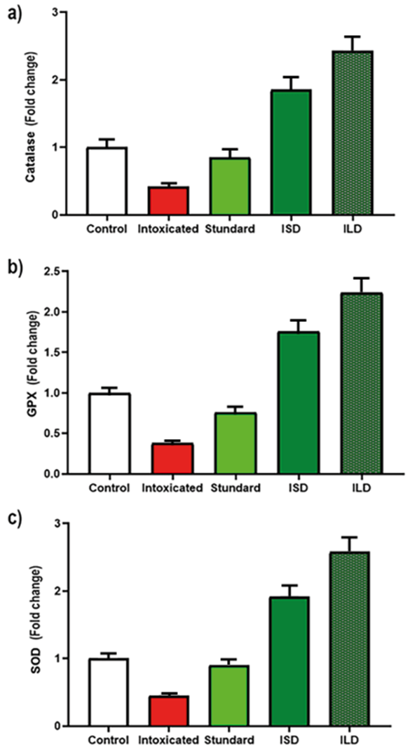
Figure 2 RT-PCR results of antioxidant markers of CSE in rats. Data are expressed as fold change of 3 observations/group. SOD, superoxidase dismutase; GPx, glutathione peroxidase; Intoxicated (TAA, thioacetamide); ISD, Intoxicated rats treated with small dose of CSE; ILD, Intoxicated rats treated with large dose of CSE.
Discussion
Liver disease and associated health problems are a global issue that requires the combined efforts of all professionals and specialists to control it. TAA has been widely used in the development of suitable animal models of acute and chronic liver injury using various doses, times and routes of administration, particularly in drinking water, due to its similarity to human liver fibrosis and cirrhosis.
The present study demonstrates the significant modulatory effects of CSE on key antioxidant enzymes in TAA-induced hepatic injury, as evidenced by oxidative stress markers and RT-PCR gene expression analysis. These results were corroborated by the histological study of the liver samples.
In the present study, TAA significantly decreased CAT, SOD and GPx activities and CSE administration restored these values in a dose-dependent manner. The effects of artichoke extract could be attributed to its antioxidant effect; however, it is necessary to investigate the active ingredients of the extract that are responsible for the effect. The antioxidant effect found in the present study is consistent with that reported by El-boshy et al., (2017), who described the protective effect of artichoke leaf extract (300 mg/kg body weight) against cadmium-induced oxidative stress (100 mg/L), hepatorenal damage and immunosuppressive and hematological disorders in rats. Artichoke leaf extract significantly improved the immune response, the antioxidant system and hepatorenal function with a significant decrease in MDA and an increase in GSH, GPx, SOD and CAT 23. Our data are also consistent with those of Colak et al., (2016), who reported that CSE(orally for 2 weeks at a dose of 1.5 g/kg) protected against CCl4 (0.2 mL/kg twice daily)-induced oxidative stress and hepatic injury in rats. Significant increases of SOD and CAT activities in the were found in the group treated with CSE. These results indicate that C. scolymus leaf extract has hepatocurative effects on CCl4-induced oxidative stress and hepatic injury by reducing lipid peroxidation 11.
However, our data disagree with those of Speroni et al. (2003), who reported the ineffectiveness of different preparations of C. scolymus (1 and 2 g/kg) in liver damage induced by CCl4 in rats. Pretreatment with either of those extracts or chlorogenic acid did not reduce MDA production 10.
Our findings also point to the modulation of the gene expression of the hepatic antioxidant enzymes following treatment with CSE in TAA-induced liver injury. In our work, TAA administration markedly suppressed the expression of key antioxidant enzymes (CAT reduced to 0.42 times, GPx to 0.38 times, and SOD to 0.45 times, compared to control values). This substantial downregulation of the antioxidant defense indicates severe oxidative stress and compromised cellular protective mechanisms in the liver tissue. The therapeutic potential of CSE was evidenced by its superior ability to restore and enhance antioxidant enzyme expression compared to the standard treatment, silymarin. Our results agree with those of Seoudi & Saleh (2018) who demonstrated that CSE significantly upregulated antioxidant enzymes in CCl4-induced liver damage, showing approximately 2-fold increases in SOD and CAT activities 24. Moreover, our study is consistent with that of El-Boushy et al (2017), who reported that artichoke extract treatment resulted in a marked elevation of hepatic antioxidant enzymes, with SOD increasing by 2.1 times and GPx by 1.9 times, in an experimental model of liver injury 23.
However, our data may be inconsistent with those of Jabbar et al. (2023), who found a lower improvement in antioxidant enzymes with CSE treatment in TAA-induced liver injury. At 200 mg/kg, CSE only increased catalase 1.3 times, GPx 1.2 times and SOD 1.4 times, substantially less than our findings4. Finally, our results are different from those of Banaee et al. (2023), who reported that silymarin has superior antioxidant enzyme modulation compared to CSE25.
Conclusion
This study provides evidence of the hepatoprotective potential of CSE against TAA-induced liver injury through the modulation of antioxidant defense mechanisms. Our findings demonstrate that CSE effectively counteracts TAA-induced oxidative stress, as evidenced by the significant restoration of key antioxidant enzymes. These findings establish CSE as a promising therapeutic agent for liver injury, mainly through its ability to improve cellular antioxidant defense mechanisms. More in-depth research into CSE active ingredients and molecular mechanisms is recommended for its potential clinical applications.

