




 Servicios personalizados
 Inglés (pdf)
 Articulo en XML
 Referencias del artículo
 Traducción automática
 Enviar articulo por email
 Citado por SciELO
  Citado por Google
  Similares en
SciELO
Similares en
SciELO  Similares en Google
 Permalink
Introduction
Sinonasal papilloma is a rare benign epithelial neoplasm arising from Schneiderian epithelium, first described by Ward in 1854 and Bilroth in 1855 [1, 2]. Inverted papilloma (IP), the most prevalent histological subtype, is associated with a high risk of local recurrence and potential malignant transformation [3, 4]. Surgical resection remains the cornerstone of treatment, with two main approaches: external (e.g., Caldwell-Luc, lateral rhinotomy, medial maxillectomy) and endoscopic [5]. Until the early 2000s, external techniques were commonly used, particularly for tumors with lateral maxillary or frontal sinus involvement. In 1992, Waitz and Wigand introduced endoscopic resection for IP, which has since become the preferred technique in most centers due to lower morbidity and improved visualization [6].
The choice of surgical approach is largely dictated by tumor extension, anatomical complexity, and available technology. Endoscopic endonasal surgery (EES) is typically recommended for tumors confined to the nasal cavity, ethmoid, and central maxillary sinus, while external approaches are generally reserved for lesions involving the lateral maxillary wall, frontal sinus, or those with malignancy suspicion [7, 8]. Notably, medial maxillectomy, historically performed via external routes, is now often conducted endoscopically, achieving similar oncologic outcomes with reduced morbidity [9].
Despite the evolution of surgical techniques, recurrence remains a significant clinical concern. Reported recurrence rates vary widely, ranging from 0 % to 50 %, and are influenced by tumor location, surgical completeness, and follow-up duration [10, 11]. A meta-analysis by Busquets and Hwang reported an average recurrence rate of 15 %, with lower rates in endoscopic (12 %) versus external (20 %) approaches [12]. Tumors originating in the nasal cavity show significantly lower recurrence risk [13]. Malignant transformation has also been documented, with metachronous carcinoma rates between 2.3 % and 11 %, and median survival of 126 months [14].
Several recent systematic reviews and meta-analyses support EES as the standard of care for most IP cases, reporting lower complication rates and equivalent or better control of recurrence [15–17]. Nevertheless, outcomes may be influenced by factors beyond the surgical technique itself, including institutional experience, technological availability, and the surgical era. For example, Bugter et al. reported a recurrence rate of 30 % despite endoscopic technique, while Sbrana et al. found recurrence exceeding 34 %, illustrating variability in clinical practice [18].
In this context, we present a retrospective analysis of patients treated over a 23-year period at a tertiary ENT (Ear-Nose-Throat) department, encompassing both external and endoscopic techniques. This study offers real-world insight into recurrence, complications, and disease-free survival across a technological transition in the surgical management of IP.
Material and Methods
Study design and setting. A retrospective study was conducted on patients diagnosed with sinonasal inverted papilloma (IP) at the ENT Department between January 1, 1995, and December 31, 2018. This tertiary-care center functions as a regional referral institution. Eligible patients were adults (≥18 years) who underwent surgical treatment via either an external or endoscopic approach. Exclusion criteria included high surgical risk, pregnancy, refusal of surgery, concomitant sinonasal malignancies, or follow-up shorter than five years.
Diagnosis and surgical approach. Diagnosis was based on clinical evaluation, including anterior rhinoscopy and flexible nasal endoscopy, supported by imaging studies (computed tomography [CT] and magnetic resonance imaging [MRI]) and histopathological confirmation by biopsy. The choice of surgical approach (external vs. endoscopic) was largely dictated by institutional capabilities and the available surgical technology at the time. Until 2008, all surgical procedures were performed using external approaches due to the lack of endoscopic instrumentation and training. From 2009 onward, endoscopic sinonasal surgery was progressively adopted as the institutional standard, following the acquisition of equipment and surgeon expertise. Therefore, the choice of surgical technique during the earlier years of the study was determined primarily by the historical context, not tumor staging.
Variables and data sources. Data were extracted from medical records and grouped into three categories:
Baseline variables: age, sex, smoking status, alcohol use, occupational exposure, inhaled drug use, allergy history, use of anticoagulant or antiplatelet therapy, and comorbid sinonasal disease (e.g., chronic rhinosinusitis with or without nasal polyposis, septal deviation, inferior turbinate hypertrophy, rhinitis). Infectious history included human immunodeficiency virus (HIV), hepatitis C virus (HCV), Epstein–Barr virus (EBV), and other sexually transmitted infections. Presenting symptoms included nasal obstruction, rhinorrhea, epistaxis, anosmia, and headache.
Perioperative variables: date of diagnosis and surgery, laterality (right, left, bilateral), radiological staging according to the Krouse classification (T1–T4), with T1–T2 categorized as localized disease and T3–T4 as extensive disease; and type of surgery (Caldwell–Luc procedure, lateral rhinotomy with medial maxillectomy, Lynch frontal-ethmoidectomy, or endoscopic sinonasal resection).
-
Postoperative variables: acute complications (epistaxis, orbital complications, cerebrospinal fluid leak), chronic complications (facial hypoesthesia, synechiae, septal perforation, lacrimal dysfunction, mucocele, atrophic rhinitis), aesthetic sequelae (visible scars), recurrence (yes, no), number of recurrences, number of reinterventions, malignant transformation, and viral markers (human papillomavirus via p16 immunohistochemistry, and EBV via EBER in situ hybridization). EBER (Epstein–Barr virus-encoded RNA) in situ hybridization is a technique used to detect latent EBV infection in tissue samples.
Statistical analysis. Statistical analysis was performed using STATA version 18. Continuous variables were summarized as means and standard deviations or medians and interquartile ranges, depending on distribution. Categorical variables were expressed as frequencies and percentages. Comparisons between groups were conducted using Student’s t-test or the Mann–Whitney U test for continuous variables, and the Chi-squared test for categorical variables. Disease-free survival was analyzed using Kaplan–Meier curves and compared with log-rank tests. A p-value < 0.05 was considered statistically significant.
Study limitations. This study has several limitations. Its retrospective design introduces risks of selection and information bias, especially in earlier cases with handwritten documentation. The modest sample size reflects the rarity of IP but limits statistical power. Surgical decisions were not randomized and were strongly influenced by technological availability across treatment eras. The absence of standardized intraoperative margin control (e.g., frozen section analysis) may have affected recurrence outcomes. Finally, the limited number of events precluded multivariate analysis, restricting adjustment for potential confounding factors such as tumor extent or surgical era.
Results
Baseline or preoperative variables
Sex, age, and type of surgery
A sample of 50 patients with sinonasal inverted papilloma was divided into two groups based on the surgical approach (Table 1). The first group included 22 patients treated with an external approach, including Caldwell-Luc, lateral rhinotomy for maxillectomy and/or ethmoidectomy, and Lynch frontal-ethmoidectomy. This group had a mean age of 60.73 years (SD ± 11.87), with 6 women and 16 men. The second group comprised 28 patients treated endoscopically, with a mean age of 56.36 years (SD ± 14.59), including 12 women and 16 men. While sex distribution was relatively balanced, there was a trend toward younger age in the endoscopic group.
Risk Factors
Risk factors showed no statistically significant differences between groups in terms of tobacco use (p = 0.283), alcohol consumption (p = 0.137), or occupational exposure to inhaled chemicals (p = 0.477). Smoking was reported by 45.5 % of the external group and 63.6 % of the endoscopic group. Alcohol use was less frequent overall, with 31.8 % in the external group and 14.3 % in the endoscopic group. Occupational exposure was noted in 13.6 % of external and 21.4 % of endoscopic patients.
Symptoms and medical conditions
Nasal obstruction was the most frequent symptom (90.0 %), slightly more prevalent in the endoscopic group (96.4 %) than in the external group (81.8 %) (p = 0.087). Other symptoms, including rhinorrhea (26.0 %), epistaxis (18.0 %), headache (10.0 %), and anosmia (4.0 %), showed no statistically significant differences. Allergies were significantly more frequent in the endoscopic group (39.3 %) than in the external group (13.6 %) (p = 0.045). Anticoagulant or antiplatelet therapy was used by 24.0 % of patients (p = 0.852). Most patients (66.0 %) had no associated sinonasal condition, and no significant differences were observed in the prevalence of chronic rhinosinusitis with polyps (20.0 %), without polyps (8.0 %), or rhinitis (6.0 %) (p = 0.616). Infectious diseases, such as hepatitis C (8.0 %) and HIV (2.0 %), were rare and equally distributed (p = 0.210). There were no reports of inhaled substance use.
Perioperative variables
Affected side and radiological classification
Most tumors in the external group were located on the left side (63.6 %), while the endoscopic group showed equal distribution between right and left sides (50.0 % each) (p = 0.335). The Krouse classification by surgical approach is presented in Table 2. T1 tumors were exclusively treated endoscopically, while T2 and T3 tumors were managed with both techniques. No T4 tumors were identified.
Table 2. Krouse classification distribution by surgical approach
|
Krouse Stage |
External (n = 22) |
Endoscopic (n = 28) |
Total (n = 50) |
|---|---|---|---|
|
T1 |
0 (0.0 %) |
14 (50.0 %) |
14 (28.0 %) |
|
T2 |
13 (59.1 %) |
9 (32.1 %) |
22 (44.0 %) |
|
T3 |
9 (40.9 %) |
5 (17.9 %) |
14 (28.0 %) |
|
T4 |
0 (0.0 %) |
0 (0.0 %) |
0 (0.0 %) |
|
Total |
22 (100 %) |
28 (100 %) |
50 (100 %) |
It is important to clarify that until 2008, all cases were treated externally due to the lack of endoscopic equipment and training. The implementation of endoscopic surgery after that year coincided with improved imaging and diagnostic techniques, which may have led to increased detection of localized tumors. Consequently, T1 tumors are only found in the endoscopic group. This historical shift in institutional practice should be taken into account when comparing surgical outcomes. For stages T2 and T3, no statistically significant differences were observed between groups, suggesting that surgeon experience and technological availability also played a role in approach selection.
Postoperative variables
Acute and chronic complications
Acute complications were rare and did not significantly differ between groups (p = 0.803). The majority of patients (88.0 %) had no acute complications. Epistaxis occurred in 4.0 % of cases, orbital complications in 6.0 % (7.1 % in the endoscopic group, 4.5 % in the external group), and one cerebrospinal fluid leak (2.0 %) was reported in the endoscopic group.
Chronic complications were observed in 44.0 % of patients, with no significant differences overall (p = 0.854). Hypoesthesia was significantly more frequent in the external group (22.7 %) compared to none in the endoscopic group (p = 0.008). Synechiae were more frequent in the endoscopic group (32.1 %) than in the external group (9.1 %), though this did not reach statistical significance (p = 0.051). Other complications such as paresthesia, septal perforation, lacrimal dysfunction, mucocele, and atrophic rhinitis showed no significant differences. Cosmetic outcomes differed markedly: no visible scars were present in the endoscopic group, while 63.6 % of patients in the external group had paralateronasal or nasolabial scars (p < 0.001).
Recurrence and reinterventions
Fifteen patients (30.0 %) had at least one recurrence: 5 in the external group (22.7 %) and 10 in the endoscopic group (35.7 %) (p = 0.279). Most patients (70.0 %) remained disease-free, with no significant difference between groups (p = 0.320). A breakdown of recurrence episodes is presented in Table 3.
Table 3. Recurrences and reinterventions by surgical approach
|
Variable |
External (n = 22) |
Endoscopic (n = 28) |
Total (n = 50) |
p-value |
|---|---|---|---|---|
|
Recurrences |
||||
|
Yes |
5 (22.7 %) |
10 (35.7 %) |
15 (30.0 %) |
0.279 |
|
Number of Recurrences |
||||
|
0 |
17 (77.3 %) |
18 (64.3 %) |
35 (70.0 %) |
0.320 |
|
1 |
5 (18.2 %) |
10 (35.7 %) |
15 (30.0 %) |
|
|
2 |
1 (4.5 %) |
4 (14.3 %) |
5 (10.0 %) |
|
|
3 |
0 (0.0 %) |
2 (7.1 %) |
2 (4.0 %) |
|
|
Number of Reinterventions |
||||
|
1 |
5 (22.7 %) |
10 (35.0 %) |
15 (30.0 %) |
0.029 |
|
2 |
1 (4.5 %) |
4 (14.3 %) |
5 (6.0 %) |
|
|
3 |
0 (0.0 %) |
2 (7.1 %) |
2 (4.0 %) |
The recurrence distribution according to Krouse stage was as follows: 2 of 14 T1 cases (14.3 %) recurred (all treated endoscopically), 7 of 22 T2 cases (31.8 %) recurred (4 external, 3 endoscopic), and 6 of 14 T3 cases (42.9 %) recurred (3 external, 3 endoscopic). No T4 tumors were observed. These data suggest a trend toward increasing recurrence with greater tumor extension.
External group: 4 patients had one recurrence, 1 had two.
Endoscopic group: 4 had one recurrence, 4 had two, 2 had three.
All 15 patients with recurrence underwent at least one reintervention. The number of reinterventions was significantly higher in the endoscopic group (p = 0.029). Specifically, one reintervention was needed in 30.0 % of patients, two reinterventions in 6.0 %, and three in 4.0 %.
Patients with recurrence were significantly younger (mean age 51.43 ± 15.43 years) compared to those without recurrence (60.94 ± 11.87 years, p = 0.024). No significant differences were found for EBER status (p = 0.550), p16 positivity (p = 0.439), HIV or hepatitis C infection (p = 0.550), or Krouse stage (although T3 tumors were more frequent in the recurrence group, 46.7 % vs. 22.2 %, p = 0.191). Tobacco and alcohol use were also comparable between groups.
Neoplastic transformation
Neoplastic transformation occurred in 4 patients (8.0 %), with a higher proportion in the external group (13.6 %) versus the endoscopic group (3.6 %) (p = 0.193). All cases were squamous cell carcinoma, with no synchronous tumors reported. All malignancies were detected after the first reintervention. Two patients received curative radiochemotherapy, one died before treatment, and one received palliative care. Only one of the four transformed cases was p16-positive, despite a global p16 positivity rate of 52.0 %. Detailed data are shown in Table 4.
Table 4. Neoplastic transformation and immunohistochemical markers
|
Variable |
External |
Endoscopic |
Total |
p-value |
|---|---|---|---|---|
|
Neoplastic Transformation |
||||
|
Yes |
3 (13.6 %) |
1 (3.6 %) |
4 (8.0 %) |
0.193 |
|
p16 * |
0.628 |
|||
|
Negative |
8 (36.4 %) |
14 (50.0 %) |
22 (44.0 %) |
|
|
Positive |
13 (59.1 %) |
13 (46.4 %) |
26 (52.0 %) |
|
|
Not Performed |
1 (4.5 %) |
1 (3.6 %) |
2 (4.0 %) |
|
|
EBER ** |
0.861 |
|||
|
Negative |
21 (95.5 %) |
27 (96.4 %) |
48 (96.0 %) |
|
|
Not Performed |
1 (4.5 %) |
1 (3.6 %) |
2 (4.0 %) |
|
|
n |
22 (44.0 %) |
28 (56.0 %) |
50 (100.0 %) |
*p16 tumor suppressor protein.
**EBER (Epstein-Barr Virus-Encoded RNA)
Survival analysis
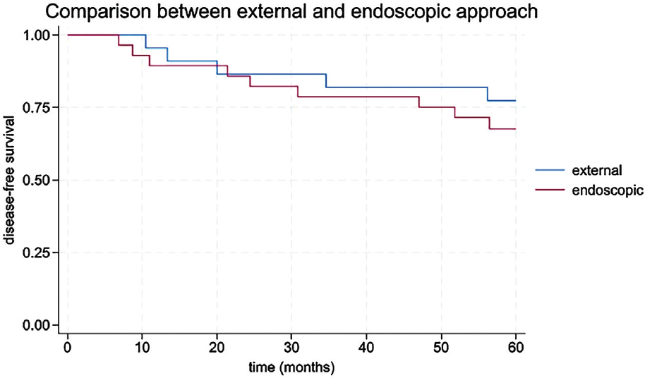
Over 60 months of follow-up, there were 5 recurrences in the external group and 10 in the endoscopic group. Disease-free survival was not significantly different between groups. Mean disease-free survival was 52.4 months (±16.41) for the external group and 48.9 months (±18.11) for the endoscopic group (p = 0.60). Survival probabilities at 60 months were 0.7727 (95 % CI: 0.5374–0.8985) for the external approach and 0.6786 (95 % CI: 0.4732–0.8180) for the endoscopic approach (Table 5). The Kaplan–Meier curve (Figure 1) showed no statistically significant difference (log-rank p = 0.325).
Incidence rate of recurrence
The incidence rate of recurrence was higher in the endoscopic group, at 0.011 recurrences per person-month, versus 0.005 in the external group. This corresponds to 1.17 recurrences per 100 patients per month in the endoscopic group versus 0.52 in the external group. Although the incidence rate ratio was 2.2, the difference was not statistically significant (p = 0.089) (Table 6).
Discussion
This retrospective analysis over a 23-year period explored outcomes of sinonasal inverted papilloma surgery, comparing external and endoscopic approaches in terms of recurrence, complications, neoplastic transformation, and disease-free survival. Our findings showed a 30.0 % overall recurrence rate, with a higher frequency in the endoscopic group (35.7 %) compared to the external group (22.7 %), although this difference was not statistically significant.
These results contrast with large series such as Chi et al., who reported a recurrence rate of 12.2 % in 574 patients undergoing endoscopic resection, emphasizing the importance of removing the tumor at its point of attachment [19]. Similarly, Ledderose et al. found a 13.9 % recurrence rate using exclusively endoscopic techniques with systematic intraoperative assessment of resection margins [20].
Conversely, other studies report rates closer to our findings. Georgalas et al. described a 33.0 % recurrence rate in a mixed cohort, especially in advanced tumors or when negative margins were not confirmed [21]. Karligkiotis et al. reported 31.4 % recurrence despite endoscopic surgery, attributing this to poor visualization or complex maxillary anatomy [22].
Our study did not routinely include intraoperative frozen section analysis, which may partially explain the relatively high recurrence rate. Multiple authors emphasize the role of margin control, such as Turri-Zanoni et al. [23], who advocated for intraoperative navigation and radical resection at the tumor origin, and Sowerby et al., who found significantly reduced recurrence rates with the use of frozen section [24].
In our cohort, recurrence appeared to correlate with tumor extension rather than surgical technique. Rates were 14.3 % for T1, 31.8 % for T2, and 42.9 % for T3 tumors. Although not statistically significant, this trend suggests that Krouse stage may be more predictive of recurrence than the approach used. Notably, endoscopic techniques were used for most reoperations, even in cases initially treated externally, reflecting institutional adaptation and the minimally invasive advantages of endoscopy.
Malignant transformation was observed in four patients (8.0 %), all squamous cell carcinomas. This aligns with published rates of 2–11 %. Notably, p16 and EBER status were not associated with transformation, suggesting limited predictive value in small cohorts.
At five years, disease-free survival was 77.3 % for the external group and 67.9 % for the endoscopic group, without significant differences. These outcomes support previous evidence showing that both techniques can offer satisfactory long-term control when appropriately indicated [25].
Overall, our findings highlight the multifactorial nature of recurrence in inverted papilloma. While endoscopic surgery offers clear advantages in terms of morbidity, visualization, and aesthetic outcomes, recurrence risk remains substantial in extensive tumors and in the absence of margin control. Long-term follow-up and careful surgical planning remain essential. Although the endoscopic approach is widely preferred due to its minimally invasive nature, our findings suggest that recurrence is more closely linked to tumor extension rather than surgical technique alone. Notably, all T1 tumors were treated endoscopically and showed the lowest recurrence rate (14.3 %), while T3 tumors, treated via both approaches, had the highest (42.9 %). Furthermore, the choice of surgical approach in our cohort was heavily influenced by historical context: until 2008, all cases were treated externally due to technological limitations. This evolution in institutional practice reflects a common learning curve in surgical management and should be considered when interpreting outcome comparisons. These observations reinforce the importance of tumor staging in guiding surgical strategy and underscore the need for tailored long-term follow-up in advanced-stage cases.
Conclusions
The analysis found no significant differences in disease-free survival, recurrence rates, or neoplastic transformation between external and endoscopic approaches for sinonasal Inverted Papilloma. While the external approach showed slightly better 5-year survival and lower recurrence, the endoscopic approach had more multiple recurrences. Neoplastic transformation was rare in both groups. These results indicate both methods are effective, with comparable long-term outcomes, underscoring the importance of individualized treatment planning.

