INTRODUCTION
Breast lymphoma (BL) is a very rare entity and accounts for less than 0.6% of breast malignancies [1, 2, 3]. It can be classified as primary breast lymphoma (PBL) in which breast is the principal site of involvement with absence of previously identified extra mammary lymphoma or secondary breast lymphoma (SBL) when there is breast involvement in a previously diagnosed case of lymphoma [4, 5]. Considering the immunophenotype, B cell lymphomas of breast are more common than T cell lymphoma [2]. The prospective diagnosis of primary and secondary breast lymphomas may be difficult as they share similar clinical and imaging features with breast carcinomas or benign entities [6, 7, 8, 9]. As previously acknowledged, fine needle aspiration cytology (FNAC) is a part of diagnostic triad for breast lesions. However, potential pitfalls cannot be overlooked and must be correlated with biopsy in suspected cases. Herein we report a case of anaplastic large cell lymphoma (ALCL) with primary involvement of breast which was misdiagnosed as suppurative mastitis on cytology.
CASE REPORT
A 20-year-old woman presented with a history of left breast lump for two weeks. It was very painful in nature and was not associated with systemic features like fever, night sweat or fatigue. On laboratory investigations, complete blood count was within normal limits. ESR was 64mm/hour. Serological test was non-reactive. Ultrasonography of breast and FNAC done at another center was suggestive of abscess.
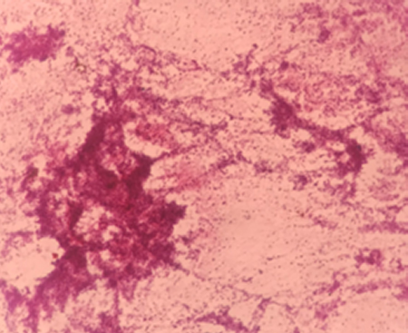
The patient had taken a course of antibiotics but was not effective. Repeat aspiration was done at our hospital, which yielded 0.5 ml of blood mixed yellowish material. The cytological smears showed acute inflammatory cells with its debris and necrotic material (Figure 1). Ziehl Nelseen stain was negative for acid-fast bacilli. Cellblock was also prepared which showed similar findings and was reported as suppurative mastitis. Excisional biopsy showed a lesion measuring 6 x 6 cm in size (Figure 2). The cut surface showed a cavity-like area containing irregular friable material and had a thickened wall. Microscopic examination demonstrated large areas of necrosis and suppuration. Also seen were sheets of monotonous atypical lymphoid cells infiltrating the breast parenchyma (Figure 3a). These cells had high N:C ratio, vesicular chromatin and brisk mitotic activity. Immunohistochemistry (Figure 3b, 3c, 3d) showed positivity for CD3, CD7, CD30, CD45RO, ALK1, BCL6, MUM1, BCL2, perforin and negative stains for p63, CK, CD4, CD5, CD8, CD10, CD20, CD138, PAX5 and cyclin D1.

Figure 3. A: H&E showing sheets of atypical lymphoid cells (400X); B: Immunopositivity for ALK1(100X); C: Immunopositivity for CD 30(100X); D: Immunonegativity for CK (100X).
Other relevant investigations like endoscopy, CT scan of chest, abdomen and pelvis were done and did not reveal any abnormalities. There was no evidence of peripheral lymphadenopathy. Therefore, this case was diagnosed as ALK positive anaplastic large cell lymphoma with primary involvement of the breast. According to the Ann Arbor staging system, the patient was staged as IE. The patient received two cycles of chemotherapy with CHOP (cyclophosphamide, doxorubicin, vincristine and prednisolone). Unfortunately, the patient lost follow-up after two months.
DISCUSSION
BL is an uncommon form of extra nodal lymphoma. This could be attributed to paucity of lymphoid cells in the breast [1]. BL are classified as primary or secondary. The reported incidence of PBL is 1% of all non-hodgkin lymphoma and 2.2% of all extra nodal lymphomas [5, 6, 9, 10]. The most accepted hypothesis for PBL is origin from intramammary lymph nodes [2]. The behavior of PBL is aggressive and has poor outcome as compared to extra nodal lymphomas of alternate sites [4]. The clinical criteria for classification of PBL was first described by Wiseman and Liao in 1972 as a) adequate pathological evaluation; b) mammary tissue in close association with lymphomatous infiltration; c) no evidence of disseminated lymphoma other than simultaneous ipsilateral lymph node involvement and d) no prior history of lymphoma [2, 7, 11, 12, 13]. All lymphomas of breast not included in these criteria are considered SBL.
BL is predominantly reported in women comprising of 95-100% of cases and is a very rare finding in men [3]. There is bimodal age distribution in BL with one group being young women with bilateral involvement and the other group being older women with unilateral involvement usually [4]. There is tendency of right breast involvement in both PBL and SBL but the reason remains unexplained [2, 9]. This finding is in contrast to our study where the left breast was involved. BL clinically manifests as painless palpable breast mass mimicking breast carcinoma but tends to be larger than carcinomas [1, 5]. This feature is different from our case as our patient presented with painful breast mass. Features of inflammatory breast carcinoma characterized by edema, erythema of the overlying skin with peau de orange appearance are less commonly seen in BL [8]. Nevertheless, such features have been reported in few cases of breast lymphomas with high grade features [12].
Majority of primary as well as secondary breast lymphomas are of B cell phenotype with Diffuse Large B cell lymphoma being the most common histologic subtype [1, 3, 10, 14]. Other less common types are burkitt lymphoma, MALT and T cell lymphomas [2, 5]. Our case is unique because of extra nodal involvement of breast by lymphoma of T cell lineage, which is extremely uncommon.
BL have variable imaging presentation with differential diagnoses ranging from benign entities such as abscess, mastitis, fibroadenoma to malignant lesions like breast carcinoma [4, 6]. Therefore, the diagnosis of BL based on clinical and radiological findings is very difficult and challenging. FNAC and biopsy techniques remain the diagnostic modality in BL. The role of FNAC in diagnosis of breast lesions is well established with sensitivity of up to 95% and diagnostic accuracy of up to 98.9% [15]. Cytological evaluation of our case revealed necrosis and inflammatory cells only. Heffernan et al reported a case of nodal ALCL misdiagnosed as abscessed metastatic carcinoma on cytology and mentioned that a neutrophil rich variant of ALCL may mimic abscess causing diagnostic difficulties [16]. Similarly, other studies have supported that suppurative ALCL can cytological mimic a wide spectrum of conditions ranging from lymphadenitis to anaplastic carcinoma, melanoma or Hodgkin lymphoma. Therefore, the potential pitfall of FNAC must always be kept in mind while dealing with breast lesions. Our case has further emphasized that biopsy should be performed for definitive diagnosis in suspected lesions. Subtyping of the lymphoma should be done by immunohistochemical studies on biopsy or cellblock material to establish the management and prognosis of the patients [4, 6, 12]. The prognosis of SBL is considered poorer than PBL and breast carcinoma owing to the advanced stage at the time of diagnosis [4].
The management strategies of BL has not been standardized yet. The treatment varies from surgical approach to chemotherapy and radiotherapy [2, 3, 5, 10]. Low-grade lymphomas are managed with surgery followed by radiotherapy [4, 14]. However, several studies have reported that only minimal surgical technique should be offered for diagnosis and mastectomy should be avoided as it neither improves survival nor reduces the risk of recurrence [5].
CONCLUSIONS
In conclusion, we report a rare case of T cell lymphoma with extra nodal involvement of the breast. Although rare, BL should be considered in differential diagnoses of breast lesions. Neutrophil rich variant of ALCL involving the breast can mimic abscesses on cytology. In suspected lesions, cytological findings should always be confirmed with biopsy techniques in conjunction with clinical and radiological features.


