


My SciELO
 Custom services
Services on Demand
Journal
Article
 text in
  English (pdf)
 Article in xml format
 Article references
 Send this article by e-mail
Indicators
-
 Cited by SciELO
 -
 Access statistics
Related links
-
 Cited by Google
 -
 Similars in
SciELO
Similars in
SciELO -
 Similars in Google
Share

 Permalink
Archivos Españoles de Urología (Ed. impresa)
Print version ISSN 0004-0614
Arch. Esp. Urol. vol.62 n.4 May. 2009
Xanthogranulomatous pyelonephritis: Review of 10 cases
Pielonefritis xantogranulomatosa: Revisión de 10 casos
Alberto Francisco Leoni, Pablo Kinleiner1, Martin Revol2, Alejandro Zaya3 and Alejandro Odicino.
Infectious Diseases Service. Aircraft Hospital of Cordoba. Cordoba.
1Urology Service. New San Roque Hospital.
2Urology Services. Aircraft Hospital and Rawson Hospital of Cordoba. Cordoba.
3Pathology Service. Rawson Hospital of Cordoba. Argentina.
SUMMARY
Objective: To report our experience with clinical presentation, appearance, diagnosis and treatment of Xanthogranulomaous pyelonephritis (XP).
Methods: Multicenter, observational, descriptive and retrosprospective study carried out during six years.
Results: We studied 10 patients, 8 women and 2 males, with an average age of 50 years. All cases presented with lumbar and abdominal pain, loss of weight, conjuntival pallor, renal lithiasis and chronic evolution. Fever and palpable abdominal mass, were present in 80% of cases and 60% presented history of urinary tract infection. Initial diagnosis, in most cases, was pyonephrosis. Two cases (20 %) were associated with cancer and other 2 (20%) with Psoriasis. Mortality was of 10%.
Laboratory hallmark were anemia, high SGV rate and leukocytosis. Urinary sediment showed pyuria. Urine culture was positive in the 50% of the patients. On the other hand urine cultures obtained from nephrostomy tube were always positive.
The onset was unilateral and diffuse in all cases without predominance in the location. Direct abdominal x-ray showed lithiasis, ultrasound showed increased renal size, with a pattern of hydronephrosis and/or intrapa-renchymatous abscesses. CT scan was useful to demonstrate disease extension.
Conclusions: Xanthogranulomatous pyelonephritis (XP) is a chronic and unusual inflammatory-infectious disease with acute episodes involving renal parenchyma. Most cases appear in medium aged women. Histopa-thologic study offers the accurate diagnosis. Antibiotic therapy avoids septic complications. Total or partial nephrectomy is the definitive treatment. We propose nephrostomy because it facilitates the microbiological diagnosis and surgery (nephrectomy).
Key words: Xanthogranulomatous pyelonephritis. Clinical presentation. Diagnosis. Treatment. Nephrostomy.
RESUMEN
Objetivo: Comunicar nuestra experiencia en Pielonefritis Xantogranulomatosa (PX) forma de presentación, manifestaciones clínicas, diagnóstico, seguimiento y tratamiento.
Método: Se efectuó un estudio multicéntrico, observa-cional, descriptivo y retrosprospectivo, durante un período de 6 años.
Resultado: Estudiamos 10 pacientes, 8 femeninos y 2 masculinos, con una media de 50 años. El dolor lumbar y abdominal, pérdida de peso, palidez conjuntival, litiasis renal y evolución crónica, se presentaron en todos los casos. El 80% presentaron fiebre con masa abdominal palpable, y el 60 % antecedentes de infección del tracto urinario. El diagnóstico inicial, en la mayoría de los casos, fue de uropionefrosis. Dos casos (20 %), se asociaron a cáncer y otros 2 (20%) con Psoriasis. La mortalidad fue del 10%.
En el laboratorio general el hallazgo más común fue anemia, eritrosedimentación muy acelerada y leucocitosis. En el sedimento urinario la piuria. El urocultivo convencional fue positivo en el 50% de los pacientes. Por el contrario, el cultivo de orina obtenido en oportunidad de la nefrostomía, siempre presentó desarrollo. En todos los casos, la forma de presentación fue unilateral y difusa, sin predominancia en la localización.
La radiografía abdominal directa mostró la presencia de litiasis, la ecografía aumento del tamaño renal, con un patrón de hidronefrosis y/o abscesos intraparenquimatosos y la TAC fue útil para demostrar la extensión lesional.
Conclusiones: La PX, es una enfermedad poco frecuente, del tipo inflamatoria-infecciosa crónica, con brotes agudos de origen infeccioso, del parénquima renal. La mayoría de los casos se presentan en mujeres de edad media. El diagnóstico de certeza es histopatológico. El tratamiento con antibióticos no soluciona el problema, pero es útil para el control del proceso infeccioso y evita las complicaciones sépticas. No obstante el tratamiento definitivo es quirúrgico, realizando nefrectomía total o parcial según corresponda. Se propone la nefrostomía, como acción facilitadora para el diagnóstico microbiológico y la cirugía (nefrectomía).
Palabras clave: Pielonefritis xantogranulomatosa. Presentación clínica. Diagnóstico. Tratamiento. Nefrostomía.
Introduction
Xanthogranulomatous pyelonephritis (PX) is a rare variant, atypical and severe fall to less than 1% of chronic pyelonephritis (1-7). This condition was first described in 1916 by Schlagenhaufer (4,5), Osterlin in 1 944 called Xantogranuloma (4) and Avnet and colleagues described in 1963 the first pediatric case (8).
Usually occurs in adults, being more common in younger women (4,8,9) and is associated in 2/3 of the cases with kidney stones infected (3,4,10-12). It is common involvement of a single kidney, although it is possible bilateral involvement (2,4).
Is accompanied by partial destruction (6,13) or total renal parenchyma, this being the most common form. Computed Tomography (CT) is the ideal diagnostic method because it not only determines the extent of parenchymal involvement, but its size and its association with extra renal neoplasia (2,7-11,13 ).
Despite that this disease presents with concomitant infection, where mentioned the recovery of microorganism alone or as partners: E. coli, Proteus mirabilis, Klebsiella spp, Staphylococcus aureus, Enterococcus spp, Pseudomonas spp, Streptococcus spp (5,11,12,14), including anaerobic (14,15); the use of antibiotics does not resolve the problem. Nephrectomy either partial or total is the final resolution (2,5), and the diagnosis of certainty is always Histopathological (10,1 6-1 8).
The xanthogranulomatosis is a particular type of inflammation. This may be due to a defect in the degradation of bacteria in the macrophages, especially when added to infection, obstruction by stones. The factors responsible for the accumulation of lipids and cholesterol in the lesion, are not defined (1-6,8).
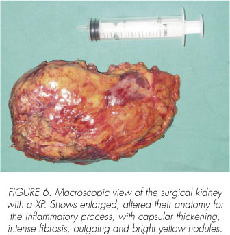
The pathology on macroscopic examination shows usually a kidney yellow, enlarged in size, and with stones inside. Microscopic examination macrophages are loaded with fat (histiocytes sparkling), explaining his color, in addition to necrosis and infiltration with leukocytes and plasmatic cells (10,11).
This chronic and destructive process can affect other organs such as gallbladder (19.20), appendix (21), bone (22), ovarian (23), bladder (24), rectum (25), prostate (26), epididymis (27) and endometrium (28).
Amyloidosis (29), although rare, is one of the forms of expression as a complication of a long evolution of disease.
We update and communicate our experience in the care of patients suffering from this rare disease: form of presentation, clinical manifestations, diagnosis, treatment and follow-up. Highlight the benefits of nephrostomy prior to nephrectomy.
Materials and methods
We conducted an observational study, descriptive and retrosprospective. Covered the total population of patients over a period of 6 years (February/2002 to January/2008). Was conducted in 3 centers throughout the city of Cordoba - Argentina - (Rawson Hospital, Aircraft Hospital, New San Roque Hospital). The study sample consisted of 10 selected medical records of patients with histopathologic diagnosis of PX. We analyzed demographic data, history, clinical and laboratory, diagnostic imaging, development and treatment.
Results
We analyzed 10 patients, 8 females (80%) and 2 men (20%), between 22 and 74 years, with an average of 50 years, and a rank of 52. The initial diagnosis suspected was in 80% of cases pyonephrosis and 30% cancer. Six patients (60%) had a history of recurrent UTI, 50% family diabetes (only one suffering from diabetes) and 3 (30%) were hypertensive. The most frequent reason for consultation was low back with abdominal pain (100%) and fever (80%) (Table I).
The clinical presentation of patients on admission in all cases (100%) was: lumbar and abdominal pain, weight loss, pale conjunctiva, kidney stones and chronic evolution of the disease. High fever and palpable abdominal mass in 80% of cases. Two patients (20%) found association with cancer (Epidermoid pyelocalicial system and urothelial transitional cell carcinoma invading the bladder) and other 2 (20%) with Psoriasis. None of our patients was found hepato and / or splenomegaly (Table II).
The laboratory showed anemia in 100% of cases, erythrocyte sedimentation rate of 100 mm or more in 90% and 70% with leukocytosis. Urine sediment pyuria presented in 100%, hematuria in 80% and bacteriuria in 60% of cases. The urine culture, collected by the technique of midstream specimen showed development in 5 cases (50%) and obtained by nephrostomy in 4 cases that took place (100%). The crops recovered were similar microbial (cultivation midstream nephrostomy) in 1 case (Enterobacter spp) in the others there were no coincidence. Were recovered various microorganisms: Escherichia coli, Proteus mirabilis, Enterobacter spp. Morganella morgani, Enterococcus faecalis, Staphylococcus coagulase negative, anaerobic Streptococcus, Citrobacter freundii, Strepretococcus viridans. In two cases (20%) developed many microbes. Liver enzymes alkaline phosphatase was altered in 6 cases which took place in 8 (75%). The Glutamic pyruvate transaminase(GPT) was increased in 2 cases (20%), the glutamic oxaloacetic transaminase (GOT) in 1 (10%), on 9 patients who were testing. All patients had negative hepatitis serologies and no background with toxic or ingestion of drugs. The gamma globulin was increased in 4 of 6 cases that took place (66%). Studies value of serum creatinine and urea showed figures within normal in all cases (Table III).
In all cases (100%) had unilateral commitment, 6 (60%) undertook the right kidney and 4 (40%) left. Plain film of the abdomen was performed in 8 cases, 7 showed a kidney stone (87%). In 4 of them staghorn calculi (57%). The ultrasound showed in all cases (100%) increase in the size kidney, stones in 9 cases (90%), hydronephrosis pattern in 8 (80%), liquid intraparechymatous images in 5 cases (50%) and alteration in the medulla-cortex ratio in 5 (50%). In 2 cases with suspected cancer, one of them was positive. CT showed enlarged kidney in 9 cases (100%) that was held, kidney stones in 8 opportunities (88.8%), alteration in the elimination of the contrast medium in 7 (77.7%), obstructive hydronephrosis in 3 cases and lesional extension to perirenal fat in 7 opportunities (77.7%). In all cases (100%) commitment was throughout the renal parenchyma (global) and in 7 patients (70%) staging was stage 2, with commitment of renal parenchymal and space Gerota (Table IV).
All patients underwent nephrectomy and made 4 of them after nephrostomy. Surgical complications were presented in 5 cases (50%): sepsis and abdominal wall abscess in 2 cases, Psoas abscess and bleeding in 1 case. Mortality was 10% (1 case) for sepsis, in a patient who had a concomitant squamous cell carcinoma of renal pelvis.
Were used in treating various types of antibiotics, initially empirically or as the result of susceptibility testing and microbial identification. In 7 cases were prescribed more than one type of antibiotic (70%) (Table V).
Discussion
We present our experience of 6 years in our country since there are no records of previous publications, except one case associated with polymicrobial flora (14) also reported by us, we are encouraged in the pursuit of this disease, so rare.
Since Schlagenhaufer described for the first time in 1916 the PX, the disease remains rare (4,5). The literature makes little mention of this inflammatory process and in general, case studies, not signing up too many patients. These communications, except for isolated cases, are made by pathologists (11,17,21-23,28).
Our findings are not significantly different to that observed in the majority of the series (17,9,1 1,30) that refer to this disease. The vast majority of cases involve women, usually middle age, with global commitment unilateral renal parenchyma associated with stones and lack of function of the organ involved. All our patients had a chronic high fever, abdominal and back pain, palpable abdominal mass, weight loss and conjunctival pallor.
The laboratory, in most cases, revealed anemia, leukocytosis, very increased erythrocyte sedimentation rate, with a pathological urinary sediment showed that, in most cases, pyuria, hematuria and bacteriuria. Liver function tests showed essentially the increase in alkaline phosphatase and transaminase in few cases, simulating a cholestatic process, too, highlighted in other publications (2,3,6,7,9,11).
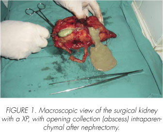
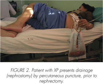
The urine culture was positive in half the cases (50%), unlike the literature that mention a more general recovery in about 75% (1,6,7,9,11). Negative cultures are understandable due to the association of infection with obstructive uropathy. Microbiological findings showed no predominance of any organism. We emphasize different isolates polymicrobial and recoveries, including midstream urine cultures and material collected on the occasion of the completion of the nephrostomy. These discrepancies are also observed by Malek RS and Elder, JS. (6). The material obtained by nephrostomy, unlike the conventional urine culture, provided it is done (4 cases) presented microbial growth. This explains the negative cultures, as these patients truly have encapsulated renal abscesses and / or obstruction of the urinary tract by calculi (Figure 1). We believe the nephrostomy as facilitator action before nephrectomy (Figure 2). This drains, although it applies in patients with renal or perirenal collections, are not described in the literature, prior to nephrectomy in this type of pathology. This therapeutic approach was proposed by us (33).
Highlights in 5 patients a family history of diabetes (50%), but only one of them had the disease. Other authors have similar findings, with percentages ranging from 5 to 40% (31). To Berrah-Bennaceur, Farad AB et al (4) is a predisposing factor. Highlight the suffering of Psoriasis in 2 (20%) of our patients. In our literature review could not find the association of this disease and PX. Psoriasis, similar to the PX, is a disease in which it described in its pathogenesis multifactorial causes. Is triggered or exacerbated by various environmental factors, which are referred to as suffering from infections our patients. Invoking also defects in the immune system (2,4,11), which is another of the hypotheses on the origin of the PX, in addition to genetic predisposition.
In contrast to what was mentioned in some studies for review of this disease (2,3,6,7,9,11), none of our patients, we found hepatomegaly and / or splenomegaly.
At variance with what is mentioned in the literature (2-5,7-9,14,16,17) 5 times (50%), postoperative complications were presented. These complications are understandable when one considers that removes a septic focus and generally adhesions to neighboring structures (7 / 10 cases), which implies an additional surgical trauma. One patient died of sepsis, a predisposing factor as having squamous cell carcinoma of the renal pelvis (Figure 4). In accordance with the majority of authors (1-9) treatment with antibiotics before, during and in the immediate postoperative period is essential to control the infection and essentially contributes to avoid commitments systemic or remote, but is unable, by itself, to eradicate the infection.

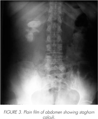
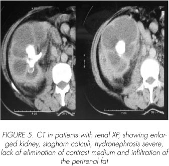
According to other publications (1-3,6,7,9,1 1,16), radiological findings that stood out was the increased size of the kidney, the presence of stone and the exclusion of the kidney. Plain film of the abdomen can view lithiasis usually staghorn calculi (Figure 3). Ultrasound can demonstrate the loss of parenchymal structure with hydronephrosis pattern (Figure 4). The CT, similar to ultrasound, said the diagnosis and extent of lesion (Figure 5).
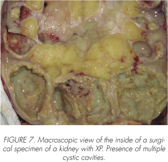
When you have previous experience, it is possible with the help of clinical, laboratory and complementary methods (especially CT), assume that condition before having pathological studies (2,13). The diagnosis of certainty (Gold Standard) is histopathological. As evidenced by our figures, the kidney is macroscopically increased in size with a poor demarcation cortex and medulla, replacing his usual color for a yellow type adipose tissue, in addition to cystic cavities, that is a complete loss of kidney structure (Figures 6, 7). Microscopically, as described by pathologists (6,9-11,14,17), there is a diffuse inflammatory reaction (chronic pyelonephritis) who infiltrates the kidney with lipid-laden histiocytes called foamy cells, which are arranged in bands or blocks, giant cells, white cells neutrophils, lymphocytes and plasma cells with little functioning parenchyma (Figures 8, 9).


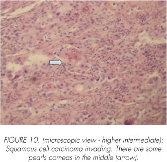
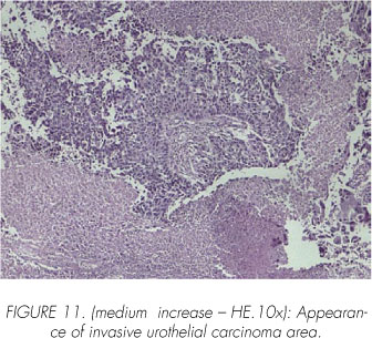
The discovery of the association PX with cancer deserves special mention, which was presented in 2 patients (20%), both with cancer of the urinary tract, a squamous cell carcinoma of the renal pelvis and another with the bladder urothelial carcinoma (Figures 10, 11). The PX associated with renal tumors are rare, Sampol J. Ballesteros (31) mentioned in the revised 1.7%, being the most frequent kidney carcinomas and urothelial carcinomas with a much lower incidence. Suspected in the preoperative is virtually impossible because they are masked by this type of pyelonephritis and they are finding in the histopathological study (32). These tumors were associated in our patients with long-standing kidney stones and chronic urinary infection, the predisposing factors (31).
In total agreement with the majority of authors (2-7,9,11,12,14-18), nephrectomy becomes the primary indication, either total or partial, mainly in children or focused on processes. But we recommend percutaneous drainage before nephrectomy as the surest way to check the type of bacteria that commits the kidney. It also allows better access to surgical nephrectomy (14,33).
Mortality was 10% (1 case) by sepsis associated with cancer as a predisposing factor. Ballesteros Sampol J. (31) in a review article mentions an overall mortality from this disease of 24% and specificity of 4%.
Conclusions
Our findings in general were not too different from other communications. Our case essentially was in middle aged women with fever, abdominal and back pain, palpable abdominal mass, weight loss and prolonged disease evolution.
The most frequent in the laboratory were anemia, leukocytosis and erythrocyte sedimentation rate accelerated. In the analysis of urine were hematuria and pyuria.
Bacterial recovery by conventional urine culture is low, due to obstruction associated process.
Ultrasound and CT can be very suggestive of the diagnosis, even when there is prior experience with this disease. Sonographically a renal mass, with evidence of stone and hydronephrosis could suggest the disease. The computed tomography is always useful to determine the extent of the lesion, especially when it involves neighboring organs. This disease should be taken into account before any verification of diffuse or localized renal mass. We emphasize the importance of percutaneous drainage prior to surgery, which in our view, is a facilitator for nephrectomy. Decrease the volume up to the kidney removal of the body, and thus facilitates the surgical process, especially when there are processes with perirenal invasion of tissues. Wherever possible this drainage, septic acute tempered table facilitates the surgical approach and technique in a second time. In addition, microbial enhanced recovery. Therefore we recommend, wherever possible, all crops preoperatively at our disposal, either by the method of midstream specimen or percutaneous puncture. Similarly, crops should be made during surgery. Treatment with antibiotic/s, but by itself is not able to solve the problem, it is useful in controlling the infection and prevents septic complications. For the effectiveness of this therapy is vital to the well-identified bacterial agents, with their sensitivity. But the definitive treatment is always surgical, with total or partial nephrectomy as appropriate.
Obstruction and chronic urinary infection are predisposing factors for recurrent urinary tract tumors, making it understandable association with cancer of the PX. On the contrary we did not find a clear explanation of its association with Psoriasis. Although we know that both diseases could be due to immune disorders may suffer from chronic infection that these patients could be a trigger of Psoriasis, in genetically predisposed individuals.
Septic surgical complications were more frequent due to several factors, but stress the concomitant infection and adherence to adjacent structures affected kidney, which involved a laborious and traumatic surgery.
Whenever the diagnosis of certainty is Histopathological either macro or microscopic, which is essential not only to confirm the diagnosis of PX, but also its association with cancer.
We consider this disease as a chronic inflammatory infectious process, with acute outbreaks of infectious origin.
The mortality was associated with concurrent diseases, mainly cancer.
Acknowledgments
Pathology Service. New San Roque Hospital.
 Correspondence:
Alberto Francisco Leoni
Servicio Infectología
Hospital Aeronautico Cordoba
Cordoba. (Argentina)
afleoni@hotmail.com
Accepted for publication: May 18th, 2008
References and recomended readings (*of special interest, **of outstanding interest)
1. Sobel JD, Kake D. Urinary Tract Infection. Chapter 66 In: Mandell, Bennett y Dolin. Principles and Practice of Infectious Diseases. Sixth edition (Vol. 1) Elsevier Churchill Livingstone, 2005. Pag 395. [ Links ]
**2. Zorzos I, Moutzouris V, Korakianitis G, Katsou G. Analysis of 39 cases of xanthogranulomatous pyelonephritis with emphasis on CT and findings. Disponible en: www.ecr.or/t/ECRO2/sciprg/abs/pc/0378.htm; 2000. [ Links ]
**3. Van Vlem B, Billiouw JM. Xantothogranuloma-tous Pyelonephritis. N Engl J Med. 2000; 342: 1572. [ Links ]
4. Berrah-Bennaceur, Farad AB et al. La Pyelonéphrite Santo-Granulomateuse. Approche Etiopathogénigue á propos de duex cas. Sem Hop Paris 1999;75: 1362-7. [ Links ]
*5. Cabrera G, Fernández Maldonado J, Mijares M, Gallego E, Roa Marquez J. Pielonefritis Xantogranulomatosa. Rev Ven Urol 1997; 29: 93-103. [ Links ]
**6. Malek, RS, Elder, JS. Xanthogranulomatous pyelonephritis. A critical analysis of 26 cases and of the literature. J Urol 1978; 119:589. [ Links ]
**7. López de Meza B, Gómez E, Gómez P. Pielonefritis Xantogranulomatosa: reporte de 15 casos y revisión de la literatura. Urol Coloma 1996; 5: 15-24. [ Links ]
8. Matthews GJ, McLoire, GA, Churchill BA, Steckler RE, Khoury AE. Xantothogranulomatous Pyelonephritis in Pediatric Patiens. Ped Urol 1995; 153: 483-8. [ Links ]
**9. Chuang, CK, Laim K, Chang PL, et al. Xantothogranulomatous pyelonephritis: Experience in 36 cases. J Urol 1992; 147:337. [ Links ]
10. Tiguert R, Gheiler EL, Yousif R, Tefilli MV, Mills K, Grignon DJ et al. Focal Xantothogranulomatous Pyelonephritis presenting as a Renal tumor with vena cava thrombus. J urol 1998; 160:117-8. [ Links ]
**11. Parsons, MA, Harris, SC, Longstaff AF, Granger, RG. Xantothogranulomatous pyelonephritis: A pathological, Clinical and etiological analysis of 87 cases. Diag Histopathol 1983; 6: 203. [ Links ]
12. Moriya A, Kubota K, Morita N. A case emphysematous pyelonephritis combined with emphysematous xantothogranulomatous pyelonephritis. Hinyokika Kiyo1989; 35:295-300. [ Links ]
13. Alam A, Chander BN, Xanthogranulomatous Pyelonephiritis: Diagnosis using Computed Tomography. MJAFI 2004; 60: 86-88. [ Links ]
14. Leoni A.F, Luque A, Sambuelli R, Valverde J, Pielonefritis Xantogranulomatosa Asociada a Flora Polimicrobiana. Rev Pan Infec 2004; 6: 23 -27. [ Links ]
15. Langale LA, Rice CL and Brown N. Emphysematous pyelonephritis in a xanthogranulomatous kidney. An unusual cause of pneumoperitoneum. Arch Surg 1980; 123: 153. [ Links ]
16. Oosterhof, GO, Delaere, KP. Xantothogranulomatous pyelonephritis. Urol Int 1986; 41:180. [ Links ]
*17. Watt, I, Kristesen, IB. Xantothogranulomatous pyelonephritis. Arch Path Microbial Scand Sect A 1980; 89:89. [ Links ]
18. Pérez, LM, Thrasher, JB, Anderson, EC. Successful management of bilateral Xantothogranulomatous pyelonephritis by bilateral parcial nephrectomy. J Urol 1993; 149:100. [ Links ]
19. Reaño, Gustavo; Sánchez Lihon, Juvenal; Ruiz Figueroa, Eloy Francisco; Celis Zapata, Juan; Payet Meza, Eduardo; Berrospi Espinoza, Francisco; Chávez, Iván; Young Tabusso, Frank; Doimi, Franco. Colecistitis xantogranulomatosa: Análisis de 6 casos. Rev gastroenterol Perú; 2005; 25: 93-100. [ Links ]
20. Silva O., Verónica; Arístides F., Guillermo; Órdenes V., Manuel; Moya P., Edisson; Cassalino F., Renato. Colecistitis xantogranulomatosa. Rev. Chil. Cir 2004; 56: 178-181. [ Links ]
21. Tardío JC, Burgos F, Riupérez J, Inflamación xantogranulomatosa del apéndice. Patología 1996; 29: 135-137. [ Links ]
22. Cozzuto D, Xanthogranulomatous osteomielitis. Arch Pathol Lab Med 1984; 108: 973-976. [ Links ]
23. Pace EH, Voet RL, Melacon JT, Xanthogranulomatous oophoritis: An inflammatory pseudotumor of the ovary. Int J Gynecol Pathol 1984; 3: 398-402. [ Links ]
24. Walther M, Glenn FJ, Vellios F, Xanthogranulomatous cistitis. J Urol 1985; 134: 745-746. [ Links ]
25. Davis M, Whitley ME, Haque HK, et al. Xanthogranulomatous abscess of a mullerian Duch remmant. A rare lesiono f the rectum and anus. Dis Colon Rectum 1986; 29: 755-759. [ Links ]
26. Miekos E, Wlodarczyk W, Szram S. Xanthogranulomatous prostatitis. Int Urol Nephrol 1986; 18: 433-437. [ Links ]
27. Wiener LB, Riehl PA, Baum N. Xanthogranulomatous epididymitis: A case report. J Urol 1987 138: 621-622. [ Links ]
*28. Russack B, Lammers RJ. Xanthogranulomatous endometritis. Reporto six cases and a proposed mechanism of development. Arch Pathol Lab Med 1990; 114: 929-932. [ Links ]
29. Bronsoms JM, Vallés M, Mas H, Sant F y Llobet M. Pielonefritis xantogranulomatosa y amiloidosis sistémica; Nefrología 1995; XV: 371-374. [ Links ]
30. Quinn FM, Dick AC, Corbally MT, McDermott MB, Guiney EJ. Xanthogranulomatous pyelonephritis in childhood; Arch Dis Child 1999; 81: 483-486. [ Links ]
**31. Ballesteros Sampol J. Inusuales formas clínicas de presentación y asociaciones patológicas raras de la pielonefritis xantogranulomatosa. Arch Esp Urol 2002; 55(2) 119-130. [ Links ]
*32. Rizzi A O, Alvarez P D, Muro F y col. Carcinoma de células escamosas de la pelvis renal. Presentación de un caso clínico. Rev Arg de Urol 2005; 70 (4): 262-5. [ Links ]
33. Revol M, Arribillaga L, Paladini M, Valverde j, Leoni A, Manavella H. Pielonefritis Xantogranu-lomatosa: Enfoque Terapéutico a Propósito de dos casos. XXXVI Congreso de la Federación Argentina de Urología, XLII Congreso de la Sociedad Argentina de Urología. 2006; Sesión Casos Clínicos TL 34. [ Links ]

