


 Servicios personalizados
 Inglés (pdf)
 Articulo en XML
 Referencias del artículo
 Enviar articulo por email
 Citado por SciELO
  Citado por Google
  Similares en
SciELO
Similares en
SciELO  Similares en Google
 Permalink
INTRODUCTION
Obesity is a multifactorial disease 1 and its prevalence is increasing quickly, some authors consider it as one of the worst problems in public health 2. Some biological processes such as oxidative stress and inflammation are closely related to obesity development 3. Recently, some authors highlighted the role of oxidative stress on the chronic inflammation status of obesity 4 and even on the progression of comorbidities as hypertension, diabetes, and atherosclerosis 5. Some studies have shown that a high fat diet 6 and high body mass index (BMI) are related to increased levels of oxidative stress biomarkers 7. Furthermore, Remla et al. (2016) 8 have shown that both IL-6 and oxidative stress biomarkers levels are higher in obese than in normal weight subjects.
Chronic inflammation is a condition intrinsically related to obesity. Some authors have shown significant differences between obese and normal weight women cytokines pattern 9. Some events, together, contribute to the maintenance of chronic inflammation in obese: adipocyte hypertrophy, hypoxia, angiogenesis, macrophage and lymphocyte infiltration, and inflammatory cytokine secretion 10. Inflammatory cytokines, like tumoral necrosis factor alpha (TNF-α), C-reactive protein (CRP) and interleukin-6 (IL-6) are some of the most important cytokines that are directly related to BMI, visceral adiposity, obesity development (Park et al., 2004) and insulin resistance 11. It has been demonstrated that IL-6 is directly related to glucose tolerance and triacylglycerides (TG) enhancing, and this might happen because IL-6 secretion may inhibit glycogenesis and stimulate gluconeogenesis and glycogenolysis in the liver 4.
Oxidative stress is characterized by an imbalance in the generation of reactive oxygen species (ROS) and antioxidant reactions. Free radicals are very reactive species and may cause damage to many tissues and macromolecules such as DNA, lipids and proteins 12. Traditionally, malondialdehyde (MDA) has been used as a biomarker for oxidative damage 13, and reflects the lipid peroxidation status. Trolox equivalent antioxidant capacity (TEAC) is an essay largely used to evaluate total antioxidant capacity, and reflects the potential of eliminating or combating free radicals 14,15,16. Studies have shown that the imbalance of such biomarkers is associated with the chronic inflammatory state, which is associated with obesity and other comorbidities 17.
Natural products are increasingly being valued as complementary treatment for chronic diseases traditional therapies, mainly because they might cause fewer side effects than conventional and synthetic drugs. Gautam and Jachak (2009) 18 described anti-inflammatory and antioxidant effects related to molecules that are plant secondary metabolites such as alkaloids, terpenoids and mainly polyphenols. Wang Shu et al. (2013) described that many classes of polyphenols are related to antioxidant and anti-obesity effects in animal studies 19. Rice-Evans et al. relate those antioxidant positive effects to the polyphenols capacity of hydrogen donating, which acts as free radical scavengers 20. Camellia sinensis, also known as green tea (GT), is a popular plant species that is largely consumed and is rich in catechins, which are part of a well-known class of polyphenols, from the flavonoids group. The most important catechin is the epigallocatechin gallate (EGCG). Recently, some authors described the anti-obesogenic effect of GT due to the regulation of adiponectin 21. Furthermore, in obese animals, catechins administration reduced serum levels of leptin and decreased the gene expression of TNF-α and IL-6 22.
The hypothesis of this study is that decaffeinated green tea extract supplementation is capable of ameliorating inflammatory and oxidative stress biomarkers in women with grade III obesity. Thus, understanding the responses triggered by green tea consumption and associating them with biomarkers modulation might contribute to the elucidation of the anti-inflammatory and antioxidant effects of green tea catechins and possibly, in the future, these compounds would be used as a complementary agent in weight loss therapies.
In this context the purposes of this study were: a) accessing oxidative stress biomarkers in both obese and normal weight women; and b) evaluating if green tea supplementation has an impact on these variables and on inflammatory cytokines biomarkers in women with severe obesity (BMI ≥ 40 kg/m2). This study was the first study that evaluated decaffeinated GT supplementation effects on oxidative stress and inflammatory biomarkers in Brazilian obese subjects.
MATERIALS AND METHODS
SUBJECTS AND TREATMENT
This study was carried out at the Clinical Hospital of Ribeirao Preto Medical School - University of São Paulo (Brazil) from July 2014 to March 2016. Severely obese (BMI ≥ 40 kg/m2) and normal weight women (BMI between 18.5 and 24.9 kg/m2) aged between 18 and 60 years old were invited to participate in the study. Patients were in fasting state before blood sample collection. Subjects were excluded if they were suffering from chronic diseases such as hypertension, diabetes mellitus, dyslipidemias, hyper or hipo-thyroidism and cancer, as well as if they were pregnant, alcoholics and smokers. For GT supplementation program, a smaller group of obese women were randomly selected from the major group and assigned for treatment. The patients received GT capsules for eight weeks and the daily dosage of EGCG was 450 mg. The standardization of the supplementation program, as well as the complete quantitative analysis of GT extract used, were performed according to previous data from our research group 23. The safety of the treatment was based on studies that pinpointed appropriate doses that did not lead to hepatotoxicity 24,25. The study was conducted in accordance with the Declaration of Helsinki and approved by the Ethics Committee (CAAE: 30247414.6.0000.5440). All women voluntary consented in participating. Regarding the sample size, all patients who filled the inclusion and exclusion criteria were selected within a given period. Therefore, a sample held for convenience/availability.
SAMPLE PREPARING
Total blood was collected in non-heparinized serum tubes with separation gel. Then, it was centrifuged at 7,000 rpm, 23 °C for 15 minutes. Serum samples were frozen immediately and stored at -80 °C.
OXIDATIVE STRESS ASSESSMENT
TEAC was measured based on the method purposed by Gomes et al. (2015) 26. A 2.5 mM solution of trolox (6-hydroxy-2, 5, 7, 8-tetramethylchromane-2-carboxylic acid; Sigma® Aldrich, St. Louis, Missouri, USA) was prepared using phosphate buffer saline (PBS) and was the standard for the reaction. The 2, 2'-azinobis (3-ethylbenzothiazoline-6-sulfonic acid) diammonium salt, also known as ABTS, and potassium persulfate (di-potassium peroxdisulfate) were also obtained from Sigma® Aldrich (St. Louis, Missouri, USA). Both ABTS solution and potassium persulfate were diluted in PBS solution, pH 7.2; the final concentration was 7 mM and 140 mM, respectively. The method is based on the reaction of ABTS with persulphate potassium. The radical cation ABTS●+ is formed and its green chromophore feature allows it to be read spectrophotometrically at 735 nm. Both trolox and sample addition reduce some ABTS●+ species into ABTS. Two readings were performed per sample with interval of five minutes after each addition; the observed bleaching is equivalent to the total antioxidant capacity.
The malondialdheyde (MDA) essay performed in this study was modified by Percário et al. (1994) 27. The basis of this method is that MDA, product of lipid peroxidation mainly from cellular injury, forms complexes with thiobarbituric acid (TBA; Sigma® Aldrich, St. Louis, Missouri, USA), and the chromophores can be read in UV-VIS spectrophotometer. In this essay, 1,1,3,3 tetrahydropropane (Sigma® Aldrich, St. Louis, Missouri, USA) were used as standard. A 10 nM solution of TBA was prepared in a 75 mM monobasic phosphate solution (Sigma® Aldrich, St. Louis, Missouri, USA) and adjusted to pH 2.5 with acetic acid (CH3COOH); then, 250 µl of sample were added to 500 µl TBA solution and placed in water bath (95 °C , 60 minutes). An extraction was performed using 2 ml of butanol (C4H10O) and centrifuged at 175 g for 15 minutes. The supernatant was read in the spectrophotometer (BECKMAN DU640) at 535 nm. This essay was performed in duplicates.
INFLAMMATORY CYTOKINES
Serum concentrations of TNF-α, IL-6, IL-10, IL-1β and IL-17α were determined using the Panel I HCYTOMAG-60K-08 kit (Merck Millipore(®). Data was processed and analyzed according to the manufacturer's instructions using the software Multiplex System manager. This method uses magnetic beads technology, which improves detection sensitivity by the Luminex xMAP® platform, due to the sensitivity and cost of the technique. The experiments were done in monoplicate.
STATISTICS
Normality was determined by the Shapiro-Wilk test and parametric data were analyzed with paired and an independent t-test. The Wilcoxon test was used to evaluate non-parametric data and p < 0.05 was considered as statistically significant. The software SPSS (version 20.0. Armonk, NY: IBM Corp.; 2011) was used to analyze data.
RESULTS
SUBJECTS
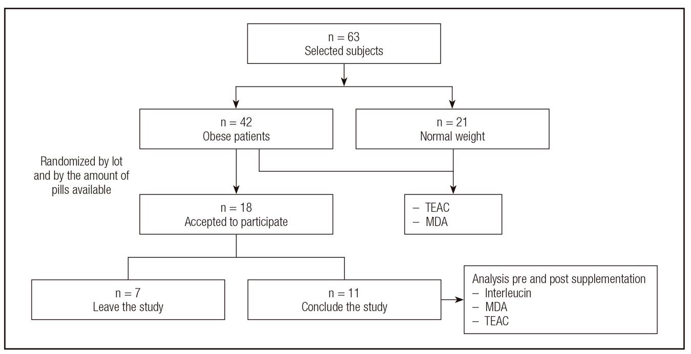
A total of 42 obese (BMI: 48.2 ± 9.3 kg/m²; age: 37.2 ± 11.17 years old) and 21 normal weight (BMI: 22.5 ± 2 kg/m²; age: 34.45 ± 7.22 years old) women were recruited and completed the whole program. A subgroup of 18 obese women were chosen randomly from the total of 42 obese women in order to receive green tea supplementation; eleven women completed the program (Figure 1). Patients did not show significant weight loss and did not note adverse effects associated with the treatment; this data was from another study of our research group 23.
OXIDATIVE STRESS BIOMARKERS COMPARATIVE EVALUATION
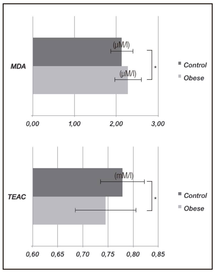
Serum TEAC and MDA levels are represented in Figure 2. The serum levels of MDA were higher in obese (2.52 ± 0.31 µmol/l) than in normal weight women (2.13 ± 0.26 µmol/l; p = 0.042). On the other hand, TEAC values were lower in the obese group (0.75 ± 0.06 mM/l) than in the eutrophic group (0.78 ± 0.04 mM/l; p = 0.017). These results were expected once MDA and TEAC have opposite relationship to oxidative stress status.
OXIDATIVE STRESS AFTER GREEN TEA SUPPLEMENTATION
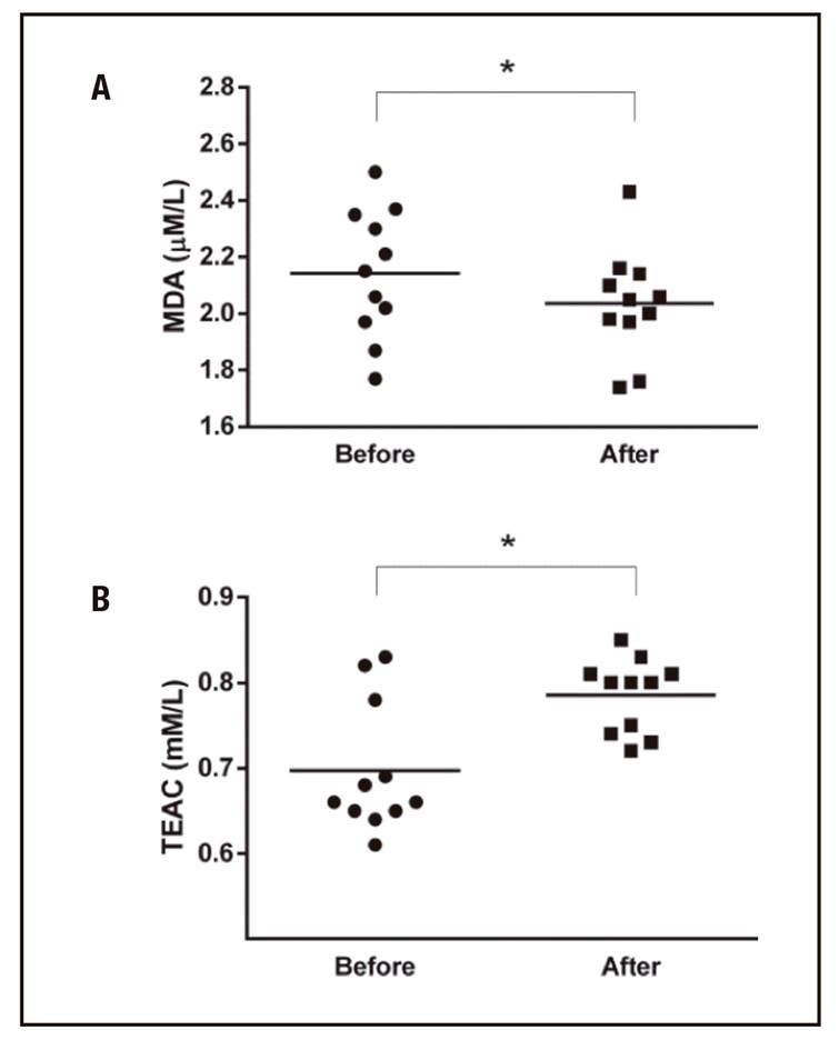
GT supplementation was performed in eleven obese women. The treated group had lower MDA levels after the supplementation (2.04 ± 0.18 versus 2.14 ± 0.23 µM/l; p = 0.01) and the TEAC level was higher after the treatment (0.78 ± 0.04 versus 0.70 ± 0.08; mM/l; p = 0.009) (Fig. 3).
INFLAMMATORY CYTOKINES
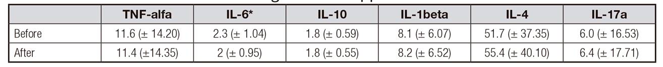
Interleukins evaluation revealed that the concentration of IL-6 decreased 12.7%, after the treatment (p = 0.03) (2.3 ± 1 versus 2 ± 0.9 pg/ml), and the levels of TNF-α, IL-10, IL-1β and IL-17A were similar before and after the intervention (Table 1).
DISCUSSION
In this study we found that obese women had lower capacity based on TEAC and MDA biomarkers. Obese women that received GT supplementation seemed to have their antioxidant and anti-inflammatory capacity improved, once TEAC, MDA and IL-6 levels significantly changed after the treatment. Since MDA is related to lipid peroxidation in cellular membranes and TEAC reveals the total antioxidant capacity, it can be assumed that after treatment obese subjects improved their oxidative stress status and also ameliorated the inflammatory response, which can be reflected by IL-6 decreasing.
Serum levels of MDA and TEAC are indicators of lipid peroxidation and capacity of fighting free radicals, respectively. In the present study, serum MDA levels were higher in obese than in non-obese women, corroborating previous studies that have first described that relation 7,28. Compared to the normal weight group, our data revealed lower TEAC values for obese; this result allows the inverse relation of TEAC and BMI to be considered. Bloomer and Fisher-Wellman (2009) 6 reported significant differences in TEAC between eutrophic and obese women after high fat diet ingestion. This study reflects how BMI is probably related to differences in the total antioxidant capacity; TEAC was also reduced in obese subjects after the diet intervention. Overall results obtained in the present study suggest that normal weight women have better antioxidant ability and less macromolecules damage caused by lipid peroxidation when compared to obese subjects. Olusi (2002) describes that it might happen because obese people normally have a high lipid and low antioxidant diet ingestion 7.
In addition, some authors believe that increased oxidative stress biomarkers in obese are directly related to a chronic low-grade inflammation status 16. Green tea was chosen for the treatment because it has both antioxidant and anti-inflammatory properties already described 29. However, there is no consensus in the literature regarding the optimum dose of polyphenols. Daily supplementation of 379 mg of GT had positive effects on oxidative stress and inflammatory biomarkers 30. Polyphenols may affect oxidative stress by enhancing antioxidant enzyme production, such as glutathione, which is rich in thiol groups that are well known reducing agents and thanks to their chemical structure they are able to directly inhibit ROS generation 31.
Our data revealed that 450 mg of EGCG supplementation leads to an increase in TEAC, corroborating the literature. Henning et al. (2004) 32 observed that TEAC was increased after 386.5 mg GT consumption in non-obese subjects. Rice-Evans (1999) 33 reported that EGCG has a good ability in scavenging free radical species, especially in the ABTS●+ radical test. Capodice et al. (2015) 34 results showed no modifications in TEAC with treatments during four weeks. Therefore, our results showed a MDA decrement after GT administration and this result corroborates some studies from colleagues. Spadiene et al. (2014) 35 also reported that MDA decreased after a 9-18 months treatment with 400-500 mg of GT, as well as other research groups that also observed this results using different therapeutic approaches 36. Our results suggest that oxidative stress biomarkers were sensitive to GT supplementation, and it is important to emphasize that further studies are needed to pinpoint the best treatment parameters for obese supplementation.
The interleukin analysis showed that the serum levels were similar in obese before and after treatment. This results corroborate Basu and Sánchez (2010) findings, which described a discrete effect on plasma cytokines after eight weeks and emphasized a decrease trend in IL-6 level 37. However, our data showed that IL-6 decreased significantly after GT treatment, and this is in accordance with Lu et al. (2012), who showed a reduction in IL-6 gene and protein expression in mice, after GT supplementation 38. Jung et al. (2008) reported that a 12-week GT treatment led to the reduction of both TNF-α and IL-6 in obese subjects 39. Bogdanski et al. (2012) showed that a three-month treatment with 379 mg of GT resulted in decreased levels of TNF-α (30). Maybe the differences between observed effects on TNF-α and other cytokines are due to the daily dosage and duration of the GT supplementation program.
Cytokines play an important role in signaling and mediating inflammation in obesity and in other inflammatory chronic diseases. Some authors describe that greater IL-6 levels are related to an antioxidant unbalance 40 and even may contribute to the disrupting signal transduction in insulin pathways, leading to insulin resistance and contributing to the development of type 2 diabetes and obesity 11.
There is a wide discussion about the administration of GT and its effects on antioxidant systems and inflammatory cytokines and about their efficacy for obese treatment. Further studies should be done to elucidate mechanisms of action and the best dose, extraction method and administration period. The GT effects described in this study may contribute to drug discovery, alternative and complementary therapies to obesity control.
STUDY LIMITATIONS
The GT intervention did not have an associated control or a placebo group; furthermore, no other dosage was tested (greater or smaller) in different periods. Financial limitations prevented GT supplementation for all subjects involved in this study. In addition, the sample size was selected according to the number of clinical appointments performed at the outpatient clinic who met the inclusion criteria.
CONCLUSION
In conclusion, the supplementation of decaffeinated GT extract in women with grade III obesity can improve the biomarkers of oxidative stress and serum level of IL-6. Therefore, based on these results, it is recommended that the intake of GT could be an adjunct practice to the treatment of obesity.

