


My SciELO
 Custom services
Services on Demand
Journal
Article
 text in
  English (pdf)
 Article in xml format
 Article references
 Send this article by e-mail
Indicators
-
 Cited by SciELO
 -
 Access statistics
Related links
 Cited by Google
 -
 Similars in
SciELO
Similars in
SciELO  Similars in Google
Share

 Permalink
Revista Española de Enfermedades Digestivas
Print version ISSN 1130-0108
Rev. esp. enferm. dig. vol.96 n.11 Madrid Nov. 2004
| CLINICAL NOTE |
Amebic liver abscess with bacterial superinfection in a patient with no epidemiologic
risk factors
P. Sánchez-Pobre, S. Saénz-López, E. Salto1, R. Sanjuán1, C. Ibero2 A. Masedo and J. A. Solís Herruzo
Department of Medicine. Service of Digestive Diseases. 1Department of Microbiology and Service of Infectious Diseases.
2Department of Internal Medicine. Hospital Universitario 12 de Octubre. Madrid, Spain
ABSTRACT
The amebic liver abscess is uncommon in developed countries like Spain, but the incidence is increasing probably due to the migratory movements of the population. We report a case of an amebic abscess, initially unsuspected due to the absence of epidemiologic risk factors and the negative serology for amebiasis, in the early stages of the disease.
Key words: Amebic liver abscess. Liver abscess. Amebiasis. Entamoeba histolytica.
Sánchez-Pobre P, Saénz-López S, Salto E, Sanjuán R, Ibero C, Masedo A, Solís-Herruzo JA. Amebic liver abscess with bacterial superinfection in a patient with no epidemiologic risk factors. A prospective study. Rev Esp Enferm Dig 2004; 96: 796-800.
Recibido: 12-11-04.
Aceptado: 16-11-04.
Correspondencia: Pilar Sánchez-Pobre. Departamento de Medicina Aparato Digestivo. Hospital Universitario 12 de Octubre. Avda. de Córdoba, s/n. 28041 Madrid. e-mail: psanchezp@meditex.es
INTRODUCTION
The amebic liver abscess (ALA) has a worldwide incidence of 50 cases per year (1). In developing countries like Vietnam, were infection by Entamoeba histolytica is endemic, the annual incidence of amebic liver abscess is 21 cases per 100,000 habitants. In developed countries such as the United States, it is unusual and is most commonly seen in immigrants from and travelers to developing countries (2). It has also been reported in imunosuppressed patients, HIV patients, patients on corticosteroids or suffering from chronic infections like tuberculosis or syphilis and patients who have undergone post-traumatic splenectomy. It has also been associated with acute hepatitis A or B (HBV) (3,4).
Entamoeba histolytica is a protozoa that infects humans by fecal-oral contact. It usually inhabits the colon, where it can cause an asymptomatic infection or it can become invasive and cause amebic colitis. After invasion into the intestinal mucosa, it can disseminate to other organs, predominantly the liver, leading to an amebic abscess.
The epidemiology in Spain is unknown. There are small series of cases and isolated cases described in the literature (5-10). Some of these cases lacked history of exposure and happened in groups at risk or were considered autochthonous, as the case that we report, that developed on the background of hepatitis B surface antigen (HBsAg) carrier, with no other factors associated.
CASE REPORT
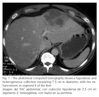
48-year-old man, active cigarette smoker and light drinker. Asymptomatic carrier of hepatitis B surface antigen (HBsAg), with normal liver profile. He denied having travelled abroad or any contact with immigrants from developing countries. He did not take any medication. He was admitted to our hospital with a 5-day history of dull and continuous abdominal pain in epigastrium and mesogastrium, sweating, chills and a temperature of up to 39ordm;-40ordm; C. He denied respiratory symptoms or changes in bowel habit. On admission, the temperature was 39ordm; C, his abdomen was tender in the right upper quadrant, with voluntary guarding but negative Murphy and Blumberg signs. The laboratory investigations revealed leucocytosis of 15,300 leuc/µl with left shift, with no anemia or thrombocytosis; the erythrocyte sedimentation rate was 93 mm/h; Other laboratory results were as follows: glucose 145 mgr/dl; creatinine 1 mg/dl; albumin 3,3 gr/dl; bilirubin 1,2 mgr/dl; serum glutamic oxalacetic transaminase 30 UI/L; serum glutamate pyruvate transaminase, 47 UI/L; gamma glutamyl transferase, 93 UI/L; alkaline phosphatase, 149 UI/L, fibrinogen 1710 mgr/L, prothrombin activity 78%, cephaline time 30 s; HBV serology: positive HBsAg, positive AntiHBe, negative antiHBs, negative AntiHB-core IgM and positive AntiHB-core IgG; positive PCR for HBV DNA, HBV viral load 2.378.000 copies/ml; negative HCV serology; negative HIV serology; negative serology for Entamoeba histolytica on admission (6 days after clinical presentation); determined by the rapid slide test for the detection of amoebic antibodies in serum (Bicho-Latex r-Amibe, Fumouz); sterile blood cultures, negative stool cultures for bacteria and parasites. Abdominal ultrasonography and computed tomography showed a hypodense collection, 7,5 x 5 cm in diameter, in segment II of the left lobe of the liver, heterogeneous and with rim enhancement, protruding from the liver surface. There were also radiologic findings of chronic liver disease without portal hypertension (Fig 1).
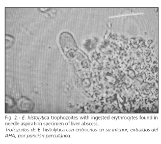
A diagnosis of pyogenic abscess was initially entertained, due to the absence of epidemiologic risk factors and the negative serology for Entamoeba histolytica. Antibiotic therapy was initiated with piperaciline-tazobactam and a percutaneous drainage was performed. Aspirate from the abscess was thick in consistency and reddish, "chocolate" coloured, with negative microscopic examination for organisms. A sample of the aspirate was cultured. The abdominal pain improved, but the patient remained with high temperature. The antibiotic regimen was changed to imipenem. The diagnosis of ALA was confirmed after identifying motile forms of Entamoeba histolytica in the liver aspirate, with negative culture for bacteria (Fig. 2). Treatment with metronidazole was initiated with disappearance of fever. The patient remained afebrile for 48 hours with subsequent recurrence of the high temperature. A second blood cultures were sterile. Piperaciline-tazobactam was added, with resolution of fever and marked improvement in the patientacute;s condition. There was no need for a second percutaneous drainage. The patient was discharged with levofloxacin for six weeks and radiologic studies obtained one month after discontinuation of antibiotic, showed decrease in the size of the abscess.
DISCUSSION
The ALA is uncommon in developed countries like Spain in the absence of an epidemiologic history of exposure, such as travelling to endemic countries, contacting with immigrants from regions of endemicity, or belonging to a risk groups. The reported case was an asymptomatic carrier of hepatitis B surface antigen (HBsAg), but had no epidemiologic risk factors for ALA. A case of a HBsAg carrier with simultaneous amebic liver abscess has been reported in an area of endemicity for amebiasis and on the background of hepatitis E infection (11). The case that we describe could be considered an autochthonous case of ALA, related to the increasing number of cases diagnosed in Spain.
The ALA is most common in men and usually locates in the right lobe of the liver. In more than 60% of cases, like in the case that we describe, the stool examination is negative for parasites. The microbiologic diagnosis is based on the detection of the parasite in the abscess aspirate, although this is an uncommon finding due to the necrotic nature of the abscess (12). The lack of findings consistent with pyogenic abscess (negative gram staining and culture) leads to the diagnosis of amebic abscess. Diagnostic percutaneous aspiration is rarely needed. In the case that we report the percutaneous aspiration was required because of false negative serology for amebiasis and the absence of epidemiologic risk factors. Serum tests are usually very helpful in the diagnosis of the disease. Although indirect hemagglutination assay (IHA) is the most sensitive test (90-100%), the latex agglutination test (13) is rapid and the results correlate with those of IHA, but they can be negative in the first week. The sensitivity increases in the second to third week of the infection. In the present case, the early determination, in the sixth day of the disease, could be the cause of the initial negative result.
The symptoms of ALA are acute and intense in 80% of cases and consist of epigastric pain and high fever. There is often leukocytosis and abnormal liver profile. The radiological findings are those of a liver abscess and consist of a low density collection on CT or a hypoechogenic lesion on ultrasonography, with posterior acustic shadowing and inner echos that mobilize with postural changes. Nevertheless, there are no pathognomonic findings for ALA (2). Gammagraphy with gallium-67 reveal "cold" lesions with peripheral rim enhancement, unlike in the case of a pyogenic abscess that appear as a capturing lesion. Nevertheless, this technique lacks the specificity that was initially believed (14).
With early diagnosis and treatment, mortality from uncomplicated liver abscess is less than 1%, ranging from 1-34%, and prevents surgery. The complications of ALA are: rupture into the peritoneum, pleural space and pericardium. The risk of rupture is higher in left-lobe abscesses, due to the smaller size of this lobe and the lack of space for a growing mass (2).
The aim of the treatment is to treat invasive liver infection and eradicate colonic colonization. Metronidazole is the drug of choice for the treatment of ALA and amebic colitis (adult dosage of 750 mg orally three times a day for 7-10 days). This therapeutic regimen should be followed by a luminar agent like paramomycin for a period of seven days (2). Most ALA respond to metronidazole therapy. The percutaneous aspiration and drainage of ALA is controversial. Indications for aspiration of liver abscesses are the need to rule out a pyogenic abscess; bacterial coinfection of ALA, large abscesses with a diameter of more than 5 cm, the prevention of rupture of left-lobe abscesses, the failure to respond clinically to drug therapy within 5 to 7 days and the threat of imminent rupture (2,15,16). The percutaneous aspiration can cause the superinfection of the ALA, so it should be avoided in those cases with no indication. This is probably what happened in our case, when the negative serology associated to the absence of epidemiologic risk factors misled to the diagnosis of pyogenic abscess, and subsequently, an early percutaneous drainage was performed. Surgical treatment should be reserved for instances of rupture of the abscess or coinfection that is not solved with medical treatment (2,10).
The increase of the number of cases in the absence of epidemiologic risk factors in our country, as derived from the increasing number of cases lately reported, and the present case, lead to consider the existence of autochthonous cases. The possibility of ALA should be entertained even in the absence of history of exposure, and serology should be repeated in suspected cases with an initially negative result. An early and accurate diagnosis avoids a higher morbi-mortality, as the treatment for ALA differs from that of the pyogenic abscess. It would be convenient to design epidemiologic studies to know the real prevalence of amebiasis in Spain.
REFERENCES
1. Ayeh-Kumi PF, Petri WA. Diagnosis and management of amebiasis. Infect Med 2002; 19: 375-82. [ Links ]
2. Rasidul Haque MB, Huston CD, Hughes M, Houpt E, Petri WA. Amebiasis. NEJM 2003; 348:1565-73. [ Links ]
3. Hughes M, Petri W. Amebic liver abscess. Infectious Clinics of North America 2000; 14: 565-82. [ Links ]
4. Amarapurkar DN, Patel N, Amarapurkar AD. Amoebic liver abscess. J Hepatol 2003; 39: 291-6. [ Links ]
5. Garré C, Morán A, Albaladejo A, Garcia J, Mercader J. Absceso hepático amebiano. Rev Esp Enferm Dig 2002; 94: 564-9. [ Links ]
6. Hidalgo ME, Rodríguez JC, Vizoso F, Díez MC. Absceso hepático amebiano de origen autóctono: presentación de un caso. Enferm Infecc Microbiol Clin 2000; 18: 145-6. [ Links ]
7. Lomas JM, Alcoucer R, Saavedra J, Pujol E. Infestación humana por Entamoeba Histolytica. ¿Una enfermedad autóctona de nuestro medio? A propósito de un caso. Rev Clin Esp 2000; 200: 399. [ Links ]
8. Perez E, Cilla G, Urbieta M, Muntilde;oz I. Infeccones autóctonas por Entamoeba Histolytica. Med Clin (Barc) 1985; 85: 254. [ Links ]
9. Rodríguez J, Canut A, Brezmes MF, de Fuentes I. Implicaciones clínico-epidemiológicas de la infección autóctona por Entamoeba Histolytica. Rev Esp Enferm Dig 1995; 87: 835-6. [ Links ]
10. Martín Ezquerro A, González Quijada S, Duentilde;as Gutiérrez C, Grande Sáez C. Absceso hepático amebiano autóctono. Presentación de un caso. Rev Clin Esp 2004; 204: 43-7. [ Links ]
11. Jain A, Kar P. HBs Ag carrier with simultaneous amebic liver absceso and acute hepatitis E. Ind J Gastroenterol 1999; 55: 179-84. [ Links ]
12. Li E, Stanley SL. Protozoa. Amebiasis. Gastroenterol Clin North Am 1996; 25: 471-92. [ Links ]
13. Robert R, Mahaza C, Bernard C, Buffard C, Senté JM. Evaluation of a new bicolores latex agglutination test for immunological diagnosis of hepatic amebiasis. J Clin Microbiol 1990; 28: 1422-44. [ Links ]
14. Asorey A, Algusacil A, Guerrra JM, Vilalta E. Amebiasis invasiva (II): formas extraintestinales y complicaciones. Diagnóstico parasitológico y serológico. Tratamiento. Rev Esp Enferm Dig 1985; 176: 271-80. [ Links ]
15. Garcia-Forcada A, Sans M, Gascon J, Valls E, Bru C, Corachan M. Absceso hepático amebiano: revisión de 13 casos. Med Clin 1995; 105: 537-40. [ Links ]
16. Rendón Unceta P, Macías Rodríguez MA, Correro Aguilar F, et al. Abscesos hepáticos: ¿es la punción aspiración simple con control ecográfico una alternativa al drenaje con catéter? Gastroenterol Hepatol 2000; 23: 470-3. [ Links ]

