


My SciELO
 Custom services
Services on Demand
Journal
Article
 English (pdf)
 Article in xml format
 Article references
 Send this article by e-mail
Indicators
-
 Cited by SciELO
 -
 Access statistics
Related links
-
 Cited by Google
 -
 Similars in
SciELO
Similars in
SciELO -
 Similars in Google
Share

 Permalink
Revista Española de Enfermedades Digestivas
Print version ISSN 1130-0108
Rev. esp. enferm. dig. vol.104 n.6 Madrid Jun. 2012
https://dx.doi.org/10.4321/S1130-01082012000600007
PICTURES IN DIGESTIVE PATHOLOGY
Usefulness of endoscopic ultrasound in the evaluation of a lymphoma with multiple gastric and pancreatic lesions
Utilidad de la ecografía en la evolución de un linfoma con múltiples lesiones gástricas y pancreáticas
Ramiro Rodríguez-Pérez1, Julio Iglesias-García2,3, Matilde Álvarez-del-Castillo2,3, José Lariño-Noia2,3, José Iglesias-Canle2,3 and J. Enrique Domínguez-Muñoz2,3
1Department of Gastroenterology. Hospital Universitario de Gran Canaria "Doctor Negrin". Las Palmas de Gran Canaria, Spain
2Department of Gastroenterology. Hospital Universitario de Santiago de Compostela. Santiago de Compostela, A Coruña. Spain
3Foundation for Research in Digestive Diseases. Hospital Universitario de Santiago de Compostela. Santiago de Compostela, A Coruña. Spain
Case report
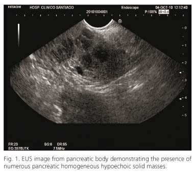
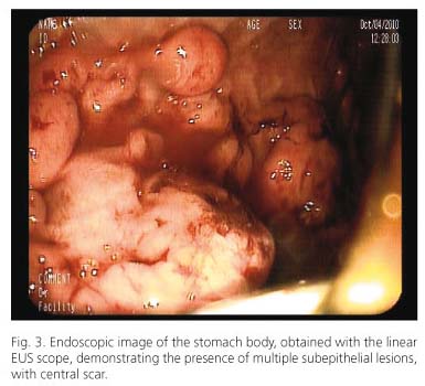
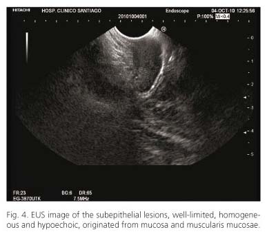
A 22-years-old woman, without relevant medical history, was hospitalized with a 10-days history of jaundice and epigastric pain. Laboratory test revealed 5,920 leukocytes/mm3, 448,000 platelets/mm3, bilirubin 3.4 mg/dL, aspartate aminotransferase 153 IU/L, alanine aminotransferase 405 IU/L, gamma-glutamyl transferase 188 IU/L, alkaline phosphatase 486 IU/L. A linear EUS was performed with Pentax EG-3870UTK® and HITACHI-Preirus®, demonstrating numerous pancreatic homogeneous hypoechoic lesions (Fig. 1), measuring from 4 to 20 mm, with a homogenous blue elastographic pattern and a strain ratio of 35.33 (Fig. 2). Microcholedocholitiasis and an upstream dilated common biliary duct due to an extrinsic compression by a pancreatic mass were also detected. Several subepithelial gastric lesions, with a central scar (Fig. 3), well-limited, homogeneous and hypoechoic, originated from mucosa and muscularis mucosae layer were also identified (Fig. 4). Biopsies were taken from these subepithelial lesions, giving the diagnosis of a diffuse large B-cell lymphoma. A multidetector-CT-scan confirmed previous finding. An endoscopic retrograde cholangiopancreatography (ERCP) was performed, and a 5 cm-10Fr plastic biliary stent was placed. The patient was submitted to Hematology Department, and started with a specific chemotherapy strategy.

Discussion
Endoscopic ultrasound (EUS) is considered as the most accurate method for the diagnosis and staging of pancreatic and gastro-esophageal lesions. EUS can also guide tissue sampling from both gut wall (by using standard forceps biopsies) and pancreatic solid lesions (by guiding fine needle aspiration (FNA) (1-3). Nowadays, new tools associated to EUS, like elastography, add relevant information to the standard B-mode image, helping in the differential diagnosis of solid pancreatic masses (4,5). In the present case, biopsies were obtained from a gastric lesion, giving the final diagnosis of a diffuse large B-cell lymphoma. When analyzing the multiple pancreatic solid masses, the strain ratio measured by EUS-guided elastography was 35.33, which has been associated in our previous studies to malignancy (5).
References
1. Dye CE, Waxman I. Endoscopic Ultrasound. Gastroenterol Clin North Am 2002; 31:863-79. [ Links ]
2. Iglesias-García J, Lariño-Noia J, Domínguez-Muñoz JE. Endoscopic ultrasound in the diagnosis and staging of pancreatic cancer. Rev Esp Enferm Dig 2009;101:631-8. [ Links ]
3. Dumonceau JM, Polkowski M, Larghi A, Vilmann P, Giovannini M, Frossard JL, et al. Indications, results, and clinical impact of endoscopic ultrasound (EUS)-guided sampling in gastroenterology: European Society of Gastrointestinal Endoscopy (ESGE) clinical Guideline. Endoscopy 2011;43:897-912. [ Links ]
4. Iglesias-García J, Lariño-Noia J, Abdulkder I, Forteza J, Domínguez-Muñoz JE. EUS elastography for the characterization of solid pancreatic masses. Gastrointest Endosc 2009;70:1101-8. [ Links ]
5. Iglesias-García J, Lariño-Noia J, Abdulkader I, Forteza J, Domínguez-Muñoz JE. Quantitative endoscopic ultrasound elastography: an accurate method for the differentiation of solid pancreatic masses. Gastroenterology 2010;139:1172-80. [ Links ]

