


Meu SciELO
 Serviços customizados
Serviços Personalizados
Journal
Artigo
 texto em
  Inglês (pdf)
 Artigo em XML
 Referências do artigo
 Enviar este artigo por email
Indicadores
-
 Citado por SciELO
 -
 Acessos
Links relacionados
-
 Citado por Google
 -
 Similares em
SciELO
Similares em
SciELO -
 Similares em Google
Compartilhar

 Permalink
Revista Española de Enfermedades Digestivas
versão impressa ISSN 1130-0108
Rev. esp. enferm. dig. vol.104 no.6 Madrid Jun. 2012
https://dx.doi.org/10.4321/S1130-01082012000600011
LETTERS TO THE EDITOR
Laparoscopy-assisted ERCP. Technical aspects
ERCP asistida por laparoscopia. Aspectos técnicos
Key words: Laparoscopy. Choledocholithiasis. Endoscopic retrograde cholangiopancreatography.
Palabras clave: Laparoscopia. Coledocolitiasis. Colangiopancreatografía retrógrada endoscópica.
Dear Editor,
When Itaru Oi et al. (1), in 1970, began using the ERCP and made the first cholangiography with an fiberduodenoscope introduced by the mouth to access the Vater's papilla, they probably could not imagine that, a few decades later, through to the development of laparoscopy, other approaches could be used for this procedure.
In patients with surgery for morbid obesity those who have been made a biliopancreatic bypass, with or without gastrectomy, and in those patients who have been made a gastroenterostomy for pyloric stenosis, it is not possible to do the conventional ERCP when they present biliopancreatic pathology.
With the arrival of laparoscopy, is not necessary that the anatomy of the upper digestive tract is intact to perform an ERCP.
We report the case of two patients with severe biliary tract disease that required to carry out an ERCP, but who had undergone several interventions for morbid obesity that prevent access to the Vater's papilla through the mouth, and was successfully resolved by laparoscopic assistance.
Case report 1
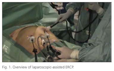
A 58-year-old woman underwent laparoscopic bilio-pancreatic diversion with distal gastric preservation for morbid obesity in April 2007. The patient was admitted to hospital with right upper quadrant and epigastric pain, fever, and jaundice. Abdominal US and computed tomography scan showed dilatation of intrahepatic biliary tract and common bile duct (CBD) (15 mm Ø) and a 10-mm diameter CBD stone. Endoscopic access to the Vater's papilla was not possible due to previous surgery. A transabdominal laparoscopic-assisted ERCP to treat choledocholithiasis was attempted (Fig. 1). A 15-mm trocar with a valve inside was placed on the greater curvature of the excluded stomach. A therapeutic duodenoscope (TJF 145, Olympus, Tokio, Japan) with an outer diameter of 12 mm was disinfected with 2% glutaraldehyde. The endoscopist advanced the duodenoscope through the gastrotomy to the major papilla. A deep cannulation of the CBD with an ERCP catheter (FSOMNI, Wilson-Cook Medical, Winston-Salem, NC, USA) was performed. Cholangiography demonstrated a dilated common bile duct and intrahepatic ducts as well as the presence of one 8-mm Ø CBD stone. Biliary sphincterotomy was performed which allowed decompression of the biliary tree. The stones were then extracted with a 12-15-mm catheter (Extractor Rx. 12- or 15-mm Retrieval Balloon, Boston Scientific, Boston, MA, USA).
Case report 2
A 35-year-old male patient operated for morbid obesity (Scopinaro technique) by open laparotomy in 2001. In May 2011, he was admitted to the hospital for severe acute cholangitis, with fever of 38.5 oC, jaundice and intense pain in the epigastrium and right upper quadrant. The ultrasound and abdominal CT showed multiple choledocholithiasis. Because of the previous gastrectomy, we performed a laparoscopic-assisted ERCP. Through a 15 mm Ø trocar, implanted directly at the angle of Treitz by the surgeon, a conventional gastroscope (Olympus GIF-Q165 videogastroscope of 9 mm Ø) was introduced to the papilla. The CBD was cannulated in a selective and deep way. The CBD was 15 mm Ø and was fully occupied by multiple filling defects, the most distal of 10 mm Ø (Fig. 2).

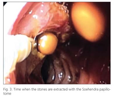
A sphincterotomy was performed by a reverse cut using a Soehendra papillotome for Billroth II (BII) (Cook, PTG-20-6-BII-NG). Then, using a 12-15 mm Ø Fogarty balloon (Boston Scientific; Extractor Retrieval), between 10 and 12 yellowish rounded calculus, similar to the soybeans, were extracted (Fig. 3).
Discussion
Due to the current development of laparoscopy, in those cases where it is required to perform an ERCP and it cannot be done orally because a gastrectomy has been performed to the patient, depending on the type of surgery performed, it can be done using a transgastric approach (2-6) or through the jejunum (7,8) , with a high level of safety and efficacy.
In the first case, it is advisable to use a duodenoscope. In the second case, a gastroscope can be used as first intention. If this one is not useful because the cannulation of the main bile duct is not possible, or very difficult, we can use a duodenoscope. For this, during laparoscopy, it is always advisable to use a large caliber trocar, of 15 mm Ø, which permits the passage of any instrument.
In the case of cannulate using a gastroscope, sphincterotomy should be performed with a sphincterotome for Billroth II, with the technical implications involved with this.
Of course, it is important that there is good collaboration between surgeons and endoscopists.
Juan J. Sebastián-Domingo, Mara Charro-Calvillo, Mónica Navarro-Dourdil, Isabel Aured-de-la-Serna,
Rafael Uribarrena-Amézaga, Tomás Cabrera-Chavesand Gloria Ceña-Lázaro
Department of Digestive Diseases. Unit of Endoscopy. Hospital General Royo Villanova. Zaragoza, Spain
References
1. Oi I, Kobayashi S., Kondo T. Endoscopic pancreatocholangiography. Endoscopy 1970;2:103-6. [ Links ]
2. Peters M, Papasavas PK, Caushaj PF, Kania RJ, Gagné DJ. Laparoscopic transgastric endoscopic retrograde cholangiopancreatography for benign common bile duct stricture after Roux-en-Y gastric bypass. Surg Endosc 2002;16:1106. [ Links ]
3. Pimentel RR, Mehran A, Szomstein S, Rosenthal R. Laparoscopy-assisted transgastrostomy ERCP after bariatric surgery: case report of a novel approach. Gastrointest Endosc 2004;59:325-8. [ Links ]
4. Nguyen NT, Hinojosa MW, Slone J, Lee J, Khiatani V, Wilson SE. Laparoscopic transgastric access to the biliary tree after Roux-en-Y gastric bypass. Obes Surg 2007;17:416-9. [ Links ]
5. Tercio L. Lopes, Ronald H. Clements, C. Mel Wilcox. Laparoscopy-assisted ERCP: experience of a high-volume bariatric surgery center. Gastrointest Endosc 2009;70:1254-9. [ Links ]
6. Bertin PM, Singh K, Arregui ME. Laparoscopic transgastric endoscopic retrograde cholangiopancreatography (ERCP) after gastric bypass: case series and a description of technique. Surg Endosc 2011; DOI 10.1007/s00464-011-1593-5 [ Links ]
7. Lopes TL, Clements RH, Wilcox CM. Laparoscopy-assisted transjejunal ERCP in a patient with Roux-en-Y reconstruction following partial gastrectomy. J Laparoendosc Adv Surg Tech A. 2010;20:55-8. [ Links ]
8. Mutignani M, Marchese M, Tringali A, Tacchino RM, Matera D, Foco M, et al. Laparoscopy assisted ERCP after biliopancreatic diversion. Obes Surg 2007;17:251-4. [ Links ]

