


Mi SciELO
 Servicios personalizados
Servicios Personalizados
Revista
Articulo
 Inglés (pdf)
 Articulo en XML
 Referencias del artículo
 Enviar articulo por email
Indicadores
-
 Citado por SciELO
 -
 Accesos
Links relacionados
-
 Citado por Google
 -
 Similares en
SciELO
Similares en
SciELO -
 Similares en Google
Compartir

 Permalink
Revista Española de Enfermedades Digestivas
versión impresa ISSN 1130-0108
Rev. esp. enferm. dig. vol.104 no.6 Madrid jun. 2012
https://dx.doi.org/10.4321/S1130-01082012000600016
LETTERS TO THE EDITOR
Double pylorus; a complication of gastric ulcer
Doble píloro: una complicación de úlcera gástrica
Key words: Double pyloric canal. Gastric ulcer. Endoscopy.
Palabras clave: Doble canal pilórico. Úlcera gástrica. Endoscopia.
Dear Editor,
Double pylorus is a rare pathological condition, which develops with fistulization between gastric antrum and duodenal bulb (1-4). Its incidence changes between 0.02 and 0.6% (5-7). It can be congenital or acquired (2). Gastric or duodenal duplications are in the etiology of congenital ones, whereas peptic ulcer diseases cause acquired ones (1-3).
Case report
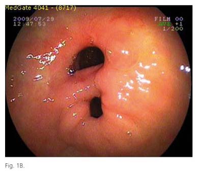
A 75-years-old, female patient, who takes non-steroidal anti-inflammatory drugs (NSAIDs) frequently due to her arthralgia symptoms, applied to our clinic with the complaints of abdominal pain in the epigastric region. She had endoscopy, which revealed a region with a depressed center at the small curvature side, distal to antrum in the prepyloric region. When this region was forced mildly by the endoscope, an ulcer mouth with a necrotic base and covered by off white exudate was detected (Fig. 1A). Biopsy samples were obtained from this region. Passing through the pylorus canal, it was reached to the bulb, which was normal, but mucosa was edematous and rough at the level of ampulla of Vater in the second region of the duodenum. Although lumen diameter was observed normal, the distal part could not be reached. Malignity was not detected in the biopsy specimens and the patients were positive for Helicobacter pylori (HP), so she received the HP treatment. Proton pump inhibitors (PPIs) are given for 2 months after the HP treatment and the patient had the control endoscopy after the 2nd month. In the endoscopy, ulcer was recovered with a large fistula mouth instead of its place (Fig. 1B). As a result of passages from fistula to the bulb directly through pylorus canal, fistula mouth opening to the bulb was observed at the roof of the bulb. Patient is still using PPI and in her control endoscopy, fistula has been observed to be open at the 5th month.

Discussion
Double pylorus is a rare condition, which was first described by Schmit and Tuttle (8). It is most frequently presented by ulcer complications (1-4). Our case had peptic ulcer like complaints for about 5 months. Double pylorus is most frequently located in the small curvature side in the antrum as it has been observed in our case. This location area is consistent with the most common localization of gastric ulcers (6-10).
Double pylorus is commonly encountered in people generally over 50 years of age with concomitant systemic diseases like diabetes mellitus, chronic renal failure, chronic obstructive pulmonary disease and cardiac failure, which delay wound healing and deteriorates microcirculation, and in people, who use NSAIDs for long terms (6-10). Our case was 76 years old and was using NSAIDs for a long time.
HP eradication is important for HP (+) cases to prevent ulcer recurrence and to relieve the symptoms in the treatment of double pylorus (9,10). Patients are advised to use PPI for long periods of time. Despite this, it is not expected that fistula is closed (1-3,5). In a study, in which 20 patients with double pylorus were followed up for 10 years, fistula was observed to have been closed in one patient (5). In our case, complaints of the patient were relieved but the fistula was not closed in 5-month follow up period. Although when compared to 10-year follow up period, this follow up period was a shorter one, we have believed that it would be difficult for the fistula to be closed.
Muzaffer Erturk
Department of Gastroenterology. SEV American Hospital. Gaziantep, Turkey
References
1. Chithriki M, Sadiq S, Jaibaji M. Double pylorus. Applied Radiology 2004;33:5-47. Available at: http://www.appliedradiology.com [ Links ]
2. Kwan WH, Yeung WH, Chan TM, Cheng CS. Double pylorus with occult gastrointestinal bleeding. J HK Coll Radio 2001;4:157-9. [ Links ]
3. Safatle-Ribeiro AV, Ribeiro Júnior U, Habr-Gama A, Gama-Rodrigues JJ. Double pylorus: Case report and review of the literature. Rev Hosp Clin Fac Med Sao Paulo 1999;54:131-4. [ Links ]
4. Lee S-Y, Kim E-S, Cho Y-S. Gastrointestinal: Acquired double pylorus; long term endoscopic observation. J Gastroen Hepatol 2012;27:413. [ Links ]
5. Hu TH, Tsai TL, Hsu CC, Lu SN, Hsiao M, Changchien CS. Clinical characteristics of double pylorus. Gastrointest Endosc 2001;54:464-70. [ Links ]
6. Peixoto P, Saido A, Cancela E, Castanheira A, Ministro P, Silva A, et al. Acute upper bleeding due to an unusual complication of peptic ulcer disease - double pylorus. Rev Esp Enferm Dig 2010;102:450-3. [ Links ]
7. Pollani A, Marchi S, Bellini M, Costa F, Bounifazi V, Tumino E, et al. Double pylorus: report of two cases and review of the literature. Ital J Gastroenterol 1991;23:360-3. [ Links ]
8. Smith VM, Tutle KW. Gastroduodenal (pyloric) band. Gastroenterology 1969;56:331-6. [ Links ]
9. Sayilir A, Kurt M, Önal K, Beyazit Y, Suvak B. An unusual complication of peptic ulcer disease: Double pylorus. Gastroenterol Nurs 2011;34:401. [ Links ]
10. Arhan M, Oztas E, Ibis M, Sezgin S, Ozin Y. A rare endoscopic finding: acquired double pylorus. Surg Endosc 2010;24:244-5. [ Links ]

