


My SciELO
 Custom services
Services on Demand
Journal
Article
 text in
  English (pdf)
 Article in xml format
 Article references
 Send this article by e-mail
Indicators
-
 Cited by SciELO
 -
 Access statistics
Related links
-
 Cited by Google
 -
 Similars in
SciELO
Similars in
SciELO -
 Similars in Google
Share

 Permalink
Revista Española de Enfermedades Digestivas
Print version ISSN 1130-0108
Rev. esp. enferm. dig. vol.105 n.4 Madrid Apr. 2013
https://dx.doi.org/10.4321/S1130-01082013000400014
LETTERS TO THE EDITOR
Hemangioendothelioma and other small bowel tumors as a cause of obscure gastrointestinal bleeding
El hemangioendotelioma y otros tumores de intestino delgado como causa de hemorragia digestiva de origen oscuro
Key words: Small bowel disease. Intestine neoplasms. Vascular tissue. Capsule endoscopy.
Palabras clave: Enfermedades del intestino delgado. Neoplasia intestinal. Enfermedad vascular. Cápsula endoscópica.
Dear Editor,
Vascular and neoplastic lesions of the small intestine are the most common cause of obscure gastrointestinal bleeding. But sometimes, in adults, we can find intermediate malignancy lesions whose management can be a challenge.
Case report
We present a 78-year-old woman with episodes of hematochezia of 4 months duration without other symptoms. She had a painful right hemiabdomen without masses at palpation, ferropenic anemia (hemoglobin 6,6 g/dL, transferrin saturation index 4 %) and normal level of carcinoembryonic antigen.
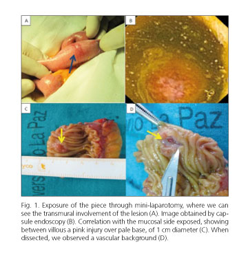
There was not any bleeding lesion in gastroscopy or colonoscopy. But the barium X-ray showed a filling contrast defect in the last ileal loop. An endoscopic capsule was performed, in which we could recognize a lesion that appeared to be submucosal, of 1 cm in size, eroded, with minimal bleeding, approximately 10 minutes before the first image of the cecum.
Because of this finding, a surgical exploration was done. A 1 cm pink lesion over a pale base, was identified, 70 cm proximal to ileocecal valve. No other lesions or lymphadenopathy were found on parietal surface of the bowel, mesentery or peritoneum. A 7 cm enterectomy was performed with safety margins of 3 cm and terminal anastomosis.
Histological analysis described a epithelioid eosinofilic cells proliferation, with irregular sized cells and prominent nucleoli, forming a kind of lobes and small blood vessels with erythrocytes inside them. Immunohistochemistry CD34+ (hematopoietic stem-cell) and proliferation index of 20 % (intermediate malignancy) was compatible with a hemangioendothelioma.
Staging PET-TC did not show pathologic uptake. Patient is in clinical remission at 3 years follow-up.
Discussion
Small bowel tumors account for 1-3 % of all gastrointestinal malignancies and approximately two thirds are malignant (1). They constitute a finding in 3 % of scans with capsule endoscopy (2,3). Among the clinical manifestations, hemorrhage is the second symptom in frequency after obstruction. Bleeding occurs in 20-50 % of the benign tumors and most of malignant (4).
If associated with anorexia and weight loss, it should suspect malignancy, although only half of malignancies presents this triad (5).The most common benign tumors are leiomyomas (40 %), adenomas (30 %), and lipomas (20 %). Among the malignant, the more frequent are adenocarcinomas (40 %), carcinoid (25-30 %), lymphomas (15-20 %), sarcomas (12 %), and metastasis (melanoma, breast cancer and hypernephromas) (6).
The term "hemangioendothelioma", coined in 1908 (Mallory) to designate any endothelial tumor, was booked from 1988 (Enzinger, Weiss) to define an intermediate histological lesion between malformation (angioma) and malignant tumor (angiosarcoma).
Hemangioendotheliomas are rare and locally aggressive. There are four histological types: epithelioid, fusiform, kaposiform, and endovascular papillary angioendothelioma (Dabska tumor) (7). Frequency order concerning location is: Skin, liver, spleen, gastrointestinal tract (especially jejunum and ileum) (8), bone, lung and head-neck. About 50 % of patients with internal organ involvement, associate skin lesions.
In adults, fusiform and epithelioid variants are the most common, affecting nodular/diffuse, uni/multi-organ. Usually are diagnosed in women at 6th decade of life. They usually manifest as iron deficiency anemia when it is located in the small intestine (9), or anemia, weight loss, and even painful hepatomegaly, portal hypertension or platelet sequestration (Kasabach-Merritt syndrome) when located in liver. Heart failure has been described because of massive arteriovenous shunts in case of big tumors. The development of hemangioendotheliomas has been associated with previous radiotherapy, oral contraceptives, and inhalation of vinyl chloride, none of which was present in our patient. Thirty percent of them metastasize via lymphatic, meaning 20-50 % 5-years survival. Although spontaneous remissions have been described, they usually require surgical excision. However, those located in the liver usually relapse and require corticosteroids, interferon or arterial embolization, with conflicting results among different reports.
Given their low frequency, we must not forget the differential diagnosis with other neoplasias. Aggressive surgery (with later histological confirmation) and early detection of multifocality are the keys to determine prognosis.
Lucía Tortajada-Laureiro1, Joaquín Poza-Cordón1, Consuelo Froilán-Torres1, Silvia Gómez-Senent1,
Eun Jin Han1, Juan Pascual-Turrión1, Fernando Luca-de-Tena-Díaz-Agero1,
Santiago Valderrábano-González2, Fernando Martínez-Ortiz3 and José María Segura-Cabral1
Departments of 1Gastroenterology, 2Surgery and 3Pathology
Hospital Universitario La Paz. Madrid, Spain
References
1. Jensen DM. Current diagnosis and treatment of severe obscure GI hemorrhage. Gastrointest Endosc 2003;58:256-66. [ Links ]
2. Romero-Vázquez J. Caunedo-Álvarez A, Herrerías-Gutiérrez JM. Cápsula endoscópica. En: Vázquez-Iglesias JL. Endoscopia Digestiva: diagnóstica y terapéutica. Madrid: Médica Panamericana; 2008. p.101-5. [ Links ]
3. Caunedo A, Rodríguez-Téllez M, García-Montes JM, Gómez-Rodríguez BJ, Guerrero J, Herrerías JM Jr, et al. Usefulness of capsule endoscopy in patients with suspected small bowel disease. Rev Esp Enferm Dig 2004;96:10-21. [ Links ]
4. Anil K. Rustgi. Small Bowel Neoplasms. En: Feldman M, Friedman LS, Brandt LJ, editors. Sleisenger & Fordtran's Gastrointestinal and Liver Diseases. 8th ed. Elsevier España 2008. p. 2706-7. [ Links ]
5. DiSario JA, Burt RW, Vargas H, McWhorter WP. Small bowel cancer: Epidemiological and clinical characteristics from a population-based registry. Am J Gastroenterol 1994;89:699-701. [ Links ]
6. Blanchard DK, Budde JM, Hatch GF, Wertheimer-Hatch L, Hatch KF, Davis GB, et al. Tumors of the small intestine. World J Surg 2000; 24:421-9. [ Links ]
7. Alvarez Sánchez JA, Fernández Lobato R, Coba Ceballos J, Fradejas López JM, Marín Lucas J, Moreno Azcoita M. Epithelioid hemangioendothelioma localized in the small intestine. Gastroenterol Hepatol 1995;18:464-7. [ Links ]
8. Yasuda S, Hashimoto T, Kanaizumi T, Kuwata H, Matsumoto I, Shiratori T. A case of hemangioendothelioma of the small intestine. Jpn J Surg 1989; 19:67-9. [ Links ]
9. Yoshida R, Takada H, Iwamoto S, Mouri T, Uedono Y, Kawanishi H, et al. Malignant hemangioendothelioma of the small intestine: Report of a case. Surg Today 1999;29(5):439-42. [ Links ]

