Mi SciELO
Servicios Personalizados
Revista
Articulo
Indicadores
-
 Citado por SciELO
 -
 Accesos
Links relacionados
-
 Citado por Google
 -
 Similares en
SciELO
 -
 Similares en Google
Compartir
Revista Española de Enfermedades Digestivas
versión impresa ISSN 1130-0108
Rev. esp. enferm. dig. vol.105 no.10 Madrid nov./dic. 2013
https://dx.doi.org/10.4321/S1130-01082013001000008
PICTURES IN DIGESTIVE PATHOLOGY
Skin metastases as initial manifestation of esophageal squamous-cell carcinoma
Metástasis cutáneas como primera manifestación de carcinoma epidermoide de esógafo
Marta Rivas-Rivas1, David Jiménez-Gallo2, Natalia Navas-García3, Cristina Albarrán-Planelles2 and Claudio Rodríguez-Ramos1
1UGC of Digestive Diseases, 2UGC of Dermatology and 3UGC of Pathology. Hospital Universitario Puerta del Mar. Cádiz, Spain
Esophageal cancer is the sixth most common cause of cancer-related deaths worldwide. Patients with esophageal cancer usually present with locally advanced disease with presence of metastases at diagnosis (1). Squamous-cell carcinoma is the most common histologic subtype in the proximal and middle thirds of the esophagus. Skin metastases from visceral tumors are rare, with a prevalence of up to 2 % (2). An association between esophageal cancer and skin metastases, as in the present case, has only been exceptionally described (3).
Case report
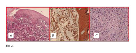
A 71-year-old male had a physical exam which revealed an ulcerated, hard tumor outgrowth, approximately 3 cm in size, on his right flank (Fig. 1A), in association with a similar 2.5 cm lesion in the left subclavicular area (Fig. 1B). He had dysphagia to both solids and liquids a few weeks after the development of said lesions, as well as 20 kg weight loss. The endoscopic study revealed a protruding, ulcerated neoplasm at 22 cm from the dental arch, which almost completely occludes the esophageal lumen (Fig. 1C) -histology found it to be an infiltrating squamous-cell carcinoma. A pathological examination of skin lesions revealed cutaneous metastatic infiltration by a squamous-cell carcinoma, with positive immunohistochemical staining for high molecular weight cytokeratine and vimentin (Fig. 2). Additional lab tests found the presence of metastatic bone disease involving the left ribs, D9 vertebral body, and right iliac bone. Treatment with a prosthesis and chemotherapy was ultimately selected.
Discussion
Skin manifestations from esophageal cancer are rare, less than 1 %, and were primarily described for adenocarcinoma (1). The presence of skin metastases from an esophageal squamous-cell carcinoma is extremely rare, and its review in the literature is limited by its uncommon nature. A study by Lookingbill et al., in 7,316 patients with skin cancer and metastases, found no individuals with a primary esophageal cancer (4). Another study by this same author in 4,020 patients only found 3 cases of skin metastasis from a primary esophageal squamous-cell carcinoma (5). Cutaneous metastases from gastrointestinal tumors usually manifest as ulcerated plaques or nodules, and their predominant site is the abdomen (6). The diagnosis of a skin metastatic malignancy relies on its histopathology, and often represents a challenge, most particularly when no clinical history or complaint is present. Immunohistochemistry is required for a proper diagnosis (7). In this case report we find the presence of skin metastases, their coexistence with a bone spread < 9 %, and their presentation as the primary tumor's initial manifestation of special interest. Importantly, this unusual tumor behavior with distant spread prior to local dissemination and clinical dysphagia must be borne in mind, and a biopsy of suspicious skin lesions should be considered.
References
1. Quint LE, Hepburn LM, Francis IR, Whyte RI, Orringer MB. Incidence and distribution of distant metastases from newly diagnosed esophageal carcinoma. Cancer 1995;76: 1120-5. [ Links ]
2. Nashan D, Muller ML, Braun-Falco M, Reichenberger S, Szeimies RM, Bruckner-Tuderman L. Cutaneous metastases of visceral tumours: A review. J Cancer Res Clin Oncol 2009;135:1-14. [ Links ]
3. Fereidooni F, Kovacs K, Azizi MR, Nikoo M. Skin metastasis from an occult esophageal adenocarcinoma. Can J Gastroenterol 2005;19:673-6. [ Links ]
4. Lookingbill DP, Spangler N, Sexton FM. Skin involvement as the presenting sign of internal carcinoma. A retrospective study of 7316 cancer patients. J Am Acad Dermatol 1990;22:19-26. [ Links ]
5. Lookingbill DP, Spangler N, Helm KF. Cutaneous metastases in patients with metastatic carcinoma: A retrospective study of 4020 patients. J Am Acad Dermatol 1993;29:228-36. [ Links ]
6. Rendi MH, Dhar AD. Cutaneous metastasis of rectal adenocarcinoma. Dermatol Nurs 2003;15:131-2. [ Links ]
7. Nashan D, Meiss F, Braun-Falco M, Reichenberger S. Cutaneous metastases from internal malignancies. Dermatol Ther 2010;23:567-80. [ Links ]


 texto en


