


Mi SciELO
 Servicios personalizados
Servicios Personalizados
Revista
Articulo
 texto en
  Inglés (pdf)
 Articulo en XML
 Referencias del artículo
 Enviar articulo por email
Indicadores
-
 Citado por SciELO
 -
 Accesos
Links relacionados
-
 Citado por Google
 -
 Similares en
SciELO
Similares en
SciELO -
 Similares en Google
Compartir

 Permalink
Revista Española de Enfermedades Digestivas
versión impresa ISSN 1130-0108
Rev. esp. enferm. dig. vol.106 no.2 Madrid feb. 2014
https://dx.doi.org/10.4321/S1130-01082014000200010
Transoral endoluminal approach to Zenker's diverticulum using LigasureTM. Early clinical experience
Tratamiento endoluminal transoral del divertículo de Zenker con LigasureTM. Experiencia clínica inicial
José Noguera-Aguilar1, Carlos Dolz-Abadía2, Angels Vilella2, José M. Muñoz-Pérez3, Héctor Julián Canaval-Zuleta2 and Leopoldo Salvatierra-Arrieta4
1Department of General Surgery. Consorcio Hospital General Universitario. Valencia, Spain
2Department of Digestive Diseases. Hospital Son Llàtzer. Palma, Spain
3Department of General Surgery. Hospital Son Llàtzer. Palma, Spain
4Department of Surgery. Hospital Universitario La Paz. Madrid, Spain
ABSTRACT
The development of minimally invasive procedures has rekindled interest in endoluminal techniques for the management of Zenker's diverticulum. Tissue sealers as employed in laparoscopic surgery have not been previously used for Zenker's diverticulum septotomy. Supported by the established safety of linear cutters, bipolar forceps, and ultrasonic scalpels, we have started a procedure using the Ligasure 5TM tissue sealer.
Safety and efficacy results in our early clinical experience are shown for a prospective series of 5consecutive Zenker's diverticulum cases that were perorally managed with tissue sealing. The procedure was quickly and safely performed in the endoscopy room under sedation. Mean number of seals per patient was2, and mean procedure duration was 33minutes. No complications developed during or after the procedure, and patients were discharged with immediate dysphagia relief and adequate oral tolerance. No diverticular relapses occurred after a mean follow-up of 21months (range18-30).
This procedure may be repeated as often as desired with no need for hospital admission. Safety should be prospectively assessed by further studies using a higher number of procedures.
Key words: Zenker's diverticulum. Flexible endoscopy. Septotomy. Endoscopic surgery.
RESUMEN
El desarrollo de los procedimientos mínimamente invasivos ha reavivado el interés por las técnicas endoluminales para el tratamiento del divertículo de Zenker. Los selladores titulares empleados en cirugía laparoscópica no han sido empleados previamente en la septotomía del divertículo de Zenker. Avalados por la seguridad previa del empleo de las cortadoras lineales, de las pinzas bipolares y de los bisturís por ultrasonidos, hemos iniciado dicho procedimiento mediante el empleo del sellador tisular Ligasure 5TM.
Se muestran los resultados de seguridad y eficacia en la experiencia clínica inicial de una serie prospectiva de 5 casos consecutivos de divertículo de Zenker tratados por vía peroral con el sellador tisular. El procedimiento se realizó en la sala de endoscopia, con sedación, de manera rápida y segura. La media de sellados por paciente fue de 2 y la duración media del procedimiento de 33minutos. No se presentaron complicaciones durante el procedimiento ni derivadas del mismo, siendo dados de alta los pacientes con desaparición inmediata de la disfagia y correcta tolerancia oral. Con un seguimiento medio de 21meses (rango 18-30), no existió recidiva del divertículo en ningún caso.
Este procedimiento puede ser repetido tantas veces como se desee y ser realizado sin ingreso hospitalario. La seguridad mostrada deberá ser evaluada prospectivamente en estudios posteriores con mayor número de procedimientos.
Palabras clave: Divertículo de Zenker. Endoscopia flexible. Septotomía. Cirugía endoscópica.
Introduction
Zenker's diverticulum originates in a spot of muscle weakness at the Killian triangle on the posterior hypopharyngeal wall, the diverticular neck being located proximal to the cricopharyngeal muscle. Obstructive pathophysiology at the upper esophageal sphincter seems a key factor for its development, in association with increased bolus pressure and resistance to deglutition arising from sphincter abnormalities.
Hypopharyngeal diverticulum management has obviously progressed over time since Wheeler's first successful operation in 1886, followed by Mosher in 1917 (1). Minimally invasive surgery was also used for the treatment of Zenker's diverticulum in a gradual attempt to minimize surgical aggression. Rigid endoscopic surgery was initially implemented, followed by an endoesophageal approach with flexible endoscopy (2). The use of laser and argon coagulation following septal section, and the more recent addition of endoscopic stapling techniques (2) have been instrumental in the management of this condition not only because of their low morbidity but also due to a reduction in surgery duration, postoperative pain, and hospital stay, and a faster return to oral ingestion (3). However, treatment limitations linger on despite leading-edge techniques. Among diverticular septal section techniques using a flexible endoluminal approach the following should be highlighted: Needle-knife sphincterotome (4,5), monopolar or bipolar coagulation forceps (6,7), and argon plasma coagulation (8,9). These septal section modalities have been combined with various technical modifications to improve septum visibility and exposure, including the use of nasogastric tubes, a transparent hood at the tip of a flexible endoscope, or an ad-hoc diverticulotomy overtube (10,11). Various types of flexible endoscope have also been used, including conventional (9-10mm in external diameter) and pediatric flexible endoscopes. The latter, with its smaller diameter, allows improved maneuverability.
Likewise, diverticular septal section may also be carried out using a rigid diverticuloscope. This modality is used by surgeons in the ENT field who usually employ linear cutter-staplers as in laparoscopic surgery (2). Harmonic scalpels have also been used in this approach with good reported outcomes (12,13). The shortcomings of ultrasonic energy, which results in light tissue sealing and high collateral heat dispersion, and of endostaplers, which have an excessive diameter and will not staple to the device end, may be alleviated by tissue sealers such as LigasureTM. The strengths of the peroral endoesophageal technique using a conventional endoscope and a 5-mm Ligasure -which allows repeat short sections with exquisite level control- include its performance under sedation in the endoscopy room and on outpatient basis.
The results of a short prospective series of Zenker's diverticulum cases perorally treated with a Ligasure 5TM tissue sealer are described below.
Patients and methods
This was an observational study using a prospective case series of 5consecutive Zenker's diverticulum patients who were treated with a peroral endoscopic approach combining flexible endoscopy and laparoscopic tissue sealing (Ligasure 5TM).
Patients were recruited at the gastroenterology clinic, which they had visited for assessment. The study was approved by the local Ethics Committee, and patients were informed prior to their consent. Symptoms included mild-moderate dysphagia (3cases) according to Karnell's swallowing scale (14), and food regurgitation (2cases). In one case several aspiration pneumonia/pneumonitis events were associated with regurgitation. After a careful anamnesis an upper flexible endoscopic procedure was first performed, which revealed a Zenker's diverticulum. No associated esophageal disease was found in the series, and studies performed included upper endoscopy and contrast-enhanced X-rays (barium exam or CT with an oral contrast) in all cases. A manometric exploration was performed when associated symptoms required that an esophageal motor disorder be excluded. Patient characteristics are listed in table I.

The therapeutic procedure was performed in the endoscopy room, in the presence of an anesthesiologist and under deep sedation with endovenous 1% propofol, no orotracheal intubation, oxygen therapy, and non-invasive monitoring. A flexible gastroscope (Olympus GIFQ165, Olympus Optical Co., Tokyo, Japan) with just one channel and 9.2mm in external diameter was used to appropriately visualize the diverticulum and esophageal lumen. A guidewire (0.035inch wide, 450cm long Jagwire, Boston Scientific, Natick MA, USA) was then passed along the esophagus to the stomach in order to withdraw the endoscope. Using the guidewire and under endoscopic vision an overtube-diverticuloscope was introduced, which was 30 cm long and 22mm in outer diameter (Cook Medical, Limerick, Ireland), and had a bivalve tip with a hole in the greater blade for the guidewire to be passed (Fig. 1). This maneuver helped us guide the overtube and place the greater blade in the esophagus and the smaller one inside the diverticulum, with good diverticular septum exposure on the medial work field.

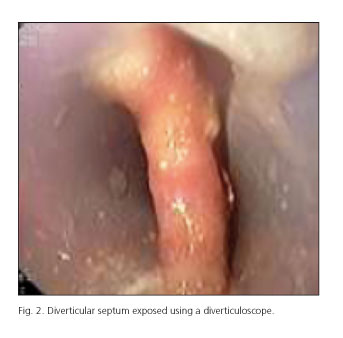
Then the conventional gastroscope was replaced by one with a smaller diameter (Olympus GIF-XP150N, 5.5mm in outer diameter). When an adequate view was obtained (Fig. 2), the 35-mm long, 5-mm thick laparoscopic tissue sealer (LigasureTM, Covidien, Massachussets, USA) was introduced in parallel with the endoscope, both of them traveling inside the overtube.
The septum was then sealed and subsequently sectioned using the sealer device, with a sealing power of 2 over 3 and in cycles covering approximately 1 cm at a time (Fig. 3). In every cycle absence of bleeding was acknowledged following tissue resection, and a new assessment of the septum's length was made by slowly advancing the overtube. After all necessary sessions, when only about 5mm remained to completely set the diverticular lumen level with the esophageal lumen the section procedure was ended and two hemostatic clips (Resolution Clip, Boston Scientific, Natick MA, USA) were used to help in the sealing of sectioned esophageal layers. Procedure ending at less than 1cm to the septal end was a safety measure to avoid perforating complications. Once the procedure was completed hemostasis was checked, devices were withdrawn, and the patient was transferred to the ward upon recovery from sedation.

Oral ingestion of fluids was restarted at 4 hours after the procedure, and patients were discharged at 24 hours after the procedure with dietary counseling similar to that of anti-reflux surgery: Ground, mashed food in the first week and avoidance of difficult-to-chew food for one month. Follow-up ensued with clinical interviews and both contrast-enhanced and endoscopic exams at 3 and 6 months.
Results
We report the early results from a prospective series of 5 consecutive patients with Zenker's diverticulum -regardless of size and with typical symptoms- who received peroral diverticular septotomy with flexible endoscopy and endoesophageal use of Ligasure 5TM for septal resection.
Mean number of sealings per patient was 2 (only one case needed 3 sealing-cutting cycles), and mean procedure duration was 3minutes. No complications arose during or after the procedure, albeit a patient developed a self-limited fever episode of unknown origin that spontaneously subsided but prolonged hospital stay to 48hours after the procedure. All remaining patients were discharged at 24 hours with immediate dysphagia relief and adequate oral tolerance.
With a mean follow-up of 21 months (range 18-30) no diverticular relapses occurred. Of all 5 patients, 3 are perfectly well with no residual symptoms. A patient with advanced age developed further dysphagia 12months after the procedure, and clinical testing ruled out diverticular relapse. This patient died of pneumonia after 13months, one month following symptom relapse. Another patient, also with advanced age, developed dysphagia at 24 months with no diverticular relapse, and refused to undergo further explorations in search of a new etiology.
Patient follow-up after the procedure was uneven as the series included individuals with highly variable ages, comorbidities, and therapy intents. Most were followed using clinical interviews at 1week after discharge, and both contrast-enhanced and postoperative endoscopy at 3 and 6 months after the procedure. A clinical interview was then performed every 6 months.
Discussion
The application of minimally invasive surgical techniques for Zenker's diverticulum has been quite delayed. While bile duct and intestinal tract conditions have witnessed an exponential growth of minimally invasive endoscopic approaches in the last 20years, cervical digestive diseases have become stragglers to this vigorous progression.This is due to difficulties in the implementation of endoscopic surgery concepts within a limited space such as the neck, and to the challenges involved in the setup of collaborative multidisciplinary teams to manage one given condition. In Zenker's diverticulum case, its low incidence among the general population must also be taken into account, which hinders the advancements that are based on the results from extensive series of cases.
Attempts at replacing the classic approaches of open surgery by less invasive techniques for the management of Zenker's diverticulum have been made, whose effectiveness is now being ascertained. The presence of very low morbidity and mortality rates, and the potential for repeat procedures over time without the difficulties entailed by open surgery reintervention, drive the gradually increasing use of novel peroral minimally invasive techniques in various sites worldwide.
The results of open surgery for Zenker's diverticulum were considered appropriate over time, but morbidity rates of up to 27% in our setting are far from negligible (15).The results of peroral endoscopic techniques carry lower morbidity and mortality rates with a comparable relapse rate, but procedures may be repeated with virtually the same safety as by first intention, which is not the case with open surgery. By perorally managing Zenker's diverticulum the opportunity arises to use rigid esophagoscopy or flexible endoscopy to perform septal resection using a number of equipments and energy sources. Regarding endoscopy type, whether rigid or flexible, it should be noted that rigid techniques call for general anesthesia, orotracheal intubation, the use of a rigid diverticuloscope -which is potentially more damaging- and forced hyperextension to the neck. This modality is usually performed by surgical teams in the ENT sphere within a surgical area, whereas flexible endoscopy is habitually carried out under sedation in an endoscopy room by a team of endoscopist gastroenterologists. According to our experience, the setup of a multidisciplinary team including endoscopists and surgeons represents a major breakthrough for the management of this condition. Procedures are performed in an endoscopy room under deep sedation in the presence of an endoscopist, surgeon, and anesthetist. The endoscopist provides exquisite control and a view unknown to surgeons when cutting the diverticular septum, whereas the surgeon contributes the potential to resect the septum using surgical equipment, that is, a safety and speed endoscopic instrumentation cannot afford.
The use of the Ligasure 5TM tissue sealer has not been previously reported in the literature in connection with this disease. Prior descriptions regarding harmonic scalpels, linear endostaplers, and other instruments allowed to ultimately understand that septal resection via a peroral route may provide symptom relief and solve the condition; however, the use of a tissue sealer seems to provide the procedure with increased safety. This type of sealer offers several benefits: It takes up little space in a situation -such as parallel flexible endoscopy with a peroral approach- where lack of room is significant; limited, controlled septal cuts may be safely made using consecutive 1-cm long sections, with sealing and subsequent cutting, in just one go, and electrocoagulation can potentially be used with the same tool should bleeding occur along the sealing/cutting line (available for current Ligasure ImpactTM versions).
Our initial experience with this tissue sealer has been highly satisfactory, with higher speed and safety levels as compared to our previous use of endoscopic instrumentation such as the needle-knife and coagulation with various terminals. Two major maneuvers endow the procedure with additional safety: The septum is not fully resected and a few distal millimeters (3-5mm) are left untouched to prevent an esophageal opening into the mediastinum, and endoscopic clips are placed upon septal cutting completion to secure the sealing of layers in the resected septum. These maneuvers do not seem to subsequently have any clinical relevance, and do not affect symptom resolution or relapses because of the millimeters of preserved septum or the placement of clips that fall off on their own and are not present at the time of follow-up endoscopy.
From all the above we believe that the technique discussed may represent a good approach to the peroral, minimally invasive treatment of Zenker's diverticulum, and one that may be carried out on an outpatient basis and with immediate oral tolerance following the procedure. Its proven safety must be prospectively assessed in further studies with a higher number of procedures.
References
1. Mosher HP. Webs and pouches of the esophagus, their diagnosis and treatment. Surg Gynecol Obstet 1917;25:175-87. [ Links ]
2. Collard JM. Endoscopic stapling technique of esophagodiverticulostomy for Zenker's diverticulum. Ann Thorac Surg 1993;56:573-6. [ Links ]
3. Bonavina L. Long-term results of endosurgical and open surgical approach for Zenker diverticulum. World J Gastroenterol 2007;14:2586-9. [ Links ]
4. Al-Kadi AS, Maghrabi AA, Thomson D, Gillman LM, Dhalla S. Endoscopic treatment of Zenker diverticulum: Results of a 7-year experience. J Am Coll Surg 2010;211:239-43. [ Links ]
5. Repici A, Pagano N, Romeo F, Danese S, Arosio M, Rando G, et al. Endoscopic flexible treatment of Zenker's diverticulum: A modification of the needle-knife technique. Endoscopy 2010;42:532-5. [ Links ]
6. Christiaens P, De Roock W, Van Olmen A, Moons V, D'Haens G. Treatment of Zenker's diverticulum through a flexible endoscope with a transparent oblique-end hood attached to the tip and a monopolar forceps. Endoscopy 2007;39:137-40. [ Links ]
7. Rieder E, Martinec DV, Dunst CM, Swanström LL. Flexible endoscopic Zenker's diverticulotomy with a novel bipolar forceps: A pilot study and comparison with needleknife dissection. Surg Endosc 2011;25:3273-8. [ Links ]
8. Rabenstein T, May A, Michel J, Manner H, Pech O, Gossner L. Argon plasma coagulation for flexible endoscopic Zenker's diverticulotomy. Endoscopy 2007;39:141-5. [ Links ]
9. Pech O, May A, Gossner L, Mayer G, Abdollahnia R, Ell C. Endoscopic therapy for Zenkers's diverticulum by means of argon plasmacoagulation. Z Gastroenterol 2002;40:517-20. [ Links ]
10. Costamagna G, Mutignani M, Tringali A, Perri V. Treatment of Zenker's diverticulum with the help of a plastic hood attached to the endoscope. Gastrointest Endosc 2002;56:611-2. [ Links ]
11. Costamagna G, Iacopini F, Tringali A, Marchese M, Spada C, Familiari P, et al. Flexible endoscopic Zenker's diverticulotomy: Cap-assisted technique vs. diverticuloscope-assisted technique. Endoscopy 2007;39:146-52. [ Links ]
12. Whited C, Lee WT, Scher R. Evaluation of endoscopic harmonic diverticulostomy. Laryngoscope 2012;122:1297-300. [ Links ]
13. Hondo FY, Maluf-Filho F, Giordano-Nappi JH, Neves CZ, Cecconello I, Sakai P. Endoscopic treatment of Zenker's diverticulum by harmonic scalpel. Gastrointest Endosc 2011;74:666-71. [ Links ]
14. Karnell MP, MacCracken E. A database information storage and reporting system for videofluorographic oropharyngeal motility (OPM) swallowing evaluations. Am J Speech Lang Pathol 1994;3:54-60. [ Links ]
15. Cañete-Gómez J, Ramírez-Plaza CP, Rueda BL, Ibáñez-Delgado F, Vázquez-Medina A, Bondia-Navarro JA, et al. Diverticulectomía y miotomía del cricofaríngeo para el tratamiento del divertículo de Zenker. Presentacion de una serie de 33 casos. Cir Esp. 2012;90:233-7. [ Links ]
![]() Correspondence:
Correspondence:
José Noguera Aguilar
Department of General Surgery
Consorcio Hospital General Universitario de Valencia
c/ Tres Cruces, n.o 2
46014 Valencia, Spain
e-mail: drjfnoguera@hotmail.com
Received: 19-02-2013
Accepted: 15-07-2013

