


Mi SciELO
 Servicios personalizados
Servicios Personalizados
Revista
Articulo
 Inglés (pdf)
 Articulo en XML
 Referencias del artículo
 Enviar articulo por email
Indicadores
-
 Citado por SciELO
 -
 Accesos
Links relacionados
-
 Citado por Google
 -
 Similares en
SciELO
Similares en
SciELO -
 Similares en Google
Compartir

 Permalink
Revista Española de Enfermedades Digestivas
versión impresa ISSN 1130-0108
Rev. esp. enferm. dig. vol.107 no.5 Madrid may. 2015
LETTERS TO THE EDITOR
Myeloid sarcoma of gastrointestinal tract: A rare cause of obstruction
Key words: Obstruction. Myeloid sarcoma.
Dear Editor,
Myeloid sarcoma (MS) is a rare disease presented as an extramedullary tumor composed of immature myeloid cells (1-3). Gastrointestinal involvement is rare.
Case report
A 27-year-old woman was admitted with a 10-days history of abdominal pain and intermittent vomiting. Her health was generally good, with no prior abdominal surgery, no history of weight loss, night sweats, anorexia, or family history of malignancy.
On admission, the patient was dehydrated. The abdominal examination revealed a mildly distended abdomen with generalized abdominal tenderness, with hyperactive bowel sounds. No rebound tenderness was noted.
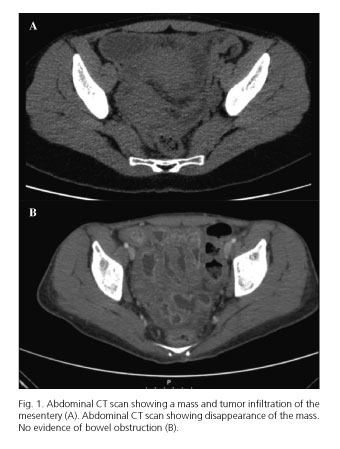
Laboratory examination was unremarkable. Abdominal X-ray revealed a few distended small bowel loops with air fluid levels. Abdominal ultrasound and enhanced computed tomography (CT) documented dilated jejunal loops attributable to parietal mass with 4.6x4.5 cm, infiltration of the mesentery and the greater omentum (Fig. 1A). The tumor markers and viral serologies were negative.
In this context, an ultrasound-guided biopsy of the lesion was performed, showing on hematoxylin and eosin staining diffuse infiltration of large cells, with occasional prominent nucleoli and moderate eosinophilic cytoplasm. Immunohistochemistry was positive for CD34, CD117, myeloperoxidase, lysozyme and Bcl-2.
The peripheral blood did not show circulating blasts, and the bone marrow aspirate excluded infiltration by malignant cells. The molecular analysis did not identify any cytogenetic abnormalities.
The patient initiated systemic chemotherapy with idarubicine and cytarabine, without response, and started mitoxantrone and etoposide with good response. The patient was proposed to allergeneic transplant by a donor family member.
An abdominal CT scan done one month later revealed the disappearance of the tumor (Fig. 1B). One year after allogeneic transplant the patient is clinically well without evidence of recurrence.
Discussion
Myeloid sarcoma (MS) is an extra medullar tumor of immature myeloid cells. More than 50% of the tumors are misdiagnosed as non-Hodgkin lymphoma. The prevalence rate is difficult to determine but it has been estimated in 2/1,000,000, with a predilection for males (2:1) (4,5).
It generally occurs in association with a myeloproliferative disorder and in this latter setting the majority (88%) of untreated patients progress to acute myeloid leukemia within 11 months (6).
MS can be present as either isolated or multiple lesions that may be synchronous or metachronous. The most common sites are the skin, bone, and lymph nodes (6,7). The synchronous involvement of the jejunum and the greater omentum is rare. Immunohistochemical stains and flow cytometry led to the correct diagnosis (2,6).
MS has been associated with various cytogenetic abnormalities, particularly t (8:21) (54%) and inv (16) (p13; q22) (25%). The prognostic significance of these chromosomal rearrangements remains uncertain (2).
The optimal therapy for primary MS still undetermined (2). Systemic chemotherapy, surgical resection, radiotherapy, bone marrow transplantation, or a combination of these approaches, are used (4).
In conclusion, this case is noteworthy because of the rarity of gastrointestinal involvement. The prompt recognition and early institution of therapy was crucial to avoid its progression to a systemic disease. The disappearance of the tumor after chemotherapy and allogeneic transplant makes this case a success.
Liliane C. Meireles1, Ana Catarina Lagos2, Inês Marques3,
Fátima Serejo1 and José Velosa1
1Centro Hospitalar Lisboa Norte. Lisbon, Portugal.
2Hospital das Forças Armadas. Lisbon, Portugal.
3Centro Hospitalar das Caldas da Rainha. Caldas da Rainha, Portugal
References
1. Álvarez P, Navascués CA, Ordieres C, et al. Granulocytic sarcoma of the small bowel, greater omentum and peritoneum associated with a CBFb/MYH11 fusion and inv(16) (p13q22): a case report. Int Arch Med 2011;4:3. [ Links ]
2. Kumar B, Bommana V, Irani F, et al. An uncommon cause of small bowel obstruction: Isolated primary granulocytic sarcoma. QJM 2009;102:491-3. [ Links ]
3. Choi EK, Ha HK, Park SH, et al. Granulocytic sarcoma of bowel: CT findings. Radiology 2007;243:752-9. [ Links ]
4. Antic D, Elezovic I, Bogdanovic A, et al. Isolated myeloid sarcoma of the gastrointestinal tract. Intern Med 2010;49:853-6. [ Links ]
5. Narayan P, Murthy V, Su M, et al. Primary myeloid sarcoma masquerading as an obstructing duodenal carcinoma Case Rep Hematol 2012;2012:490438. [ Links ]
6. Sivan-Hoffmann R, Waksman I, Cohen HI, et al. Small bowel obstruction as a presenting sign of granulocytic sarcoma. Isr Med Assoc J 2011;13:507-9. [ Links ]
7. Kohl SK, Aoun P. Granulocytic sarcoma of the small intestine. Arch Pathol Lab Med 2006;130:1570-4. [ Links ]

