


Mi SciELO
 Servicios personalizados
Servicios Personalizados
Revista
Articulo
 texto en
  Inglés (pdf)
 Articulo en XML
 Referencias del artículo
 Enviar articulo por email
Indicadores
-
 Citado por SciELO
 -
 Accesos
Links relacionados
-
 Citado por Google
 -
 Similares en
SciELO
Similares en
SciELO -
 Similares en Google
Compartir

 Permalink
Revista Española de Enfermedades Digestivas
versión impresa ISSN 1130-0108
Rev. esp. enferm. dig. vol.107 no.7 Madrid jul. 2015
LETTERS TO THE EDITOR
Pneumoperitoneum after CT colonography in a patient with ulcerative colitis
Pneumoperitoneo tras TAC-colonoscopia en paciente con colitis ulcerosa
Key words: Pneumoperitoneum. CT colonography.
Palabras clave: Pneumoperitoneo. TAC-colonoscopia.
Dear Editor,
CT colonography or virtual colonoscopy is an increasingly used test for the screening of colon cancer, its use being supported by several meta-analyses. It can diagnose polyps greater than 1 cm (1), with a sensitivity similar to that of colonoscopy for polyps 6 mm in diameter and above (2-5).
Its use entails a lower risk of complications when compared to colonoscopy. However, despite its being a mildly aggressive approach, complications have been reported that range from vasovagal illness to colon perforation (6,7).
Case report
We report the case of an 89-year-old male patient with a history of ulcerative colitis, myelodysplastic syndrome, and benign prostate hyperplasia. Because of his ulcerative colitis, the patient had been followed up by the Gastroenterology Department with serial colonoscopies, but colonography was later selected when he rejected further colonoscopy procedures.
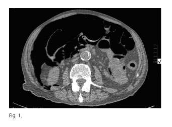
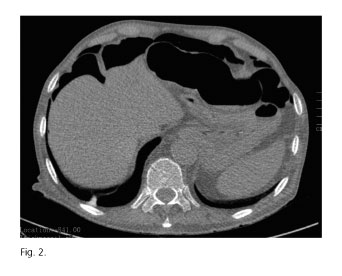
The study was performed at the Radiodiagnosis Department using manual air insufflation through a rectal catheter. During the procedure the radiologist detected the occurrence of bilateral subdiaphragmatic pneumoperitoneum (Fig. 1), as well as multiple bubbles in the mesentery, and emphysema in the greater omentum and mesoileum (Fig. 2). Two sessile polyps were identified, 9 and 7 mm in size, within the distal descending colon.
Diagnosis was only radiographic since the patients remained symptom-free. He was admitted to the General Surgery Ward and underwent clinical and laboratory monitoring, with absolute fasting and empiric antibiotic therapy. An abdominal CT was repeated at 24 hours, which found no free intra-abdominal fluid and a modestly decreased pneumoperitoneum. Laboratory tests showed no increase in acute phase reactants and water was tolerated on day 4. A week after admission, the patient was discharged with no intra-abdominal complications.
CT colonography is a diagnostic and screening method suitable for colorectal cancer. The technique is increasingly used because of its relative safety, albeit complications do arise, including vasovagal responses in 0.16% of patients (8), in contrast to 1% with colonoscopy (1-4), which likely results from colonoscopy-related sedation. Other potential complications described in the literature include intestinal perforation (as in our patient), contrast-related adverse events (renal toxicity, allergic responses, etc.), infection, and harmful radiation effects.
In various studies, the incidence of perforation after CT colonography ranges from 0.02% to 0.009% (6-8). Perforations are mostly asymptomatic, as was the case with our case report (6,7). Several factors have been associated with increased perforation risks: Insufflation method, recent colonoscopy or colonic surgery, diverticulitis, sigmoid tumors or hernias containing the sigmoid colon... In general, any disorder or disease involving the colonic or rectal wall may increase the risk for barotrauma. Regarding insufflation techniques and according to reports in the literature, the use of automated low-pressure carbon dioxide or air delivery systems reduces perforation risk (2), as does the use of flexible, atraumatic catheters. Recent colonic surgery or colonoscopy, particularly when associated with polypectomy, is also relevant for perforation risk, it is recommended that colonography be postponed for 15-30 days following colonoscopy with biopsy taking, and for 4-6 weeks following surgery.
Risk factors in the case reported herein included long-standing ulcerative colitis, manual insufflation, and a recent colonoscopy, although over 6 weeks had elapsed since the procedure.
Most complications secondary to CT colonography are mild to moderate and do not require treatment, save for perforation. Perforation management is usually restricted to digestive rest and clinical-laboratory monitoring, as in our patient. The need for urgent surgery remains exceptional.
Despite the above, the risk for perforation associated with CT colonography is considerably smaller as compared to colonoscopy (between 0.03% and 0.196%) (9,10). The fact that these percentages only refer to patients with symptoms or receiving a diagnosis during the procedure is to be taken into account; it is mentioned in the literature that up to 50% of perforations go unnoticed after colonoscopy (9), with a mortality rate of 0.07% (2).
CT colonography is an effective technique with lower risks, both in incidence and severity, compared to colonoscopy.
José Silvestre, María del Mar Sánchez-Lauro, María del Mar Callejón,
Aurora María Burgarolas, Francisco Cruz and Joaquín Marchena
Department of General Surgery and Digestive Diseases.
Hospital Universitario Dr. Negrín. Las Palmas de Gran Canaria, Spain
References
1. Johnson CD, Chen MH, Toledano AY, et al. Accuracy of CT colonography for detection of large adenomas and cancer. N Engl J Med 2008;359:1207-17. DOI: 10.1056/NEJMoa0800996. [ Links ]
2. Pendsé DA, Taylor SA. Complications of CT colonography: A review. Eur J Radiol 2013;82:1159-65. DOI: 10.1016/j.ejrad.2012.04.011. [ Links ]
3. Reuterskiöld MH, Lasson A, Svensson E, et al M. Diagnostic performance of computed tomography in symptomatic patients and in patients with increased risk for colorectal disease. Acta Radiol 2006;47:888-9. DOI: 10.1080/02841850600964966. [ Links ]
4. Laghi A, Iannaconne R, Carbone I, et al. Detection of colorectal lesions with virtual computed tomograph colonography. Am J Surg 2002;183:124-31. DOI: 10.1016/S0002-9610(01)00857-1. [ Links ]
5. Iafrate F, Hasssan C, Ciolina M, et al. High positive predictive value of CT colonography in a referral center. Eur J Radiol 2011;80:289-92. DOI: 10.1016/j.ejrad.2010.12.080. [ Links ]
6. Bellini D, Rengo M, De Cecco CN, et al. Perforation rate in CT colonography: A systematic review of the literature and meta-analysis. Eur Radiol 2014;24:1487-96. DOI: 10.1007/s00330-014-3190-1. [ Links ]
7. Sosna J, Blachar A, Amitai M, et al. Colonic perforation at CT Colonography: Assessment of risk in a multicenter large cohort. Radiology 2006;239:457-63. DOI: 10.1148/radiol.2392050287. [ Links ]
8. Iafrate F, Iussich G, Correale L, et al. Adverse events of CT colonography: An Italian National Survey. Dig Liver Dis 2013;45:645-50. DOI: 10.1016/j.dld.2013.02.020. [ Links ]
9. Gatto N, Frucht H, Sundararajan V, et al. Risk of perforation after colonoscopy and sigmoidoscopy: A population based study. J Natl Cancer Inst 2003;95:230-6. DOI: 10.1093/jnci/95.3.230. [ Links ]
10. Rabeneck L, Paszat LF, Hilsden RJ, et al. Bleeding and perforation after outpatients colonoscopy and their risk factors in usual clinical practice. Gastroenterology 2008;135:1899-906. DOI: 10.1053/j.gastro.2008.08.058. [ Links ]

