


My SciELO
 Custom services
Services on Demand
Journal
Article
 text in
  English (pdf)
 Article in xml format
 Article references
 Send this article by e-mail
Indicators
-
 Cited by SciELO
 -
 Access statistics
Related links
-
 Cited by Google
 -
 Similars in
SciELO
Similars in
SciELO -
 Similars in Google
Share

 Permalink
Revista Española de Enfermedades Digestivas
Print version ISSN 1130-0108
Rev. esp. enferm. dig. vol.107 n.8 Madrid Aug. 2015
LETTERS TO THE EDITOR
Retrorectal tumors - A diagnostic-therapeutic challenge
Tumores retrorrectales: un reto diagnóstico-terapéutico
Key words: Retrorectal. Presacral. Rectum. Kraske.
Palabras clave: Retrorrectal. Presacro. Recto. Kraske.
Dear Editor,
Retrorectal tumors are an uncommon, heterogeneous group of lesions that occur in the presacral region. Both diagnosis and treatment represent a challenge for colorectal surgeons. They must be resected because of their potential malignity and risk of infection (1).
Case report
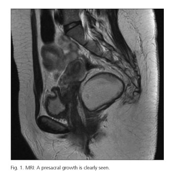
A 74-year-old male with ischemic heart disease, four bypasses, and nonspecific perianal discomfort had a growth palpated at around 5 cm from the anal margin. A CT scan found a presacral space occupying lesion without infiltration of the sacrum, rectum, or bladder. MRI revealed a well-delimited mass with no infiltration of neighboring organs and 12 cm in length (Fig. 1). A mixed (abdominal-perineal) approach was selected for surgery. Histopathology classified the lesion as a desmoid tumor. On the third day after surgery the patient had an acute myocardial infarction that required a prolonged stay in the ICU. He was discharged one month after admission. Presently he has no symptoms and no evidence of relapse.
Discussion
Retrorectal masses have an incidence of 2 cases/year. Congenital masses are most common (66%). They are usually asymptomatic or present with nonspecific manifestations; furthermore, their location in a difficult-to-access anatomical region requires expertise regarding their diagnostic-therapeutic approach in order to deliver appropriate treatment (2). Their clinical presentation results from their mass effect, superinfection (in cystic growths), or organ invasion (in malignancies) (3), with pain being the most common symptom. Digital rectal examination, which is mandatory, may identify the lesion as a mass palpated in the rectum's posterior aspect (4). Pelvic MRI is now essential, this being the study with the highest specificity. It informs on the relation of the tumor to the sacrum and neighboring structures, differentiates solid from cystic growths, and suggests the lesion's benign or malignant nature. Preoperative biopsy is advised against, given the risk for tumor spread and local infection. Management is always surgical (5). Surgical approach depends on tumor location; accessible lesions with an upper border lower than the third sacral vertebra may be approached posteriorly (transacral or Kraske posterior approach) (6). Bigger tumors and tumors above S3 may require an abdominal or combined approach. They may be approached with laparoscopy, the advantages being identical to those seen in other surgical scenarios (7). Of late, transanal endoscopic microsurgery (TEM) is used for benign, smaller tumors (8,9). Patients with complete benign tumor resection have a 5-year survival of 90%, with recurring lesions in 0-15% of cases. For malignancies, recurrence rates are higher (42-100%) and survival is lower (50-69%) (10).
To conclude, presacral tumors are rare and their diagnosis is challenging. Treatment should be delivered by a team with expertise in pelvic and cancer surgery in order to obtain the best possible outcomes with the lowest morbidity and mortality.
Joaquín Carrasco-Campos, Santiago Mera-Velasco, Iván González-Poveda,
Manuel Ruiz-López, José Antonio Toval-Mata, José Manuel Aranda-Narváez
and Julio Santoyo-Santoyo
Coloproctology Unit. Department of General Surgery, Digestive Diseases
and Transplants. Hospital Regional Universitario. Málaga, Spain
References
1. Aranda Narváez JM, González Sánchez AJ, Montiel Casado C, et al. Posterior approach (Kraske procedure) for surgical treatment of presacral tumors. World J Gastrointest Surg 2012;4:126-30. [ Links ]
2. Woodfield JC, Chalmers AG, Phillips N, et al. Algorithms for the surgical management of retrorectal tumours. Brit J Surg 2008;95:214-21. [ Links ]
3. Ghosh J, Eglinton T, Frizelle FA. Presacral tumors in adults. Surgeon 2007;5:31-8. [ Links ]
4. Canelles E, Roig JV, Cantos M, et al. Tumores presacros. Análisis de nuestra experiencia en 20 casos tratados quirúrgicamente. Cir Esp 2009;85:371-7. [ Links ]
5. Baratti D, Gronchi A, Pennacchioli E, et al. Chordoma: Natural history and results in 28 patients treated at a single institution. Ann Surg Oncol 2003;10:291-6. [ Links ]
6. Vega Menéndez D, Quintáns Rodríguez A, Hernández Granados P, et al. Hamartomas quísticos retrorrectales. Cir Esp 2008;83:53-60. [ Links ]
7. Witherspoon P, Armitage J, Gatt M, et al. Laparoscopic excision of retrorectal schwannoma. Dis Colon Rectum 2010;53:101-3. [ Links ]
8. Zoller S, Joos A, Dinter D, et al. Retrorectal tumors: Excision by transanal endoscopic microsurgery. Rev Esp Enferm Dig 2007;99:547-50. [ Links ]
9. Serra Aracil X, Gómez Díaz C, Bombardó Junca J, et al. Surgical excision of retrorectal tumour using transanal endoscopic microsurgery. Colorectal Dis 2010;12:594-5. [ Links ]
10. Wei F, Dang GT, Liu ZJ. Surgical factors underlying the recurrence of primary spine tumor. Chin J Surg 2005;43:221-4. [ Links ]

