


Mi SciELO
 Servicios personalizados
Servicios Personalizados
Revista
Articulo
 Inglés (pdf)
 Articulo en XML
 Referencias del artículo
 Enviar articulo por email
Indicadores
-
 Citado por SciELO
 -
 Accesos
Links relacionados
-
 Citado por Google
 -
 Similares en
SciELO
Similares en
SciELO -
 Similares en Google
Compartir

 Permalink
Revista Española de Enfermedades Digestivas
versión impresa ISSN 1130-0108
Rev. esp. enferm. dig. vol.107 no.9 Madrid sep. 2015
LETTERS TO THE EDITOR
Leiomyosarcoma of the ascending colon: A rare tumor with poor prognosis
Key words: Gastrointestinal stromal tumors. Colonic leiomyosarcoma. Liver metastasis.
Dear Editor,
The gastrointestinal leiomyosarcoma is a very unusual tumor that arises from smooth muscle cells of intestinal wall (1).
The most frequent anatomical locations affected by these tumors are stomach (50%); small intestine (30%); colorectal (15%); and esophagus (5%) (2).
Colorectal sarcomas represent approximately 0.1% of all the malignant tumors at this level (3).
Case report
An 89-year-old woman without any relevant medical history presented to the emergency department complaining of a 2-day evolution of abdominal pain, nausea and vomiting. There were not changes in her bowel habits but mid-term hyporexia was found. On physical examination, her abdomen was slightly swollen and the pain was dull, located in the right iliac fossa without rebound tenderness. Laboratory test results revealed elevated C-reactive protein levels, normal leucocyte count, anemia and slight hyponatremia.
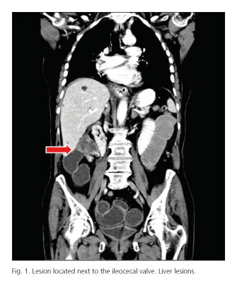
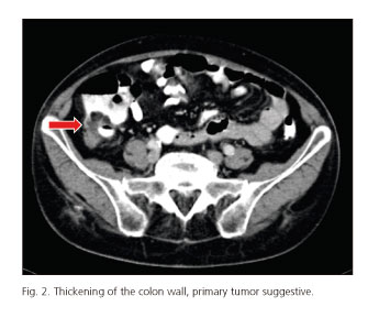
Due to a supposed subocclusive condition, a computed tomography (CT) was performed. A 4.5 cm lesion located next to the ileocecal valve compatible with a primary colonic tumor was described. Furthermore, multiple liver metastatic images were revealed (Figs. 1 and 2).
Despite a conservative management after being admitted to our institution, the patient did not improve, so she finally underwent surgery and a right colectomy was performed. In this surgery solid lesions of the liver metastases were confirmed with biopsy. The patient received adjuvant chemotherapy.
The definitive pathological assessment indicated a neoformation with several ulcerations of mesenchymal ancestry without lymph-node involvement. Immunohistochemistry tests revealed the presence of smooth muscle proteins (actin, calponin, vimentin) and Ki-67 at 30%. They were negative for C-Kit. The final diagnosis was compatible with a colonic leiomyosarcoma (LMS).
Discussion
The interest in this case report lies in a totally unexpected histology of a primary malignant tumor in colon such as LMS, which is an extremely rare entity.
In epidemiological terms, this tumor affects patients aged between 40-60, with a higher frequency among men than women (1.8:1), and also among African Americans (3,4).Its clinical presentation and endoscopic findings can be unspecific (1). Its most common symptoms and signs are abdominal pain, a palpable abdominal mass, changes in bowel habits, or typical gastrointestinal tumor complications such as bleeding, perforation or obstruction/invagination (5).
LMS is an aggressive tumor with a high rate of local recurrence as well as a significant hematogenous spread. It rarely presents with lymph node-involvement. Liver is the most affected abdominal organ for distant metastases. Lung and bone have been described as metastatic targets but they are unusual sites (5-7).
Diagnosis is based on the histologic examination and immunohistochemistry tests of the surgical specimen. The histologic grade, determined by the number of mitotic figures per field, is the most important criterion for diagnosis of LMS. Determination of c-KIT is negative, opposite to GIST, while the immunohistochemistry test is positive for smooth muscle proteins like actin, vimentin and desmin (1,6).
The surgical resection keeps being the best treatment and it should be performed so as to achieve a complete resection. The most important prognosis indicators are the integrity of surgical resection and its malignant potential (8).
Lucía Granero-Peiró, Patricia Martínez-Ortega,
Carlos Sánchez-Justicia and José Luis Hernández-Lizoaín
Department of General and Digestive Surgery.
Clínica Universidad de Navarra. Pamplona, Navarra. Spain
References
1. Pilliado Páez H, Charua Guindic L, Avendaño Espinosa O, et al. Leiomiosarcoma colorrectal. Reporte de dos casos. An Med Asoc Med Hosp ABC 2000;45:140-4. [ Links ]
2. McGrath PC, Neifeld JP, Lawrence W Jr. Gastrointestinal sarcomas. Analysis of prognostic factors. Ann Surg 1987;206:706-10. DOI: 10.1097/00000658-198712000-00004. [ Links ]
3. Tran T, Davila JA, El-Serag HB. The epidemiology of malignant gastrointestinal stromal tumors: An analysis of 1,458 cases from 1992 to 2000. Am J Gastroenterol 2005;100:162-8. DOI: 10.1111/j.1572-0241.2005.40709.x. [ Links ]
4. Bellver M, Rodríguez Lago I, Queipo F, et al. Colocolonic instussusception secondary to high grade colonic leiomysarcoma. Rev Esp Enferm Dig 2011;103:601-3. DOI: 10.4321/S1130-01082011001100013. [ Links ]
5. Mata JF, Escalante R, Linares K, et al. Leiomyosarcoma of the gastrointestinal tract. GEN 1993;47:35-44. [ Links ]
6. Flores Pastor B, Pellicer Franco E, Navarro Martínez MN, et al. Metástasis hepáticas secundarias a leiomiosarcoma de recto. Presentación de un caso. Cir Esp 2000;67:394-5. [ Links ]
7. Fallahzadeh H. Leiomyosarcoma of colon: Report of two cases. Ann Surg 1995;61:294-6. [ Links ]
8. Matushansky I, Hensley M. Leiomyosarcoma: An overview of etiology, prognosis and treatment options. Am J Cancer 2006;5:81-91. DOI: 10.2165/00024669-200605020-00002. [ Links ]

