


Mi SciELO
 Servicios personalizados
Servicios Personalizados
Revista
Articulo
 Inglés (pdf)
 Articulo en XML
 Referencias del artículo
 Enviar articulo por email
Indicadores
-
 Citado por SciELO
 -
 Accesos
Links relacionados
-
 Citado por Google
 -
 Similares en
SciELO
Similares en
SciELO -
 Similares en Google
Compartir

 Permalink
Revista Española de Enfermedades Digestivas
versión impresa ISSN 1130-0108
Rev. esp. enferm. dig. vol.108 no.8 Madrid ago. 2016
PICTURES IN DIGESTIVE PATHOLOGY
Gastric inflammatory fibroid polyp mimicking a gastrointestinal stromal tumor
Marco Silva1, Andreia Albuquerque1, Hélder Cardoso1, Jennifer Costa2 and Guilherme Macedo1
Departments of 1 Gastroenterology and
2 Pathology. Centro Hospitalar São João. Porto, Portugal
Background
Inflammatory fibroid polyp (IFP) of the gastrointestinal tract is a rare, benign neoplasm, most frequently located in the gastric antrum. Symptoms depend on the location and size of the lesion. Biopsies are limited for the diagnosis of inflammatory fibroid polyps and diagnosis may not be possible until resection.
The authors present a case of a 55-year-old woman, presenting with an upper gastrointestinal bleeding due to a large gastric inflammatory fibroid polyp imitating a gastrointestinal stromal tumor (GIST).
Case report
A 55-year-old woman was admitted to the emergency department due to abdominal pain, fever and hematemesis. The laboratory data revealed anemia (hemoglobin 11 g/dL), and the abdominal ultrasonography showed a hypoechoic mass located in gastric antrum.
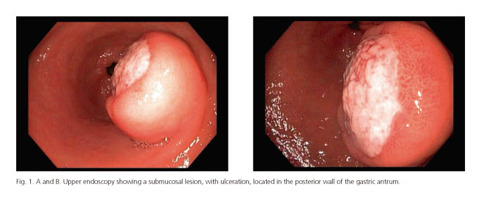
Upper endoscopy was performed revealing a 40 mm submucosal lesion with ulceration, located in the posterior wall of the gastric antrum, suggestive of a gastrointestinal stromal tumor (Figs. 1 A and B). Abdominal computed tomography confirmed a gastric polypoid lesion, with endoluminal projection and without extramural extension.
The patient had another episode of upper gastrointestinal bleeding and a laparoscopic atypical gastrectomy was performed. Histopathologic evaluation revealed a well-defined submucosal nodular lesion, with focal mucosa ulceration, formed by proliferation of spindle cells and inflammatory infiltrate of lymphocytes and eosinophils, consistent with inflammatory fibroid polyp (IFP) (Fig. 2). Immunohistochemistry showed CD34 positivity; desmin, CD117, s100, DOG1 and C-Kit were negative.

The patient was discharged, one week later, asymptomatic.
Discussion
IFPs are rare benign mesenchymal tumors that can arise in the gastrointestinal tract, more commonly occurring in the gastric antrum and normally smaller than 25 mm (1-3). They are submucosal lesions with perivascular onion skinning and prominent eosinophilic infiltrate (2). These cells are negatively stained for s100 protein and desmin, and positively for CD34 (4).
They often appear as an incidental finding on examination of the upper digestive tract, but may be associated with abdominal pain, weight loss, dyspeptic symptoms, iron deficiency anemia and intestinal obstruction, but rarely massive digestive tract hemorrhage (5). Biopsies are unhelpful for the diagnosis of IFPs and diagnosis may not be possible until resection (1). In the case of large tumors, surgical treatment is often necessary and IFPs usually do not recur or metastasize. In this case, due to the increased risk of complications associated with endoscopic removal of large lesions and the recurrence of hematemesis, a surgical approach was performed.
Gastric IFPs are rare lesions, in this case with a very atypical presentation and larger size, mimicking a GIST.
References
1. Albuquerque A, Ríos E, Carneiro F, et al. Evaluation of clinicopathological features and Helicobacter pylori infection in gastric inflammatory fibroid polyps. Virchows Arch 2014;465:643-7. DOI: 10.1007/s00428-014-1659-6. [ Links ]
2. Carmack SW, Genta RM, Schuler CM, et al. The current spectrum of gastric polyps: A 1-year national study of over 120,000 patients. Am J Gastroenterol 2009;104:1524-32. DOI: 10.1038/ajg.2009.139. [ Links ]
3. De la Plaza R, Picardo AL, Cuberes R, et al. Inflammatory fibroid polyps of the large intestine. Dig Dis Sci 1999;44:1810-6. DOI: 10.1023/A:1018886421409. [ Links ]
4. Kwiatkowski AP, Pa nik K. Large inflammatory fibroid polyp of cardia managed laparoscopically - A case report and review of the literature. Videosurgery Miniinv 2014;9:623-6. DOI: 10.5114/wiitm.2014.46448. [ Links ]
5. Zhang C, Cui M, Xing J, et al. Massive gastrointestinal bleeding caused by a giant gastric inflammatory fibroid polyp: A case report. Int J Surg Case Rep 2014;5:571-3. DOI: 10.1016/j.ijscr.2014.05.004. [ Links ]
