


My SciELO
 Custom services
Services on Demand
Journal
Article
 text in
  English (pdf)
 Article in xml format
 Article references
 Send this article by e-mail
Indicators
-
 Cited by SciELO
 -
 Access statistics
Related links
-
 Cited by Google
 -
 Similars in
SciELO
Similars in
SciELO -
 Similars in Google
Share

 Permalink
Revista Española de Enfermedades Digestivas
Print version ISSN 1130-0108
Rev. esp. enferm. dig. vol.108 n.11 Madrid Nov. 2016
https://dx.doi.org/10.17235/reed.2016.3970/2015
CASE REPORTS
Perivascular epithelioid cell tumor of the ileum. A case report
Tumor de células epitelioides perivasculares de íleon. Descripción de un caso
Rosa Virginia Acosta-Materán1, María Isabel Martín-Arribas1, Antonio Velasco-Guardado1, Cristina González-Velasco2, Ana María Mora-Soler1, Cristina Revilla-Morato1 and Antonio Rodríguez-Pérez1
Departments of 1Gastroenterology and 2Pathology. Hospital Universitario de Salamanca. Salamanca Institute for Biomedical Research (IBSAL). Salamanca. Spain.
ABSTRACT
Perivascular epithelioid cell tumors (PEComa) are tumors of perivascular epithelioid cells with immunohistochemical features of smooth muscle and melanocytic tumors. The PEComa of the gastrointestinal tract is rare. The treatment is surgical, although there are data that suggest a good response to rapamycin.
Key words: PEComa. Immunohistochemistry. Rapamycin. Ileum.
RESUMEN
Los tumores de células epiteliodes perivasculares son tumores de células epiteliales vasculares con características inmunohistoquímicas de músculo liso y células melanocíticas. Los gastrointestinales son infrecuentes. El tratamiento es quirúrgico aunque existen datos que indican buena respuesta a la rapamicina.
Palabras clave: PEComa. Inmunohistoquímica. Repamicina. Íleo.
Introduction
Perivascular epithelioid cell tumors (PEComas) are mesenchymal neoplasms that include angiomyolipoma, lymphangioleiomyoma, clear-cell myomelanocytic tumor of the falciform ligament, clear-cell "sugar" tumor of the lung and other locations (1). These tumors affect mainly young adults, particularly women. PEC in the gastrointestinal tract are exceptionally rare. These tumors are usually associated with the tuberous sclerosis (TS). Ultrasound, CT and magnetic resonance are the imaging tests used. All PEComas show immunoreactivity for both melanocytic (HMB45 and melan-A) and smooth muscle (actin and desmin) markers. PEComas are usually malignant. The diagnosis is generally confirmed after surgery (2, 3). The clinical is variable, and it ranges from asymptomatic cases to cases of acute abdomen.
Case reports
We present the case of a 19-year-old male with a family history of gastric cancer (maternal grandfather). He was admitted as an emergency with recurrent abdominal pain in the epigastrium and vomiting. The symptoms had worsened over the last 24 hours. In the physical examination he referred pain in epigastrium and mesogastrium without peritonitis. The analysis reveals leukocytosis with neutrophilia and high lactate levels. Abdominal ultrasound scan showed small bowel loops with "target" appearance, which suggest an ileal intussusception. An emergency assessment was requested to the surgeon, and a computed tomography scan (CT) was performed. It revealed a 2-cm long ileo-ileal invagination and a tumor in this area; free fluid in the pelvis. An emergency operation was decided. The patient underwent a laparotomy in which the proximal ileum was resected, which contained, in its central portion, an 11-cm intestinal segment and an adjacent mass of 2.7 x 2.7 cm. The pathologist described a tumor located on the muscle wall of the proximal ileum, made up of epithelioid cells and perivascular growth, without atypia, necrosis, and vascular lymphatic invasion. The mitotic count was 0-1/50. Markers: Actin and Desmin +; HMB45 y Melan A-. Given all these findings, a diagnosis compatible with ileal PEComa was established. The patient recovered favorably and 6 months later the patient has no recurrence.
Discussion
The articles published on PEComa are scarce y descriptive. As published, the most common location is the colon (45.7%) followed by the small intestine (28.6%), 6 tumors appeared in the ileum. With regard to monitoring, 31.4% showed a malignant behavior, with local recurrence in 4 cases and metastasis in 19 (1).
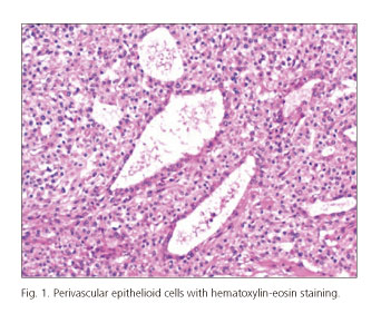
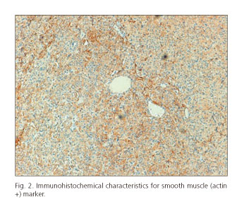
The anatomopathological diagnosis was compatible with PEComa due to the morphology of the lesion, with immunohistochemistry positive in smooth muscle markers (actin and desmin) and negative for melanocytic markers (Figs. 1 and 2).
Folpe et al. published 26 cases and proposed prognostic criteria, which can be stratified into "benign", "uncertain malignant potential" and "malignant". In the study, a significant association was found between the size of the tumor larger than 5 cm, the infiltrative growth pattern, the high nuclear grade, necrosis and mitotic activity greater than 1/50 and the subsequent aggressive behavior (4).
Surgery is the standard treatment (5). Therapeutic, there are anecdotal cases; Rapamycin seems to block signaling through cellular growth. Studies of TS in animal models have shown significant results regarding the in vivo response to rapamycin (6-8).
References
1. Lu B, Wang C, Zhang J, et al. Perivascular epithelioid cell tumor of gastrointestinal tract: case report and review of the literature. Medicine (Baltimore). 2015; 94: e393. DOI: 10.1097/MD.0000000000000393. [ Links ]
2. Al-Saleem T, Wessner LL, Scheithauer BW, et al. Malignant tumors of the kidney, brain, and soft tissues in children and young adults with the tuberous sclerosis complex. Cancer. 1998;83:2208-16. DOI: 10.1002/(SICI)1097-0142(19981115)83:10<2208::AID-CNCR21>3.0.CO;2-K. [ Links ]
3. Abdulla M, Bui HX, del Rosario AD, et al. Renal angiomyolipoma. DNA content and immunohistochemical study of classic and multicentric variants.Arch Pathol Lab Med. 1994 Jul;118(7):735-9. [ Links ]
4. Folpe AL, Mentzel T, Lehr HA, et al. Perivascular epithelioid cell neoplasms of soft tissue and gynecologic origin: a clinicopathologic study of 26 cases and review of the literature. Am J Surg Pathol. 2005;29:1558-75. DOI: 10.1097/01.pas.0000173232.22117.37. [ Links ]
5. Larbcharoensub N, Karnsombut P, Jatchavala J, et al. Primary hepatic clear cell myomelanocytic tumor. Case report and review of the literature. APMIS. 2007;115:1454-9. DOI: 10.1111/j.1600-0463.2007.00733.x. [ Links ]
6. Kenerson H, Dundon TA, Yeung RS. Effects of rapamycin in the Eker rat model of tuberous sclerosis complex. Pediatr Res. 2005;57:67-75. DOI: 10.1203/01.PDR.0000147727.78571.07. [ Links ]
7. Lee L, Sudentas P, Donohue B, et al. Efficacy of a rapamycin analog (CCI-779) and IFN-gamma in tuberous sclerosis mouse models. Genes Chromosomes Cancer. 2005;42:213-27. DOI: 10.1002/gcc.20118. [ Links ]
8. Moritz W, Karen B, Marcus F, et al. Perivascular epitheloid cell tumor (PEComa) mimicking retroperitoneal liposarcoma. World J Surg Oncol. 2014;12:3. [ Links ]
![]() Correspondence:
Correspondence:
Rosa Virginia Acosta-Materán.
Department of Gastroenterology.
Hospital Universitario de Salamanca.
Paseo San Vicente s/n. 37007. Salamanca, Spain.
e-mail: rosa.acosta27@gmail.com
Received: 28-05-2015
Accepted: 12-09-2015
