


Mi SciELO
 Servicios personalizados
Servicios Personalizados
Revista
Articulo
 texto en
  Inglés (pdf)
 Articulo en XML
 Referencias del artículo
 Enviar articulo por email
Indicadores
-
 Citado por SciELO
 -
 Accesos
Links relacionados
-
 Citado por Google
 -
 Similares en
SciELO
Similares en
SciELO -
 Similares en Google
Compartir

 Permalink
Revista Española de Enfermedades Digestivas
versión impresa ISSN 1130-0108
Rev. esp. enferm. dig. vol.109 no.3 Madrid mar. 2017
PICTURES IN DIGESTIVE PATHOLOGY
Hemolymphangioma as a cause of overt obscure gastrointestinal bleeding: a case report
Hemolinfangioma como causa de hemorragia de origen obscuro evidente. Presentación de un caso
Gerardo Blanco-Velasco, Amina Tun-Abraham, Óscar Hernández-Mondragón and Juan Manuel Blancas-Valencia
Endoscopy Service. Hospital de Especialidades. Centro Médico Nacional Siglo XXI. Instituto Mexicano del Seguro Social. Ciudad de México, México
Case report
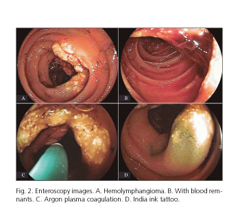
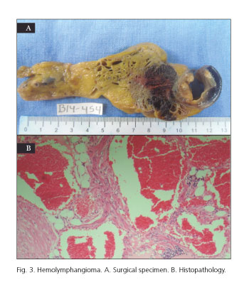
A 45-year-old woman without an important history complained of recurrent melena for about a year, with decreased hemoglobin of 4.5 g/dl, and required six blood transfusions. Gastroscopy and colonoscopy results were normal. Video capsule endoscopy showed a zone of lymphangiectasias with red blood in the proximal jejunum (Fig. 1). Double-balloon enteroscopy identified a 3-cm segment of proximal jejunum with nearly 50% of circumferential lymphangiectasias with oozing blood. It was treated with argon plasma coagulation and tattooed with India ink (Fig. 2). Later, the patient underwent an exploratory laparotomy which showed a jejunal tumor at 90 cm from the Treitz ligament. A 15-cm segment of jejunum was resected with primary anastomosis. Histological examination showed an 8-cm long mesenteric hemolymphangioma with infiltration to jejunum and free borders (Fig. 3).

Discussion
Hemolymphangiomas are rare benign tumors, composed of dilated lymphatic spaces, extravasation of red blood cells and fibrosis. These malformations are either congenital or acquired. The most common location is the mesentery (1). The clinical onset of hemolymphangiomas can vary in size and location, and can present hemorrhage, rupture and infection. Histologically, they consist of blood vessels and lymphatic channels (2). Diagnosis can be done by video capsule endoscopy. Although surgical resection appears to be the definitive treatment, double-balloon enteroscopy allowed effective treatment of bleeding small-bowel hemolymphangiomas (3).
References
1. Antonino A, Gragnano E, Sangiuliano N, et al. Int J Surg Case Rep 2014;5:118-21. [ Links ]
2. Fang YF, Qiu LF, Du Y, et al. Small intestinal hemolymphangioma with bleeding: A case report. World J Gastroenterol 2012;18:2145-6. DOI: 10.3748/wjg.v18.i17.2145. [ Links ]
3. Li F, Osuoha C, Leighton JA, et al. Double-balloon enteroscopy in the diagnosis and treatment of hemorrhage from small-bowel lymphangioma: A case report. Gastrointest Endosc 2009;70:189-90. DOI: 10.1016/j.gie.2008.09.036. [ Links ]
