


My SciELO
 Custom services
Services on Demand
Journal
Article
 text in
  English (pdf)
 Article in xml format
 Article references
 Send this article by e-mail
Indicators
-
 Cited by SciELO
 -
 Access statistics
Related links
-
 Cited by Google
 -
 Similars in
SciELO
Similars in
SciELO -
 Similars in Google
Share

 Permalink
Revista Española de Enfermedades Digestivas
Print version ISSN 1130-0108
Rev. esp. enferm. dig. vol.109 n.3 Madrid Mar. 2017
PICTURES IN DIGESTIVE PATHOLOGY
Acute esophageal necrosis resolved in 72 hours
Necrosis aguda de esófago resuelta en 72 horas
Noelia Alcaide1, Luis Fernández-Salazar1, Lourdes Ruiz-Rebollo1 and Elvira González-Obeso2
Departments of 1Gastroenterology and 2Anatomy. Hospital Clínico Universitario. Valladolid, Spain
Case report
The case is an 80 year old man with arterial hypertension under treatment with chronic kidney disease. Three months previously, the patient underwent endoscopic dilatation with the hydrostatic balloon technique by dysphagia secondary to Schatzki stenotic ring.
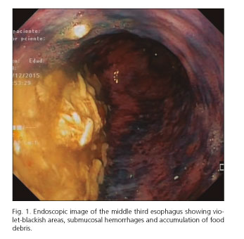
The patient was admitted with sudden dysphagia, feeling food bolus impaction and intense epigastric pain. Laboratory evaluation did not show any alteration. Upper endoscopy showed the esophageal lumen diffusely dilated, with submucosal hemorrhages and confluent violet-blackish areas. All these findings were suggestive of esophageal necrosis. It was also observed that food debris caused decubitus of the mucosa of the middle third esophagus (Fig. 1). The Schatzki ring allowed the passage of the endoscope without difficulty. A hiatal hernia was also present. The patient was treated with intravenous hydration and high-dose proton pump inhibitors. Three days later, the upper endoscopy was performed again and showed a totally normal esophageal mucosa and slightly dilated lumen (Fig. 2). The esophageal biopsy showed fragments of esophageal mucosa with micro-hemorrhages and microscopic foci of necrosis.

Discussion
Acute esophageal necrosis is an infrequent clinical entity with high morbidity and mortality. The most common presentation (> 80%) is upper gastrointestinal bleeding (1). Risk factors include cardiovacular disease, diabetes mellitus, chronic kidney insufficiency and malignancy in connection with tissue hypoperfusion, diminished immune defenses or infectious conditions (2). Esophageal perforation is rare and delayed stricture formation may occur (3).
In our case, we saw that the unusual form of presentation and the esophageal mucosa returned to its normal appearance in a brief period of time.
References
1. Talebi-Bakhshayesh M, Samiee-Rad F, Zohrenia H, et al. Acute esophageal necrosis: A case of black esophagus with DKA. Arch Iran Med 2015;18:384-5. [ Links ]
2. Gurvits GE, Cherian K, Shami MN, et al. Black esophagus: New insights and multicenter international experience in 2014. Dig Dis Sci 2015;60:444-53. DOI: 10.1007/s10620-014-3382-1. [ Links ]
3. Chugh P, Tzimas D, Gurvits GE. A rare cause of upper gastrointestinal bleeding. Gastroenterol 2013;145:e11-2. DOI: 10.1053/j.gastro.2013.07.045. [ Links ]
