


Meu SciELO
 Serviços customizados
Serviços Personalizados
Journal
Artigo
 texto em
  Inglês (pdf)
 Artigo em XML
 Referências do artigo
 Enviar este artigo por email
Indicadores
-
 Citado por SciELO
 -
 Acessos
Links relacionados
-
 Citado por Google
 -
 Similares em
SciELO
Similares em
SciELO -
 Similares em Google
Compartilhar

 Permalink
Revista Española de Enfermedades Digestivas
versão impressa ISSN 1130-0108
Rev. esp. enferm. dig. vol.110 no.2 Madrid Fev. 2018
https://dx.doi.org/10.17235/reed.2017.5183/2017
LETTERS TO THE EDITOR
Ileocecal endometriosis as an infrequent cause of intussusception
1Hospital Universitario Virgen Macarena. Sevilla. España
Key words: Intestinal endometriosis; Intestinal intussusception; Subocclusive
Dear Editor,
Ileocecal affectation by endometriosis is rare (4.1%) and generally affects the serosa. The mucosal layer is affected in only 10% of cases and alterations are identified by colonoscopy. The symptomatology is variable but rarely produces bowel obstruction or perforation. Sánchez Cifuentes et al. 1 presented in 2016 their experience with 17 cases; two of these cases had ileocecal affectation and one required surgery for obstructive symptoms.
Case report
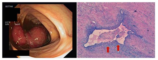
We herein present the case of a 51-year-old woman who had a simple total hysterectomy in 2009 for symptomatic uterine myomatosis. In 2016, she underwent a colonoscopy due to persistent abdominal pain. A congested and invaginated erythematous mucosa was identified that went beyond the ileocecal valve; it was not possible to delimit its extension (Fig. 1). Abdominal computed tomography (CT) showed a well-defined hypodense image of 6 x 2 cm in the cecum that acted as a head of an ileocolic intussusception that produced a subocclusion. Right hemicolectomy showed macroscopically an 8 cm fragment of invaginated mucosa in a "glove finger" form. The histology showed foci of endometriosis with adjacent ulcerated mucosa (Fig. 1). Other locations of endometriotic implants were not confirmed after follow-up by the Gynecology Service.
Discussion
Intestinal intussusception is a rare entity in adults (5% of cases) 2 and is usually located in the small intestine as benign lesions (50-70%). When this condition involves the colon or ileocecal valve, the main cause is neoplastic such as adenocarcinoma 3. Endometriosis is an estrogen-dependent disorder that affects 12-15% of women of reproductive age 4 and is usually located in the recto-sigma (85-90%) 1) (4. It presents a non-specific clinical manifestation including abdominal pain, dysmenorrhea, diarrhea, abdominal mass and infertility. MRI is the gold standard diagnostic technique, although an anatomopathological study is required for a definitive diagnosis 1. It is important to bear in mind that ileocecal endometriosis can be a benign cause of intussusception in the adult.
Bibliografía
1. Sánchez Cifuentes A, Candel Arenas M, Albarracín Marín-Blázquez A. Endometriosis intestinal. Nuestra experiencia. Rev Esp Enferm Dig 2016;108(8):524-5. [ Links ]
2. Shenoy S. Adult intussusception: A case series and review. World J Gastrointest Endosc 2017;9(5):220-7. DOI: 10.4253/wjge.v9.i5.220 [ Links ]
3. Kim JW, Lee BH, Park SG, et al. Factors predicting malignancy in adult intussusception: An experience in university affiliated hospitals. Asian J Surgery 2017;S1015-9584(16)30314-1. DOI: 10.1016/j.asjsur.2016.11.010 [ Links ]
4. Tong Y-L, Chen Y, Zhu S-Y. Ileocecal endometriosis and a diagnosis dilemma: A case report and literature review. World Gastroenterol 2013;19(23):3707-10. DOI: 10.3748/wjg.v19.i23.3707 [ Links ]
