


 Custom services
 English (pdf)
 Article in xml format
 Article references
 Send this article by e-mail
 Cited by SciELO
  Cited by Google
  Similars in
SciELO
Similars in
SciELO  Similars in Google
 Permalink
INTRODUCTION
Cutis verticis gyrata is a little-known sign, condition, clinical stigma or disease that is regarded as very rare that affects mostly males. It consists of folds in the scalp that look similar to the folds of the brain. The extent, shape and thickness may vary and it appears in the parietal and occipital areas of the scalp. The skin and subcutaneous tissue of the cranium thicken and copy the shape of the brain surface, which is why it has received other names, now no longer used, such as pachydermie vorticellee, cerebriform deformity, cutis sulcata, bulldog scalp or cutis capitis strata1-8.
The prevalence of the disease is unknown, but it is estimated that it affects 1/100,000 men and 0.26/100,000 women in the general population1,4,5. Regardless of the appearance and location of the cerebriform ridges, the consensus is that there are three different forms of CVG, each with a different degree of severity, prognosis and management:
The aetiology is considered to be idiopathic or essential (CVG primary essential) when it is not associated with any other pathology2-5. In such essential cases, no malignant disruption has been reported, and the only problem may be an aesthetic one. The cases of CVG primary non essential are much more frequent and are the ones associated with neurological and ophthalmologic conditions, especially severe mental retardation, intracranial brain disorders, convulsions, cerebral palsy, strabismus, cataracts or blindness1,3,8. Finally, the cases of CVG secondary (occasionally reversible) have not been very well clarified and have become a “catch all”, where there is confusion between associations and different causes (pachydermoperiostosis, cerebriform intradermal nevus, liver disorders, hamartomas, neurofibromas, dermatosis of the scalp, psoriasis, tumours, hypergammaglobulinemia inmunoglobulina E (IgE), endocrine disruption, syphilis, acromegalia, amiloidosis, cretinism, diabetes mellitus, fragile X chromosome syndrome, Ehlers-Danlos syndrome, Graves’ disease, Turner’s syndrome and a wide range of chromosome disorders1-8.
This study reports the first case of a patient carrying CVG in prison. The objective is to make this sign, problem or entity more public, describe initial management in consultation and establish the complementary examinations required to diagnose the sub-type, the prognosis and the therapeutic approach.
MATERIALS AND METHODS
Clinical case
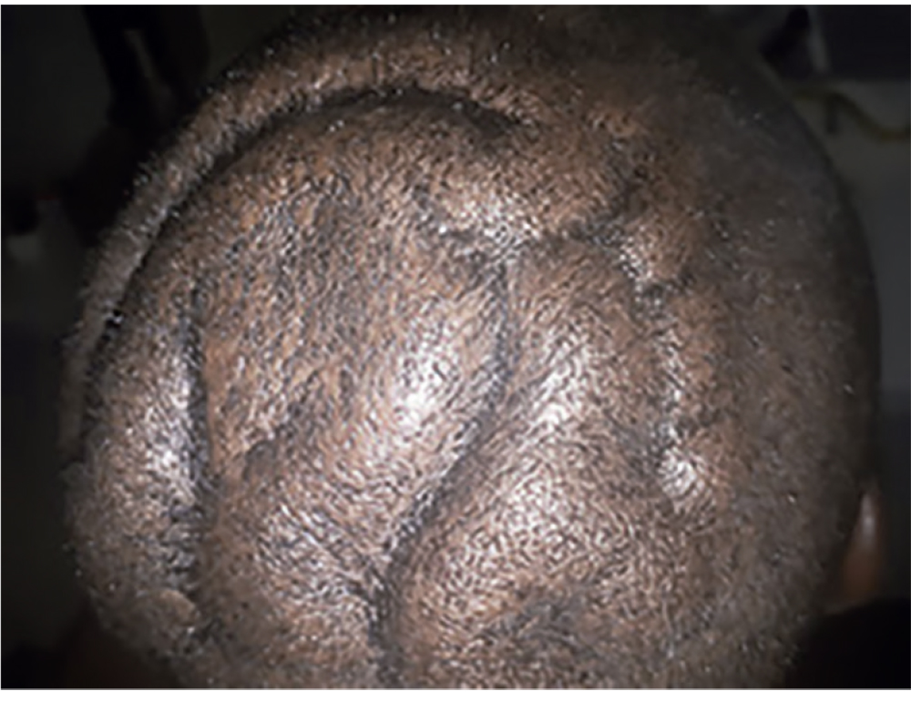
The patient was a black male of 28 years of age, native of Gambia, who was an inmate in module VII of the prison of Cuatre Camins (La Roca del Vallès, Barcelona). He came to the medical consultation saying that a series of sizeable cerebriform-shaped skin deformities had suddenly appeared on his head in the area of the crown, without any traumatic precedents (Figure 1). The patient was surprised when he came to consultation, just after having his head completely shaved, after being advised to do so by the inmate who had shaved him, and the patient affirmed that previously he did not have the deformities, although he had never completely shaved his head before. The wounds showed a slight inflammatory activity. The patient was immediately taken to the dermatology surgery, where after a simple inspection he was diagnosed with cutis verticis gyrata. The image is pathognomonic (Figure 1).
Case history, physical examination and initial approach with the patient
After diagnosis, he was once again transferred to the medical consultation of the module, where the first interview and a physical examination were carried out, with no significant results. The patient was informed in a simple and understandable manner that a series of complementary examinations were necessary in order to find out the prognosis and treatment. He stated that he was willing to undergo the specific examinations to establish the case, signing the corresponding informed consent and authorisation to transmit his images for scientific and research purposes. He was informed that he had probably had the problem for some time and that it might not disappear on its own.
Given the likelihood of CVG secondary, which could respond positively to corticosteroids, a treatment was recommended with 30 mg of prednisone after breakfast for ten days, and another follow-up appointment when the treatment ended. He was advised to let his hair grow back to hide the defect if he felt more comfortable that way.
Additional examinations
To categorise the CVG for prognostic and therapeutic purposes, the following additional diagnostic tests and consultations were programmed, based on the recommendations of other authors1-8:
Basic blood analysis, serology for hepatitis, syphilis and human immunodeficiency virus (HIV), hormone tests (growth hormone, thyroid function, androgens, oestrogens), tumour markers (carcinoembryonic antigen and alphaprotein), inflammation markers (erythrocyte sedimentation rate; C-reactive protein) and IgE analysis.
Radiography of cranium, chest and right forearm-hand, to test for possible parenchymal lung lesions, bone anomalies or possible periosteosis.
Computerised axial tomography (CAT) of cranium without contrast, to rule out possible intracranial lesions.
Biopsy of skin lesion, to rule out secondary causes and find out the anatomo-pathological nature of the lesion.
Genetic laboratory study, to determine possible altered genes associated with the diagnosis and prognosis.
Consultation with clinical psychology, to establish the IQ, and a neuropsychological analysis and personality study.
Consultation with psychiatry, to rule out possible severe psychiatric illnesses that do not appear in current diagnoses.
RESULTS
The results of the complementary examinations have not been completed, they are being researched at this moment in time, and should determine the final diagnosis. However, the available data ten days after commencing treatment with prednisone, and the data from the physical examination already give indications as to the directions the diagnosis should take (Figures 2 and 3).
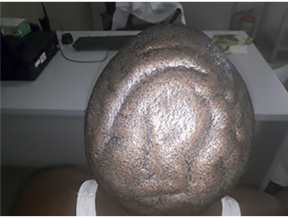
Figure 2 Cutis verticis gyrata (CVG) after ten days of treatment with prednisone 30 mg/day. Superior-posterior view of cranium.
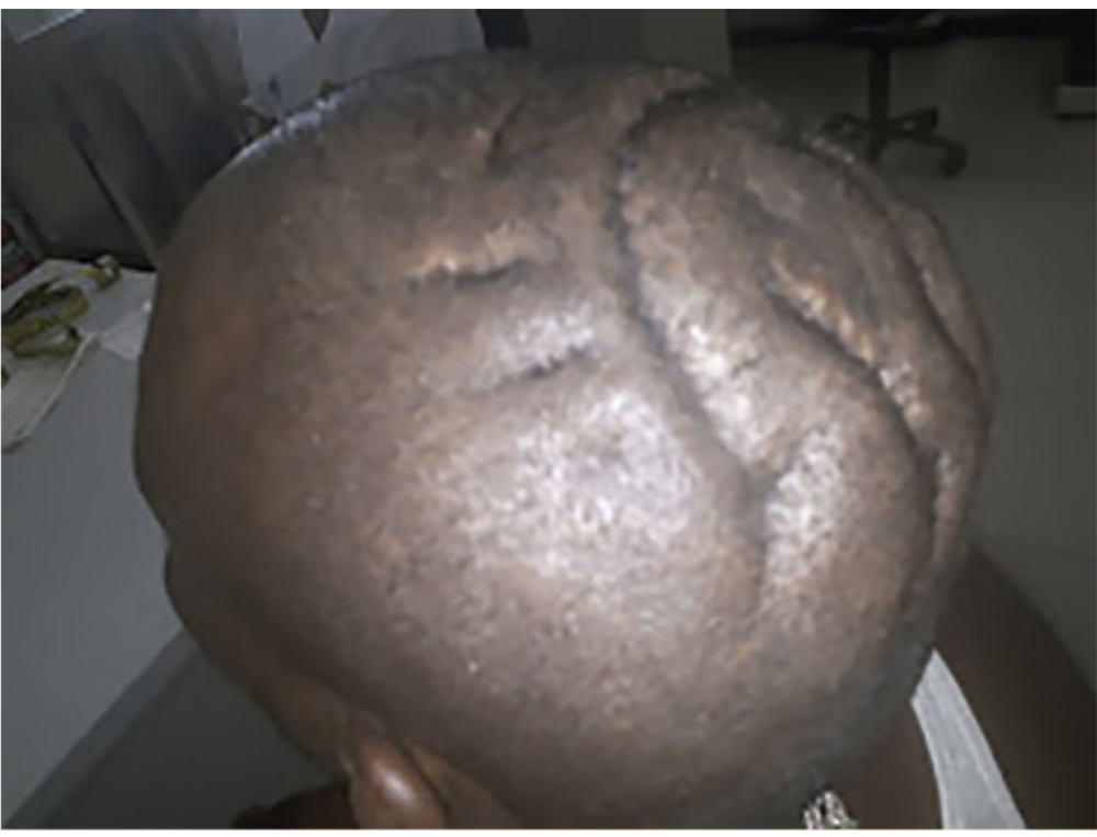
Figure 3 Cutis verticis gyrata (CVG) after ten days of treatment with prednisone 30 mg/day. Left posterolateral view of cranium.
Case history after ten days and satisfaction with the treatment
The patient said that he had greatly improved. He asked only for the treatment to be continued so as to finally eliminate all the bumps and protrusions that, according to him, had “suddenly” appeared, as he stated at the beginning, and now he stated he may have noticed something, but that he never imagined it was something like that. When they asked him about the symptoms, he made no mention of joint pains, breathing difficulties, stomach pains or signs of any other kind. He was solely concerned about the visual and distinguishing aspects of the problem.
The treatment with prednisone may have somewhat reduced the volume of the protrusions. Perhaps, they appeared to be less marked when using photographic comparisons, but without any really significant changes according to our observations. For this reason the treatment was interrupted. However, the patient repeated that he had noticed a major improvement and that the “lumps” (pachyderma) were notably smaller. The steroid treatment was terminated in descending order over one week and no other medication was prescribed (Figures 2 and 3).
The question was again asked if he noticed lumps or ridges when he had long hair, or if the scalp bothered him or itched, since these symptoms are common in CVG1,2,6. He said that ha was not aware of having any lesions, and this time he admitted that he had shaved his head 5 or 6 years previously and that then there was no visible anomaly. He did not know of any similar cases in his family or amongst his siblings. He had never noticed any irritations, stinging or discomfort in the zone, given that he was practically unaware of the lesion because he had long hair.
Details of the inspection and examination at ten days
He presented in good general condition, blood pressure of 140/80, muscular and with no notable anthropometric parameters: height = 170 cm, weight = 82 kg, body mass index (BMI) = 28.37 kg/m2, cranial perimeter (transversal greater in the eyebrow area) = 58 cm, largest perimeter of chest (when inhaling) = 111 cm, waist perimeter (1 cm above navel) = 86 cm, perimeter of hips (in area of trochanters) = 98 cm, waist-hips index (WHI) = 0.88.
In the consultation he showed himself to be of apparently normal intelligence, with fluid and coherent speech, with no psychotic, aggressive or impulsive traces, collaborative, polite and grateful to the medical staff, because he understood that they were trying to help him. He did not present any neurological alterations or evident mental retardation. There was no background of epilepsy. He did not present any dysmorphic facial features, or pachyderma on his face, or squints, or abnormal angles in the eyes. The skin was oily in appearance, with perspiration and a number of keloids on both ears. Hands and feet showed no signs of acromegaly, but there was slight brachydactyly, incipient nail clubbing and thickening of the skin of the fingers. Inspection of the oral cavity showed no notable anomalies (without macroglossia); heart and lung auscultation showed no abnormal sounds, and there were no significant findings in the neurological and other examinations.
DISCUSSION
It was found in Sweden in the 1960s that the prevalence of CVG in institutions for the mentally deficient was thousands of times higher than amongst the general public. It was found that most of the persons affected had an IQ of less than 35, and that only 38 of the cases were women. An increased incidence of convulsions and eye defects in carriers of CVG was also reported9,10. More recently, Hernández et al. published a study showing that the prevalence of CVG in a male psychiatric institution in Colombia is 5.9% (65 cases out of 1,100 institutionalised)8. The most frequent diagnoses was behavioural disorder (46% of cases), followed by impulse control disorder and schizophrenia (percentages not shown). If this study is analysed in greater depth, it can be seen that the information is confused and does allow any conclusions to be drawn other than the confirmation that most of the cases of CVG could be related to severe neuro-psychiatric conditions8. The fact that there are many more male inmates in prison, along with the fact that the neuro-psychiatric disorder is also much more prevalent, highlights the potential interest that this rare problem has in such a setting.
This is the first case of CVG described in prisons, in a patient who does not show any neuro-psychiatric disorders or any severe psychiatric illness. The preliminary results available at this moment in time (pending complementary explorations) indicate a possible case of primary pachydermoperiostosis (Touraine-Solente-Golé’s syndrome). In the review of the histories of previous cases that have been published, it can be seen that some patients labelled with CVG primary essential could correspond to incomplete forms of this very rare disease characterised by pachyderma, periostosis and nail clubbing1.
If the hair covers a large part of the defect and it is not a major aesthetic problem, the treatment for CVG is good hygiene when washing the hair, rubbing shampoo into the folds to eliminate residues and dirt. In patients with very pronounced defects, plastic surgery to improve the person’s appearance may be proposed2.
CONCLUSION
The diagnosis of CVG is by simple inspection, because all the images published are very similar to the ones shown here, pathognomonic of this strange condition. Such a diagnosis is impossible when the characteristics of the lesion are unknown, but very simple once the images provided are seen. There are no agreed protocols or guides for these cases, and so the approach presented may help in advancing towards a better classification of the different cases, with more data and better knowledge of this defect.

