


Meu SciELO
 Serviços customizados
Serviços Personalizados
Journal
Artigo
 texto em
  Inglês (pdf)
 Artigo em XML
 Referências do artigo
 Enviar este artigo por email
Indicadores
-
 Citado por SciELO
 -
 Acessos
Links relacionados
 Citado por Google
 -
 Similares em
SciELO
Similares em
SciELO  Similares em Google
Compartilhar

 Permalink
Medicina Oral, Patología Oral y Cirugía Bucal (Ed. impresa)
versão impressa ISSN 1698-4447
Med. oral patol. oral cir. bucal (Ed.impr.) vol.9 no.5 Nov./Dez. 2004
Primary MALT limphoma of the tongue
GOTERI G, ASCANI G, FILOSA A, RUBINI C, OLAY S, BALERCIA P. PRIMARY MALT LIMPHOMA OF THE TONGUE. MED ORAL PATOL ORAL CIR BUCAL 2004;9:459-63.
SUMMARY
Primitive malignant lymphoma mucosa associated lymphoid tissue (MALT) on the tongue are rare entities. We report here a case of an old woman (80 years old) with a tumor in the dorsum of the tongue, which was histologically diagnosed as an extra-nodal marginal B cell lymphoma. An inflammatory reaction resembling myoepithelial sialoadenitis was observed in the minor salivary glands adjacent at the tumour, suggesting a possible derivation of the lymphoma from a previous reactive process of unknown origin.
Key words: Salivary gland lymphoma, oral MALT lymphoma.
INTRODUCTION
The lymphomas are an own pathology of the lynphoid nodes but they can show in others sites like salivary glands, stomach, thiroid, etc. (extra-nodal lymphomas). Extra-nodal No Hodgkin lymphomas are habitually site in intestine, and cervico-facial area, (about a 34% of the cases) (1).
In the oral cavity lymphomas can be primary or, more frequently, represent a secondary involvement from a lymphomas arising elsewhere. They are localized commonly in the palatin tonsil, which is part of the Waldeyer's ring, an interesting lymphoid barrier between the lymph node system and the lymphoid system associated to the mucosa. Waldeyer's ring lymphomas constitute approximately 5-10% of malignant lymphoma (2). Less frequent site of involvement are the jaws, and the vestibular and gingival mucosa (3,4). Lymphomas of the tongue are still more uncommon (5). From a series of papers dealing with head and neck lymphomas, we can postulate that they represent approximately 3% of overall head and neck lymphomas (6,7).
In the past, lymphomas of the oral cavity as in other extra-nodal sites have been classified according to current classification for nodal lymphomas: most were B-cell lymphomas, while T-cell lymphomas and Hodgkin's disease were rarely observed. Cases with the morphologic features of mucosa associated lymphoid tissue (MALT) in the oral cavity have been described only more recently, since Isaacson and Wright in 1983 (8) studied a serie of patients with lynphoma B gastrointestinal of low grade of malignancy. Since then MALT lymphomas have been reported also in the salivary glands, frequently in association with a preexisting Sjogren's disease (9), they also can arise in the Waldeyer's ring and rare case have been reported also in the tongue (2,10,11). The last classification consider the MALT lymphomas like extranodals lymphomas of the B-cells, in the marginal area of lynphoid tissue associated to mucosa (12). We here describe a case of MALT lymphoma occurring in the dorsum of the tongue, which was primary and arised in association with a prominent lymphoid infiltrate in the minor salivary gland.
CLINICAL CASE
A 80-year-old woman without personal antecedents interesting, presented with one month history of increasing dysphagia. Clinical examination we found a partially ulcerated nodule, hard and erythematosus, with painful at the touch on the dorsum of the tongue.(Fig.1). Regional lymph nodes were not enlarged. An incisional biopsy revealed the presence of monotonous proliferation of lymphoid cells consisting with a diagnosis of lymphoma. Preoperative routine blood studies (red and lymphoid blood cells and biochemistry) and chest radiographic exploration were normal. The lesion was then excised with abundant free margins, under local anesthesia./p>

Microscopically the lymphoid infiltrate ulcerated the epithelial surface and infiltrated deeply the lingual musculature. Throughout the infiltrate, multiple lymphoid follicles were present. Some showed reactive germinal centers, well polarised and with starry-sky histiocytes. Others showed regressive changes of the germinal centers with deposits of homogenous eosinophylic substance and enlargement of mantle rings. All were surrounded and partially obliterated by a prominent peri-follicular infiltrate composed by small lymphoid cells with slightly irregular nuclei with moderately dense chromatin and moderately abundant pale cytoplasm, consistent with centrocyte-like cells of the marginal zone. Numerous histiocytes and eosinophils were also present. Occasional larger cells with 2-3 conspicuous nucleoli were intermixed with the smaller cells and in some areas formed large confluent sheets of centroblast and immunoblast-like cells (Fig. 2). A prominent plasmacytoid differentiation was evident, predominantly arranged in paraseptal and sub-epithelial bands. At the edge of the infiltrate, partially preserved lobules of minor salivary glands of mucinous type were present, showing a prominent lymphoid infiltration. Salivary ducts were dilated and infiltrated by B-cells with formation of occasional epimyo-epithelial islands. Abundant plasmacells were also seen around ducts.

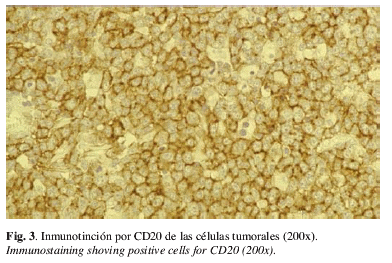
Immunophenotyping of the lymphoid infiltrate was performed on frozen sections and in formalin-fixed paraffin-embedded tissue sections according to the streptavidin-biotin method and with the following antibodies: CD20, CD3, CD5, CD10, CD23, CD43, DBA44, CD68, Immunoglobulin α, γ, µ heavy and κ, λ light chains. Neoplastic centrocyte-like cells showed a B-phenotype, with expression of CD20, CD5, CD43 (Fig. 3). CD10, CD23 and DBA44 were not expressed. A consistent amount of small T CD3+ cells were also detected. Plasmacells in the paraseptal and subepithelial bands, as well those sourrounding ducts of mucous glands, showed monotypic IgG/κ production. A diagnosis of MALT lymphoma was done. The morphologic change of the minor salivary gland near the tumor was interpreted as myoepithelial syaloadenitis.
Clinical examination excluded salivary gland enlargement and a Sjogren's disease. The patient did not suffer for other autoimmune disease like rheumatoid arthrytis, disseminated lupus erythematosus, Hashimoto's thyroiditis. The lymphoma was therefore considered in stage IE. The patient received no treatment except surgery, and is alive and well after a year of the diagnostic.
DISCUSSION
Clinically MALT lymphomas tend to have an indolent course. They tend to remain localized for years and if disseminating, they tend to relapse in other extra-nodal sites. Even when they progress to high grade, prognosis is better respect to the nodal counterpart, because the can be cured by local excision(10,13) Morphologically they repeat the architecture of normal lymphoid tissue associated to the mucosa, showing lymphoid follicles surrounded by B cell of medium size like centrocyte cell with tendency to destroy epithelial structures and to colonise germinal centers. Plasma cell differentiation can be part of the picture. The case we present has the morphological features diagnostic The immunophenotype however deserves a comment regarding expression of CD5 antigen by neoplastic cells. CD5 positive cells were in fact much more numerous than CD3 positive ones, so that we can presume that it refers to the neoplastic cells and not to the abundant T-cell reactive population. This CD5 is a marker useful in the differential diagnosis of MALT lymphomas with other types of low grade B-cell lymphomas, like chronic lymphocytic leukemia and mantle cell lymphoma. In our case, the absence od CD23 expression however points against the diagnosis of alymphocytic lymphoma/ leukemia chronic that usually presents bone-marrow and peripheral blood affectation at the moment of the diagnostic moreover, the tumor was rich of normal or regressive germinal centers The immunophenotype of mantle cell lymphomas also is as the present study C23 negative neoplasic cells transformed, that it's present in our case. CD5 expression is however not against the diagnosis of MALT lymphomas, because sporadic cases of MALT with CD5 expression have been reported (14,15). Characteristically all these cases were head and neck lymphomas (two orbital, one lingual) and showed propensity to early dissemination to other extranodal organs and to the bone marrow. On the basis of these observations, it has been suggested that CD5 expression may identify a subset of MALT lymphomas with a propensity for relapse and dissemination to the bone marrow, as the mantle cell lymphoma and the small lymphocytic B lymphoma/leukemia, which characteristically express this marker (14,15). The case of lingual MALT lymphoma we present, however, did not show any propensity to an early dissemination, despite of CD5 expression. The patient did not have any evidence of lymphoma elsewhere, particularly in the stomach, where most of MALT lymphomas arise. So we can consider it as a primary lymphoma on the tongue and this raises some questions about the etiopathogenesis of the tumor. We know in fact that MALT lymphomas frequently occurr in a background of inflammatory disorders, when B cell clones become independent in their growth. In the stomach the antigen that drives the inflammation has been recognised as Helicobacter pylori, and in early phases are treated by antibiotic therapy. Also autoimmune disorders as Sjögren's disease are prone to develop MALT lymphomas (16). In our case we observe, at the periphery of the lymphoma, a prominent lymphoplasmacellular infiltrate with an important secretion of immunoglobulin light chains and with focal tendency to invade the epithelium of the dilated ducts. We think that this picture is very similar to the myoepithelial sialadenitis observed in the Sjogren's disease and associated with a high risk of developing a MALT lymphoma. On this basis we could hypothesize that lymphoma has occurred in the background of a sialadenitis myoepithelial in the minor salivary gland. MALT lymphomas of the salivary glands is much more common in the parotid gland, but any major or minor salivary gland can be involved, sometimes with a mutifocal involvement (9,17). Most patients are over 50 years and predominantly women. However, our patient did not have any clinical evidence of Sjogren's disease, or other autoimmune disease, like rheumatoid arthritis, disseminated lupus erythematosus, Hashimoto's thyroiditis and the other salivary glands were not enlarged. We can still postulate the presence of an inflammatory unspecific process in the minor salivary gland where a MALT lymphoma has later appeared.
REFERENCES
1. Horny HP, Ferlito A, Carbone A. Laryngeal lymphoma derived from mucosa associated lymphoid tissue. Ann Otol Rhinol Laryngol 1996;105:577-83. [ Links ]
2. Menarguez J, Mollejo M, Carrión R, Oliva H, Bellas C, Forteza J, et al. Waldeyer ring lymphoma. A clinicopathological study of 79 cases. Histopathology 1994;24:13-22. [ Links ]
3. Zanakis SN, Kambas I, Gourlas PG. A non Hodgkin’s lymphoma in the buccal mucosa. A case report. Oral Surg Oral Med Oral Pathol 1992;74:340-2. [ Links ]
4. Piattelli A, Di Alberti L, Artese L. Non-Hodgkin’s lymphoma of the tongue: a case report. Oral Oncol Eur J Cancer 1996;32B:207-9. [ Links ]
5. Toubul E, Ghenim C, Chantelar JV. Lymphomes non Hodgkiniens de la tète e du cou, stades I et II. A propos de 35 cas. Rev Stomatol Chir Maxillofac 1985;86:300-9. [ Links ]
6. Fukuda Y, Ishida T, Fujimoto M, Ueda T, Aozasa K. Malignant lymphoma of the oral cavity: clinicopathologic analysis of 20 cases. J Oral Pathol 1987;16:8-12. [ Links ]
7. Wolvius EB, Van der Valk P, van der Wal JE, van Diest PJ, Huijgens PC, van der Waal I, et al. Primary extranodal non Hodgkin‘s lymphoma of the oral cavity. An analysis of 34 cases. Oral Oncol Eur J Cancer 1994;30B:121-5. [ Links ]
8. Isaacson P, Wright DH. Malignant lymphoma of mucosa associated lymphoid tissue. A distinctive type of B-cell lymphoma. Cancer 1983;52:1410-6. [ Links ]
9. Wolvius EB, van der Valk P, van der Wal JE, van Diest PJ, Huijgens PC, van der Waal I, et al. Primary non-Hodgkin’s lymphoma of the salivary glands. An analysis of 22 cases. J Oral Pathol Med. 1996;25:177-81. [ Links ]
10. Zinzani PL, Magagnoli M, Galieni P, Martelli M, Poletti V, Zaja F, et al. Nongastrointestinal low-grade mucosa-associated lymphoid tissue lymphoma: analysis of 75 patients. J Clin Oncol 1999;17:1254-58. [ Links ]
11. Paulsen J, Lennert K. Low-grade B-cell lymphoma of mucosa-associated lymphoid tissue type in Waldeyer’s ring. Histopathology 1994;24:1-11. [ Links ]
12. Jaffee ES, Harris NL, Stein H, Vardiman JW (Eds). World Health Organization Classification of Tumors. Pathology and Genetics of Tumors of Hematopoietic and Lymphoid Tissues. IARC Press: Lyon 2001. [ Links ]
13. Ferrer A, López-Guillermo A, Bosch F, Montoto S, Hernández JC, Camós M. et al. Linfomas del tejido linfoide asociado a mucosas (MALT) de localización extragástrica: análisis de 14 casos. Med Clin (Barc) 1999;112:577-80. [ Links ]
14. Ferry JA, Yang WI, Zukerberg LR, Wotherspoon AC, Arnold A, Harris NL. CD5+ extranodal marginal zone B-cell (MALT) lymphoma. A low-grade neoplasm with a propensity for bone marrow involvement and relapse. Am J Clin Pathol 1996;105:31-7. [ Links ]
15. Ballesteros E, Osborne BM, Matsushima AY. CD5+ low-grade marginal zone B-cell lymphomas with localized presentation. Am J Surg Pathol 1998;22:201-7. [ Links ]
16. Thieblemont C, Berger F, Coiffier B. Mucosa-associated lymphoid tissue lymphomas. Curr Opin Oncol 1995;7:415-20. [ Links ]
17. Gleeson M, Bennet M, Cawson R. Lymphomas of the salivary glands. Cancer 1986;58:669-704. [ Links ]

