


Mi SciELO
 Servicios personalizados
Servicios Personalizados
Revista
Articulo
 texto en
  Inglés (pdf)
 Articulo en XML
 Referencias del artículo
 Enviar articulo por email
Indicadores
-
 Citado por SciELO
 -
 Accesos
Links relacionados
-
 Citado por Google
 -
 Similares en
SciELO
Similares en
SciELO -
 Similares en Google
Compartir

 Permalink
Medicina Oral, Patología Oral y Cirugía Bucal (Internet)
versión On-line ISSN 1698-6946
Med. oral patol. oral cir.bucal (Internet) vol.11 no.2 mar./abr. 2006
Periapical surgery of maxillary posterior teeth. A review of the literature
Cirugía periapical en dientes posteriores maxilares. Revisión de la bibliografía
Berta García1, Luis Martorell1, Eva Martí2, Miguel Peñarrocha3
(1) Degree in Dental Surgery and resident of the Master of
Oral Surgery and Implantology.
Valencia University Medical and Dental School
(2) Degree in Dental Surgery. Private practice in Valencia
(3) Assistant Professor of Stomatology. Director of the
Master of Oral Surgery and Implantology.
Valencia University Medical and Dental
School. Valencia (Spain)
ABSTRACT
In recent years, periapical surgery has evolved thanks to new
diagnostic and technical advances. A review is made of the literature on
periapical surgery of the antral teeth, based on a Medline search and on the
revision of Spanish dental journals in the period between 1974 and 2003.
The anatomy of the maxillary sinus is discussed, along with
the diagnosis of periapical lesions and the relation of the maxillary sinus to
the antral teeth. The surgical technique, special considerations and prognosis
of periapical surgery in these teeth are also addressed.
Recent studies postulate that the proximity of the antral
teeth to the maxillary sinus should not be viewed as a contraindication to
periapical surgery, and recommend such surgery in teeth with chronic periapical
disease that are refractory to conventional endodontic management, despite the
proximity of the maxillary sinus.
Key words: Periapical surgery, apicoectomy, antral teeth.
RESUMEN
En los últimos años, la cirugía periapical
ha evolucionado gracias a la incorporación de avances diagnósticos y técnicos.
El objetivo del presente artículo es realizar una revisión bibliográfica de
los trabajos publicados sobre cirugía periapical en los dientes antrales; hemos
revisado el Medline y las revistas españolas de Odontología desde 1974 hasta
el 2003.
Comentamos la anatomía del seno maxilar, el diagnóstico de las lesiones
periapicales y la relación del seno maxilar con los dientes antrales; también
la técnica quirúrgica, sus consideraciones especiales, y el pronóstico de la
cirugía periapical en estos dientes.
Los trabajos recientes plantean que la proximidad de los dientes antrales al
seno maxilar, no es una contraindicación para la cirugía periapical, y
recomiendan su realización en dientes con patología periapical crónica,
refractarios al tratamiento endodóncico convencional, a pesar de la proximidad
del seno maxilar.
Palabras clave: Cirugía periapical, apicectomía, dientes antrales.
Introduction
In recent years, periapical surgery has evolved considerably thanks to new diagnostic and surgical advances (1-3). Posterior teeth with periapical pathology not amenable to endodontic treatment are candidates for periapical surgery, and only when the latter is not possible should extraction be considered.
The difficulty posed by these teeth is their location in the posterior zones of the oral cavity. This situation requires adequate evaluation of the surgical access, the relationship of the teeth to anatomical structures such as the maxillary sinus, and their proximity to the mandibular dental canal (4).
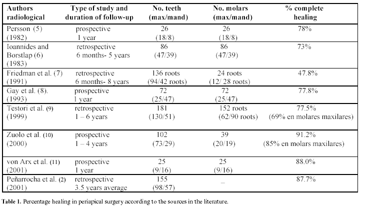
The present study reviews the literature on periapical surgery of the maxillary premolars and molars, referred to as antral teeth because of their close relation to the antrum or maxillary sinus. A Medline search has been conducted, covering the period between 1974 and 2003, together with a review of the Spanish dental literature. We evaluated the most important articles on periapical surgery of the maxillary posterior teeth, selecting those clinical series comprising over 25 teeth and involving a minimum follow-up of 6 months (2,5-11) (Table 1). From the Results obtained it is deduced that the proximity of the antral teeth to the maxillary sinus is not a contraindication to periapical surgery (12).
Anatomy of the maxillary sinus and of the antral teeth
The maxillary sinus is a hollow space lined by a membrane, and communicates with the nasal fossa. The sinus has the shape of a triangular pyramid, with an internal base and an external vertex facing towards the zygoma, and is in relation to the maxillary molars and premolars (13).
The lining sinus membrane is composed of ciliary mucosa that expels the mucosal secretions towards the antral orifice.
The vascularization of the maxillary sinus is fundamentally provided by two arteries: the sphenopalatine artery and the superior alveolar artery. Blood supply also comes from the facial artery, the anterior ethmoidal artery and the suborbital artery (14).
Sensory innervation in turn originates from the posterior dental nerve and the infraorbital nerve with its corresponding branches: mid-dental, anterior dental and small direct branches of the sinus mucosa. These fibers distribute in plexus form above the apexes to innervate both the dental roots and the maxillary sinus proper (14). Tributary vegetative innervation in turn originates almost exclusively from the Meckels sphenopalatine ganglion.
The maxillary first premolar in most cases presents two roots (vestibular and palatine), with even two canals in the vestibular root, though in some instances there may be a single root with two canals. The maxillary second premolar tends to have a single root with one canal, though in 40% of cases the tooth presents dos canals that merge to form a single apical foramen. Depending on the level of apicoectomy, we can find one or two canals. The first and second maxillary molars have three roots and three canals; in order to gain access to the palatine root we approach the tooth from the palatal side, though in some cases a vestibular approach is indicated (15).
Diagnosis of periapical lesions
The extraoral panoramic X-ray study provides general information on the oral condition, and on the existence of periapical lesions and their relation to the nearby hard structures and other anatomical elements (12,16). Intraoral periapical X-rays in turn afford greater detail, with evaluation of bone height, the number, length and shape of the roots, the possible existence of internal or external reabsorptions, periapical lesion extent, the apexes implicated in the lesion, and the relation to the maxillary sinus and dental root. Pepelassi et al. (17) found panoramic X-rays to show important distortion, while intraoral periapical X-rays proved to be more precise than extraoral panoramic imaging.
New digital radiographic techniques have also been developed. In this context, Sullivan et al. (18) used radiovisiography (RVG) in application to small radiotransparencies, allowing contrast modification and more precise visualization of the contour and size of such areas. Cotti et al. (19) prefer computed tomography (CT) for differential diagnostic purposes, definition of the treatment plan and follow-up of extensive periapical lesions. Velvart et al. (20) compared conventional radiography versus CT in application to periapical lesions in 50 patients programmed for periapical surgery of lower premolars and molars. Eighty presumed periapical lesions were evaluated by means of a periapical X-ray study and CT. Surgery diagnosed 78 lesions all of which had been identified by CT, while the periapical X-ray studies identified only 61. Moreover, while CT offered a clear image of the mandibular canal in all cases, conventional radiography did so in only 31 cases.
In recent years, new instruments have been developed, such as the surgical microscope, and endoscopy has been incorporated to periapical surgery thereby contributing to improve diagnostic performance. The microscope affords superior surgical field illumination, contributing to improve each phase of the operation and allowing the performance of smaller ostectomies. With this instrument it is possible to identify perforations, fractures and accessory canals, and different magnifications can be used. Its main inconveniences are its high cost and the prolongation of operating time (21). In turn, the endoscope offers exceptional visibility during surgery. It measures 6 cm in length and 3 mm in thickness, and has an angle of vision of 70 degrees thus making it possible to gain access to the most difficult locations. Endoscopy makes it easier to identify accessory canals, perforations, vertical and oblique fractures, and to assess marginal adaptation of retrograde filling (22).
Relation between the maxillary sinuses and the antral teeth
The posterior teeth are more difficult to treat, because of the more restricted space of the oral vestibular region, which in turn makes flap raising more difficult (8).
The relation between the roots of the maxillary molars and premolars and the sinus has been studied by different authors (12,14,23,24). The roots of the maxillary first and second molars are in intimate relation to the floor of the maxillary sinus in 40% of cases (14).
The palatine roots of these teeth are closer to the antral floor than to the palate, and in 20% of cases are in close proximity to the maxillary sinus (23). Their location complicates an approach through the sinus, and a palatine access is therefore usually adopted (24).
The vestibular roots of the upper posterior teeth are closely related to the floor of the maxillary sinus. However, root access is much easier in this case than in the case of the palatine roots, and in most cases treatment can be carried out without having to perforate the sinus wall (12). In some cases the apexes protrude into the sinus, and the sinus membrane must be raised in order to treat them.
Surgical technique and special considerations
During periapical surgery of the maxillary molars and premolars it is possible to find the same complications as in any apicoectomy, including for example damage to a neighboring tooth. The specific considerations applicable to these teeth are: careful aperture of the maxillary sinus wall or floor; avoidance of sinus membrane perforation; and care to prevent the Introduction of foreign bodies within the maxillary sinus (5,14,25,26).
The Introduction of ultrasound instruments in retrograde cavity procedures has constituted a major advance in apical surgery changing the prognosis of the operation, greatly improving healing, and ensuring improved surgical access to zones with limited possibilities for entry (27).
Regarding aperture of the wall of the maxillary sinus, Ericson et al. (28) performed periapical surgery in 159 maxillary premolars and molars, with aperture of the wall or floor of the maxillary sinus in 18% of the cases. According to these authors, the Introduction of foreign bodies within the maxillary sinus during the operation may cause thickening of the sinus mucosa with symptoms of maxillary sinusitis. Jerome and Hill (29) recommend the use of gauze to block the maxillary sinus aperture and thus avoid the penetration of foreign bodies. Friedman et al. (7) performed periapical surgery in 94 roots of maxillary teeth (12 roots corresponding to maxillary molars). In 11.8% of cases, aperture of the sinus wall or floor was carried out. According to Selden (30), pathological exposure of the floor of the maxillary sinus during periapical surgery predisposes to orosinusal communications.
Regarding sinus membrane perforation, Persson (5) carried out periapical surgery in 18 maxillary molars, with perforation of the membrane in 44% of the cases. Despite this complication, the reported surgical success rate was 78%. No relationship was observed between perforation of the membrane and surgical success. Ioannides and Borstlap (6) performed 47 operations upon maxillary molars, with perforation in 14.8%. According to these authors, perforation of the membrane had no repercussions upon the formation of periapical bone.
Regarding the possible consequences of sinus membrane perforation, Rud and Rud (31) conducted periapical surgery in 200 maxillary first molars, with perforation of the membrane in 50% of the cases. Despite this incidence, sinusitis was only recorded in two cases. Freedman and Horowitz (32), in a study involving 440 patients subjected to 472 apicoectomies of maxillary molars and premolars, perforated the sinus membrane in 10.4% of the cases (23% of 79 molars, 13% of 223 second premolars, and 2% of 170 first premolars). In no case was sinusitis or sinus membrane hyperplasia observed, though sinus membrane polyps were identified in three cases. The authors concluded that if the surgical technique is properly performed and the required postoperative care is provided, periapical surgery can be regarded as the treatment of choice for antral teeth before considering extraction.
Watzek et al. (26) recorded no significant difference in terms of healing of the sinus mucosa between patients with and without intraoperative perforation of the sinus membrane after completing 146 apicoectomies. In this context, Selden (30) found the sinus mucosa to fully regenerate within 5 months after its total surgical removal.
Prognosis
Mikkonen et al. (33) considered the following healing criteria: (a) Clinical success, defined as the absence of pain, swelling and fistulas; (b) Uncertain healing, in the presence or not of clinical symptoms when the patient shows radiological evidence of bone destruction; and (c) Failure, defined by the presence of symptoms in addition to bone destruction and root resorption.
Rud and Andreasen (34) established a series of radiological criteria for defining lesion healing classifying bone healing into three different categories (Table 2).

To assess global healing or success, Von Arx and Kurt (27) used the following criteria: (a) Success, defined by bone regeneration in excess of 90% and a pain score on the clinical scale of 0; (b) Improvement, when bone regeneration reaches 50-90% and the pain score is 0; and (c) Failure, defined by bone regeneration of less than 50%, with a pain score or ≥ 1. These authors defined the clinical scale, rating pain and swelling with ascending numerical scores.
Persson (4), in a series of 31 maxillary roots (18 molars) reported complete radiological healing in 78% of cases after a follow-up of one year. Friedman et al. (6), in 12 maxillary roots, reported a clinical success rate of 50%, after 6 months to 8 years of follow-up. Gay et al. (7) in turn conducted periapical surgery in 72 molars (24 maxillary molars), with a follow-up of one year complete healing being recorded in 77.8%. Testori et al. (8), in a series of 62 maxillary roots, reported complete radiological healing in 69% of cases, involving a mean duration of follow-up of 4.6 years. Zuolo et al. (9), in a one-year prospective study of 20 maxillary molars, recorded complete healing in 85% of cases.
Peñarrocha et al. (2), in a series of 50 upper premolars and molars subjected to periapical surgery, recorded sinus aperture in three cases, radiological healing in 46 cases, and no healing in four cases. There was no relationship between radiological healing and maxillary sinus aperture. According to Von Arx et al. (10) in a one-year prospective study of 15 maxillary roots with periapical lesions (9 molars) subjected to periapical surgery, the success rate was 88% (with complete radiological healing and no clinical signs or symptoms).
The incorporation of ultrasound to periapical surgery has made it possible to perform smaller ostectomies, and to gain access to the apexes of very long roots, with palatine or lingual angulations and in close proximity to the maxillary sinus. In recent years the percentages of complete healing after periapical surgery of the antral teeth have reached 88-91.2% (Table 1). The technique has been shown to be safe in application to maxillary molars and premolars. Periapical surgery is recommended as habitual practice in application to antral teeth before considering the possibility of removal, since the complications caused by sinus perforation are minimal.
![]() Correspondence
Correspondence
Dr. Miguel Peñarrrocha Diago
Clínica Odontológica
Gascó Oliag, 1
46021 Valencia
E-mail: miguel.Penarrrocha@uv.es
Received: 10-01-2004
Accepted: 17-12-2005
References
1. von Arx T, Walker W. Microsurgical instruments for root-end cavity preparation following apicoectomy: a literature review. Endod Dent Traumatol 2000;16:47-62. [ Links ]
2. Peñarrocha M, Sanchis JM, Gay C. Cirugía periapical con técnica de ultrasonidos y relleno con amalgama de plata. A propósito de 122 casos. Rev Eur Odont Estomatol 2001;13:181-8. [ Links ]
3. Peñarrocha M, Sanchis JM, Gay C. Periapical surgery of 31 lower molars based on the ultrasound technique and retrograde filling with silver amalgam. Med Oral 2001;6:376-82. [ Links ]
4. Gutmann JL, Harrison JW. Posterior endodontic surgery: anatomical considerations and clinical techniques. Int Endod J 1985;18:8-34. [ Links ]
5. Persson G. Periapical surgery of molars. Int J Oral Surg 1982;11:96-100. [ Links ]
6. Ioannides C, Borstlap WA. Apicoectomy on molars: a clinical and radiographical study. Int J Oral Surg 1983;12:73-9. [ Links ]
7. Friedman S, Lustmann J, Shaharabany V. Treatment results of apical surgery in premolar and molar teeth. J Endod 1991;17:30-3. [ Links ]
8. Gay C, Paredes J, Berini L. La cirugía periapical en los molares. Rev Eur Odont Estomatol 1993;2:95-102. [ Links ]
9. Testori T, Capelli M, Milani S, Weinstein RL. Success and failure in periradicular surgery: a longitudinal retrospective analysis. Oral Surg Oral Med Oral Pathol 1999;87:493-8. [ Links ]
10. Zuolo ML, Ferreira MOF, Gutmann JL. Prognosis in periradicular surgery: a clinical prospective study. Int Endod J 2000;33:91-8. [ Links ]
11. von Arx T, Gerber C, Hardt N. Periradicular surgery of molars: a prospective clinical study with a one-year follow-up. Int J Endod 2001;34:520-5. [ Links ]
12. Hauman CH, Chandler NP, Tong DC. Endodontic implications of the maxillary sinus: a review. Int Endod J 2002;35:127-41. [ Links ]
13. Bailey BJ, ed. Head and neck surgery-otolaryngology. Philadelphia (USA): Lippincott-Raven Publishers;1998. p.418-20. [ Links ]
14. Wallace JA. Transantral endodontic surgery. Oral Surg Oral Med Oral Pathol 1996;82:80-3. [ Links ]
15. Berkovitz BKB, Holland GR, Moxham BJ. Macroscopic anatomy of the oral cavity and related area. Dento-osseous structures.En: Berkovitz BKB, Holland GR, Moxham BJ, eds. A coulour atlas and textbook of oral anatomy, histology and embryology. Aylesbury: Wolfe Publishing Ltd;1992. p.18-54. [ Links ]
16. Doornbusch H, Broersma L, Boering G, Wesselink PR. Radiographic evaluation of cases referred for surgical endodontics. Int Endod J 2002;35:472-7. [ Links ]
17. Pepelassi EA, Tsiklakis K, Diamanti-Kipioti A. Radiographic detection and assessment of the periodontal endosseous defects. J Clin Periodontol 2000;27:224-30. [ Links ]
18. Sullivan JE, Di Fiore PM, Koerber A. Radiovisiography in the detection of periapical lesions. J Endod 2000;26:32-5. [ Links ]
19. Cotti E, Vargiu P, Dettori C, Mallarini G. Computered tomography in the management and follow-up of extensive periapical lesion. Endod Dent Traumatol 1999;15:186-9. [ Links ]
20. Velvart P, Hecker H, Tillinger G. Detection of the apical lesion and the mandibular canal in convencional radiography and computed tomography. Oral Surg Oral Med Oral Pathol 2001;92:682-8. [ Links ]
21. Pecora G, Andreana S. Use of dental operating microscope in endodontic surgery. Oral Surg Oral Med Oral Pathol 1993;75:751-8. [ Links ]
22. von Arx T, Hunenbart S, Buser D. Endoscope and video-assisted endodontic surgery. Quitessence Int 2002;33:255-9. [ Links ]
23. Waite DE. Maxillary sinus. Dent Clin North Am 1971;15:349-68. [ Links ]
24. Eberhardt JA, Torabinejad M, Christiansen EL. A computed tomographic study of the distances between the maxillary sinus floor and the apices of the maxillary posterior teeth. Oral Surg Oral Med Oral Pathol 1992;73:345-6. [ Links ]
25. Khongkhunthian P, Reichart PA. Aspergillosis of the maxillary sinus as a complication of overfilling root canal material into the sinus: report of two cases. J Endod 2001;27:476-8. [ Links ]
26. Watzek G, Bernhart T, Ulm C. Complications of sinus perforations and their management in endodontics. Dent Clin North Am 1997;41:563-83. [ Links ]
27. von Arx T, Kurt B. Root-end cavity preparation after apicoectomy using a new type of sonic and diamond- surfaced retrotip: a 1 year follow-up study. J Oral Maxillofac Surg 1999;57:656-61. [ Links ]
28. Ericson S, Finne K, Persson G. Results of apicoectomy of maxillary canines, premolars and molars with special reference to oroantral communication as a prognostic factor. Int J Oral Surg 1974;3:386-93. [ Links ]
29. Jerome CE, Hill AV. Preventing root tip loss in the maxillary sinus during endodontic surgery. J Endod 1995;21:422-4. [ Links ]
30. Selden HS. The interrelationship between the maxillary sinus and endodontics. Oral Surg Oral Med Oral Pathol 1974;38:623-9. [ Links ]
31. Rud J, Rud V. Surgical endodontics of upper molars: relation to the maxillary sinus and operation in acute state of infection. J Endod 1998;24:260-1. [ Links ]
32. Freedman A, Horowitz I. Complications after apicoectomy in maxillary premolar and molar teeth. Int J Oral Maxillofac Surg 1999;28:192-4. [ Links ]
33. Mikkonen M, Kullaa-Mikkonen A, Kotilainen R.Clinical and radiologic re-examination of apicoectomized teeth. Oral Surg Oral Med Oral Pathol 1983;55:302-6. [ Links ]
34. Rud J, Andreasen JO. A study of failures after endodontic surgery by radiographic, histologic and stereomicroscopic methods. Int J Oral Surg 1972;1:311-28. [ Links ]

