My SciELO
Services on Demand
Journal
Article
Indicators
-
 Cited by SciELO
 -
 Access statistics
Related links
-
 Cited by Google
 -
 Similars in
SciELO
 -
 Similars in Google
Share
Medicina Oral, Patología Oral y Cirugía Bucal (Internet)
On-line version ISSN 1698-6946
Med. oral patol. oral cir.bucal (Internet) vol.11 n.2 Mar./Apr. 2006
Sterilizing effects of the Erbium: Yag laser upon dental structures:
An in vitro study
Efectos esterilizantes del láser Erbium:
Yag
sobre las estructuras dentarias:
Estudio in vitro
Ma Isabel Leco Berrocal1, José Ma Martínez González2, Manuel Donado Rodríguez3,
Carmen López Carriches4
(1) Associate Professor of Adult Integrated Odontology,
European University of Madrid.
Collaborating Professor of the Master of Oral
Surgery, Madrid Complutense University
(2) Assistant Professor of Surgery, Madrid Complutense
University
(3) Chairman of Oral and Maxillofacial Surgery, Madrid
Complutense University
(4) Associate Professor of Adult Integrated Odontology,
European University of Madrid (Spain)
ABSTRACT
Aim: An evaluation was made of the sterilizing effects of
the Erbium:YAG laser at different power ratings upon dental structures in
vitro.
Design: An in vitro study was made of 47 single-root
teeth removed for periodontal reasons in the Oral and Maxillofacial Surgery
Teaching Unit (Department of Medicine and Orofacial Surgery, Madrid Complutense
University Dental School, Spain). The teeth were divided into three laser
irradiation groups (250, 350 and 450 mJ) and a non-irradiated control group. The
teeth were then immersed in an enrichment medium for 72 hours under conditions
of anaerobiosis, with visual controls after 24, 48 and 72 hours. Posteriorly,
microbiological cultures were made in blood agar to confirm the Results of the
visual inspections.
Results: Increased percentage sterilization of the
samples was recorded with increasing irradiation power - statistically
significant differences being observed between all irradiated groups versus the
controls.
Conclusions: The Erbium:YAG laser exerts a sterilizing
effect upon dental structures in vitro. This effect increases with
increasing laser power ratings.
Key words: Erbium:YAG laser, sterilization, dental structures.
RESUMEN
Objetivo: Comprobar el efecto esterilizante del láser
de Erbium:YAG, a diferentes potencias, en dientes in vitro
Diseño del estudio: Estudio in vitro sobre 47 dientes unirradiculares extraídos
por motivos periodontales en la Unidad Docente de Cirugía Bucal y Maxilofacial
(Departamento de Medicina y Cirugía Bucofacial) de la Facultad de Odontología
de la Universidad Complutense de Madrid. Las muestras fueron divididas en tres
grupos de irradiación con láser (250mJ, 350mJ y 450mJ) y un grupo control -no
irradiados-. A continuación se introdujeron en un medio de enriquecimiento
durante 72 horas en anaerobiosis, realizando controles visuales a las 24, 48 y
72 horas. Y posteriormente se realizaron cultivos microbiológicos en agar
sangre, para confirmar los Resultados de los controles visuales.
Resultados: Se observa como a medida que aumenta la potencia de irradiación
con el láser hay un mayor porcentaje de esterilización de las muestras,
existiendo diferencias estadísticamente significativas entre el grupo control y
cualquiera de los grupos láser.
Conclusiones: El láser de Erbium:YAG presenta efecto esterilizante sobre
las estructuras dentarias in vitro, que se va incrementando a medida que aumenta
la potencia de aplicación del mismo.
Palabras clave: Láser de Erbium:YAG, esterilización, estructuras dentarias.
Introduction
Since Maiman (1) in 1960 officially announced the Introduction of the first laser, more than a hundred different types of laser systems have been developed for different applications (2). Lasers have been used in Medicine from the early stages, and at present practically all medical disciplines benefit from the use of such systems including dental practice (3). In 1964, Stern and Sognnaes (4) vaporized caries with the ruby laser, and posteriorly found laser-treated enamel to be more resistant to dissolution as a result of acid action (5). Lhuisset (6), Melcer (7), Bonin (8) and Seux (9) in turn made use of the CO2 laser to induce structural modifications in the mineralized tissues of teeth.
In 1988, Hibst and Keller introduced the Erbium:YAG (Er:YAG) laser in dental practice (10,11). The functioning of this laser is based on a special thermomechnical removal process without heating of the underlying tissue as a result of which no damage is exerted upon enamel, dentin or pulp (12). In addition, the Er:YAG laser destroys bacteria as a result of the instantaneous vaporization of intracellular water. This capacity to sterilize the irradiated surface is one of the main advantages afforded by the Er:YAG laser in dentistry (13).
The present study explores the sterilizing effects of the Erbium:YAG laser at different power ratings upon dental structures in vitro.
Material and Methods
The study comprised 47 single-root teeth removed for periodontal reasons in the Oral and Maxillofacial Surgery Teaching Unit (Department of Medicine and Orofacial Surgery, Madrid Complutense University Dental School, Spain). The teeth were divided into four groups: group I (control: 10 non-irradiated teeth), group II (13 teeth irradiated at 250 mJ), group III (12 teeth irradiated at 350 mJ) and group IV (12 teeth irradiated at 450 mJ).
Immediately after extraction, the teeth in groups II, III and IV were irradiated with an Erbium:YAG laser (mobile KEY Laser 2 Kavo; operating frequency 10 Hz) over the entire crown-root surface under sterile conditions.
The samples were then immersed in enrichment medium (tryptone soya broth, TSB) and incubated for 72 hours at 37ºC under conditions of anaerobiosis. Visual controls were made after 24, 48 and 72 hours the growth of microorganisms being defined by the appearance of turbidity in the medium (+). The absence of growth was in turn defined by transparency of the medium (-)(Figure 1).
Posteriorly, seeding was performed on blood agar plates with hemin and menadione under the same conditions as before (i.e., anaerobiosis at 37ºC), followed by incubation for 7 days. After this period of time a stereoscopic magnifying glass and microscope were used to confirm the earlier visual control findings in enrichment medium.
A descriptive statistical analysis was made involving frequency tables to assess the response of each variable, together with contingency tables for determining the association between variables based on the chi-square test.
Results
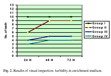
The control (group I) samples not subjected to Er:YAG irradiation all showed turbidity in enrichment medium at visual inspection after 24 hours. As expected, these Results persisted at the subsequent inspections after 48 and 72 hours, and were moreover confirmed by the blood agar plate cultures after one week under anaerobic conditions.
In turn, 46% of the 13 teeth irradiated with the Er:YAG laser at a power rating of 250 mJ (group II) were found to be contaminated i.e., 6 teeth showed turbidity in the enrichment medium, at the first visual inspection after 24 hours. This percentage was seen to increase to 69.2% after 48 hours (i.e., 9 teeth showed turbidity, while 4 retained transparency in the enrichment medium). These same Results persisted after 72 hours. In turn, the blood agar culture findings coincided with the visual control Results after 72 hours in enrichment medium i.e., 30.8% of the teeth showed no bacterial growth after one week at 37ºC under conditions of anaerobiosis (4 plates thus remained sterile).
Group III, comprising 12 teeth irradiated with the Er:YAG laser at a power rating of 350 mJ, showed a comparative decrease in bacterial growth. In effect, the first inspection after 24 hours showed 66.7% of the sample to remain transparent in enrichment medium (i.e., only 4 teeth showed turbidity). The following control after 48 hours revealed turbidity for one additional tooth (58.4% of the sample remaining transparent). Finally, examination after 72 hours yielded the same Results as after 48 hours. The microbiological culture findings in blood agar coincided with the observations in enrichment medium after 72 hours, i.e., 58.4% of the sample remained sterile, while 41.6% showed bacterial contamination.
In group IV, comprising 12 teeth irradiated with the Er:YAG laser at a power rating of 450 mJ, the first visual inspection in enrichment medium after 24 hours showed transparency in 75% of cases, with turbidity of the medium in the remaining 25% of teeth. After 48 hours one additional tooth showed contamination, i.e., turbidity was recorded in 33.3% of cases. In the same way as in groups II and III, these findings persisted at evaluation after 72 hours. The blood agar culture Results confirmed the observations in enrichment medium after 72 hours 66.7% of the teeth remaining sterile after incubation at 37ºC in anaerobiosis for 7 days.
In comparative terms, a decrease in bacterial growth was confirmed as the power rating of the Er:YAG laser was increased.
Figure 2 shows that the teeth subjected to Er:YAG laser irradiation generated less turbidity in enrichment medium after 24 hours compared with the control group (group I), in which 100% of the teeth showed contamination. After both 48 and 72 hours, the number of contaminated teeth increased in all the irradiated groups, while in the control group all teeth continued to show contamination.
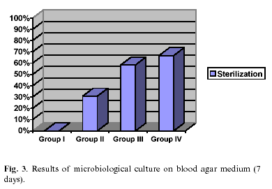
The blood agar culture findings were the same as the Results recorded after 48 and 72 hours in enrichment medium, for all four groups of teeth. Thus, 66.7%, 58.4%, 30.8% and 0% of the teeth in groups IV, III, II and I, respectively, remained sterile. These Results are reflected in Figure 3, where sterilization is seen to increase gradually from 0% in the control group with rising laser power ratings. A statistically significant relationship was observed between the treatment applied and negativity of the visual and blood agar culture evaluations made for all power ratings used i.e., regardless of the power rating employed, laser irradiation was seen to exert a significant sterilizing effect upon the dental structures (p< 0.01).
On the other hand, no significant differences were observed between groups III and IV after 24, 48 and 72 hours in enrichment medium, or after one week of blood agar culture.
Discussion
The present study was designed to evaluate the sterilizing effect of Er:YAG laser irradiation upon the dental structures reported by a number of authors in the literature. To this effect, an in vitro evaluation of single-root teeth was made, with three laser power ratings assessing the presence or absence of bacterial growth by visual inspection, and sterilizing action via microbiological culture in blood agar.
The study of the Results obtained shows that in group I (control), where none of the teeth were irradiated, bacterial growth was confirmed in all cases as soon as after 24 hours of incubation. The blood agar cultures in turn confirmed the visual findings after 24, 48 and 72 hours in enrichment medium.
These observations coincide with those reported by Del Canto et al. (14) in their study of the sterilizing effects of the CO2 laser upon dental structures. These authors detected bacterial growth in all samples not subjected to irradiation, from the first evaluation after 24 hours.
In contrast to the above, the laser-irradiated groups showed a clear reduction in bacterial growth at evaluation after 24, 48 and 72 hours particularly in the specimens exposed to the highest laser power rating. It therefore can be affirmed that bacterial presence diminishes as the laser power rating is increased.
In this sense, on comparing groups II, III and IV (i.e., the Er:YAG irradiated teeth) with the non-irradiated control teeth (group I), a statistically significant association was observed between the treatment applied and negativity of the bacterial growth controls, with any of the laser power ratings used (250, 350 or 450 mJ) i.e., regardless of the power used, the Er:YAG laser exerts significant sterilizing effects upon the dental structures (p<0.01). Nevertheless, on contrasting this effect among the three irradiated groups, the maximum power rating (450 mJ, group IV) was seen to yield the greatest sterilization percentage at all of the evaluation timepoints. On the other hand, no statistically significant differences were found between group IV and the next lower power rating (350 mJ, group III).
These observations coincide with those reported by Verdasco et al. (15), who after applying this laser system described a sterile dental surface resulting from irradiation. Padrós et al. (16) likewise reported a sterilizing effect upon the dental surface after irradiation with the Er:YAG laser, with no overheating in areas adjacent to the beam target zone.
In turn, Mehl et al. (17) reported an irradiation time-dependent decrease in the presence of microorganisms after applying the Er:YAG laser to the interior of root canals in endodontic practice. Takeda et al. (18), in application to root canal cleaning, found the Er:YAG laser to be more effective than argon and Nd:YAG lasers.
Thus, on the basis of our Results and those of other authors, it can be confirmed that the Er:YAG laser exerts a clear sterilizing effect on dental structures.
![]() Correspondence
Correspondence
Dra. Ma Isabel Leco Berrocal
C/ Clara del Rey, 44, 5°D
28002 Madrid
E-mail: maria.leco@uem.es
Received: 12-03-2005
Accepted: 30-07-2005
References
1. Maiman TH. Stimulated optical radiation in ruby. Nature 1960;187:493-4. [ Links ]
2. España AJ. Aplicaciones del láser de CO2 en Odontología. Madrid: Edit Ergon, S.A.; 1995. p. 3-13. [ Links ]
3. Martínez-González JM, Donado M. Láser en Cirugía Bucal. En: Donado M. Cirugía Bucal. Patología y Técnica. Madrid: Ed. El autor; 1990. p. 799-816. [ Links ]
4. Stern RH, Sognnaes RF. Laser effect on dental hard tissues. A preliminary report. Calif Dent Asso J 1965; 33:17-9. [ Links ]
5. Stern RH, Sognnaes RF. Laser inhibition of dental cries suggested by first test in vitro. J Am Dent Assoc 1972; 85:1087-90. [ Links ]
6. Lhuisset F (citado por Uribe Echevarría). Note à propos d´impacts de laser à gaz carbonique sur le dentine. Le Chir Dent de France 1979;31:37-9. [ Links ]
7. Melcer J, Melcer F. Rèsultats à court et moyen terme de l´utilisation du laser en Odontologie. Inf Dent de France 1982;22:2147-51. [ Links ]
8. Bonin P, Duprez J, Perol J, Vincent R. Analyse comparative de differents lasers sur les tissus durs de la dent en fonction du mode d´application. Analyse des impacts au microscope èlectronique à balayage. Rev d´Odont- Stomatol 1985;1:29-35. [ Links ]
9. Seux J, Jofre A, Bonin P, Magloire M. Effects du rayonement laser CO2 sur l´incisive de souris. Rev d´Odont- Stomatol 1987;4:231-5. [ Links ]
10. Hibst R, Keller U. Experimental studies of the application of the Er:YAG laser on dental hard substances I. Measurement of ablation rate. Lasers Surg Med 1989;9:338-44. [ Links ]
11. Keller U, Hibst R. Experimental studies of the application of the Er:YAG laser on dental hard substances II. Light microscopic and SEM investigation. Lasers Surg Med 1989;9:345-51. [ Links ]
12. Keller U, Hibst R. Ultrastructural changes of enamel and dentin following Er:YAG laser radiation on teeth. SPIE 1990; 1200:408-15. [ Links ]
13. Padrós E, Arroyo S. El láser de Er:YAG en la práctica odontológica general. Quintessence 1999;12:61- 76. [ Links ]
14. Del Canto M, Martínez-González JM, Alobera MA, Meníz C, Donado M. El láser CO2 en la esterilización de estructuras dentarias I. Estudio in vitro. Avances en Odontoestomatol 1998;14:403-9. [ Links ]
15. Verdasco M, Ortiz B. Láser de Er:YAG principios físicos y sus aplicaciones en Odontología. Quintessence 1996; 9:657-68. [ Links ]
16.-Padrós E, Arroyo S. Evaluación clínica del láser Er:YAG en odontología. Informe Dental 1998;2:428-32. [ Links ]
17. Mehl A, Folwaczny M, Haffner C, Hickel R. Bactericidal effects of 2´9 um Er:YAG laser radiation in dental root canals. J Endod 1999;25:490- 3. [ Links ]
18. Takeda FH, Harashima T, Kimura Y, Matsumoto K. Comparative study about the removal of smear layer by three types of laser devices. J Clin Laser Med Surg 1998;16:117-22. [ Links ]


 text in


