


Meu SciELO
 Serviços customizados
Serviços Personalizados
Journal
Artigo
 texto em
  Inglês (pdf)
 Artigo em XML
 Referências do artigo
 Enviar este artigo por email
Indicadores
-
 Citado por SciELO
 -
 Acessos
Links relacionados
-
 Citado por Google
 -
 Similares em
SciELO
Similares em
SciELO -
 Similares em Google
Compartilhar

 Permalink
Medicina Oral, Patología Oral y Cirugía Bucal (Internet)
versão On-line ISSN 1698-6946
Med. oral patol. oral cir.bucal (Internet) vol.11 no.4 Jul. 2006
Structural and functional salivary disorders in type 2 diabetic patients
Alteraciones salivares en pacientes con diabetes tipo 2
Carmen Carda1, Nezly Mosquera-Lloreda2, Lucas Salom3, Maria Elsa Gomez de Ferraris4, Amando Peydró5
1Catedrática de Histología, Dpto de Patología, Facultad de
Medicina y Odontología, Universidad de Valencia, España
2Servicio de Análisis clínicos del Hospital Universitario
La Fe de Valencia, España
3Servicio de Cirugía Maxilofacial del Hospital
Universitario La Fe de Valencia, España
4Catedrática de Histología y Embriología, Directora del
Departamento de Biología Bucal, Facultad de Odontología,
Universidad Nacional de
Córdoba, Argentina
(5) Catedrático de Histología, Director del Dpto de
Patología, Facultad de Medicina y Odontología, Universidad de Valencia, España
Supported by the grant: Secretaria de Ciencia y Tecnología, Universidad Nacional de Córdoba SECyT UNC Res 202/05.
ABSTRACT
Diabetes mellitus type 2 is the most common metabolic
disorder and it causes an important morbimortality. The structural modifications
in the parotid gland (sialosis) had already been described in these patients and
could result in variations in the salivary composition, as well as an increase
in periodontal and dental pathology.
Objectives:
to compare the biochemical findings in the
saliva and to correlate these biochemical disturbances with the morphologic
findings previously described.
Patients and methods: clinical information were gathered
about 33 patients, 17 had type 2 diabetes. Samples of whole saliva were obtained
for biochemical analysis and serum samples to determine metabolic control.
Results: in the diabetics saliva we found urea and total
proteins increased and reduced levels of microalbumina. Salivary glucose was
only augmented in patients with poor metabolic control. Clinical symptoms of
xerostomia were present in 76,4% and dental and periodontal disease in 100%. The
parotid gland was characterised by the presence of small acini, lipid
intracytoplasmic droplets, as well as adipose stroma infiltration. The acinar
cytoqueratins expression was heterogeneous and very positive in the hyperplasic
ducts.
Conclusions: these biochemical disorders in the saliva of
the type 2 diabetic patients would be related with the structural changes
previously observed in parotid glands.
Key words: Diabetes tipe 2-parotid gland, structural and functional modifications.
RESUMEN
La diabetes mellitus tipo 2 es el desorden metabólico más
frecuente, siendo además causante de una importante morbi-mortalidad. En estos
pacientes se han descrito alteraciones estructurales de la parotida (sialosis)
que podrían comportar modificaciones en la composición salivar, así como un
incremento de patología dental y periodontal.
Objetivos:
establecer las alteraciones bioquímicas de la
saliva y su posible correlación con los hallazgos morfológicos.
Diseño del estudio:
se realizo un estudio clínico de 33
pacientes, 17 de ellos con diabetes tipo 2. Se recogieron muestras de saliva
para análisis bioquímico y suero para control metabólico.
Resultados:
en la saliva de los pacientes diabéticos
encontramos un incremento de la urea y las proteínas totales, así como una
reducción de la microalbumina. La glucosa salivar estaba solo aumentada en los
diabéticos con mal control metabólico. Los síntomas de xerostomía se detectaron
en el 76,4% de los casos y las lesiones dentales y periodontales en el 100% de
los pacientes.
Conclusión:
estos desordenes bioquímicos en la saliva de
los pacientes con diabetes tipo 2 se pueden correlacionar con las alteraciones
estructurales descritas previamente.
Palabras clave: Diabetes tipo 2, glándula parótida, modificaciones bioquímicas, alteraciones estructurales.
Introduction
The diabetes mellitus type 2 (non insulin-dependent) or adult diabetes affects people aged over 40, frequently overweight or obese. This metabolic variety is characterized by the partial shortage of insulin that is proved by disturbances in the metabolism of glucose and therefore the normal assimilation process is affected (1,2). The most common alterations, at a stomatologic level, include periodontal diseases, caries, candidiasis, commissural quelitis and sialomegaly. All of the already mentioned are linked to the xerostomy and glandular hypofunction (1,3-6). Some authors (2,3,7) state that the decrease of the salivary stream in diabetics is caused by the increase of diuresis or poliuria, that make the extracellular liquid decrease notoriously, and as a consequence, the production of saliva.
Diabetes mellitus is one of the etiological causes of sialosis, a pathology generally characterized by a bilateral enlargement, neither neoplastic nor inflammatory, of the parotid gland (8-10). Sialosis, however, can have different origins, having been described as a consequence of hormonal, nutritional or metabolic disturbances, medicamentosus or neurohumoral alterations (3,11,12). Furthermore, the process is not exclusive to the parotid, but also affects, to diverse degrees, the other larger and smaller salivary glands (13-16). Clinically it is said that the swelling of diabetic origin frequently has a prearicular ubication (1,3,7) different from the alcoholic sialosis is located at an retromandibular level. In addition, the diabetic sialosis shows a more pronounced swelling (1,17).
The sialosis generally involves glandular hypertrophy, produced either by adipose infiltration or by acinar hypertrophy. There are authors who accept the coexistence of both modifications, while others deny such a possibility (18,19). The fact is that acinar hypertrophy is not always present in sialosis, as a consequence some authors centre their attention on the glandular dysfunction. This dysfunction is generally manifested as salivary hypofunction and xerostomia (12).
This pathology is not considered neither inflamatory nor tumoral, but a degenerative one. It is also linked to an alteration in the autonomous glandular neuroregulation (1,7,12) produced by a demielination (or sympatic denervation) and an atrophy of the mioepithelial cells. This would interfere with the secretion mechanism that is produced by the stimulation of the alpha and beta adrenergic receptors of the acinar cells, that phisiologically induce exocytosis (3,5,12).
In previous studies of parotid glands from individuals with alcoholic sialosis we described heterogeneous accumulations of secretory granules of different sizes, irregularly distributed throughout the cytoplasm of the acinar cells, unlike the Von Ebner serosa glands, where the granules were smaller, homogeneous and preferentially located in the apical region. Likewise, the alterations at the epithelial level of the ductal system were highly evident. The striate ducts exhibited an epithelium of pseudostriated appearance, with elongated nuclei of dense chromatin, together with other nuclei surrounded by loose chromatin. In the excretory ducts, of note, was the increase in ductal diameter, the stasis of the secretory material with desquamated cells, and epithelial atrophy, immunohistochemically heterogeneous for cytokeratins (13,14, 20, 21).
With respect to its function there have been described, other than the flow disturbances, modifications of the salivary biochemistry in type 2 diabetic patients: disturbances in the glucose concentration, total protein count, albumin, lisozymes, peroxidase, electrolytes (sodium, potassium, chloride, phosphorus, magnesium, calcium), amylase, IgA, and in its buffer capability. Although these findings have not been related by all the studies. Therefore Ben-Aryeh et al. (22) studied 35 type 2 diabetics and they compared them with a control group. The results of this study found increased levels of glucose, total protein, and potassium, normal levels of amylase, IgA and sodium and a reduced salivary flow, not finding any correlation between blood and salivary glucose levels.
Dodds et al. (23) studied the effects of metabolic control in salivary flow, protein concentration, and salivary amylase activity in type 2 diabetics, finding a reduced salivary amylase activity, but no significant difference in the protein concentration or in the salivary flow. Reuterving et al. (24) studied the influence of the degree of severity of diabetes in the salivary flow and in the glucose concentration in 11 patients with type 1 and 2 diabetes, not finding any significant difference in pH, buffer capacity, total proteins, electrolytes, lysozymes, peroxidases, or metabolic control. They concluded that the degree of metabolic control doesnt have a great influence in salivary composition, except in the salivary concentration of glucose. Forbat et al. (25) measured the concentration of blood and salivary glucose in 31 patients with type 2 diabetes mellitus. They concluded that salivary glucose levels dont reflect blood glucose levels.
In type 1 and 2 diabetic patients it has also been tried to correlate salivary composition with the presence of oral pathology, finding dental caries in 100% of diabetic patients and an overall increase in periodontal disease (26-28).
The objectives of our study were: to compare the biochemical findings in the saliva in a sample group of diabetics against the saliva of a control group; to establish if the salivary biochemical disturbances are related with metabolic control; to determine if the variables of the oral and periodontal findings are related with the salivary biochemical disturbances; and to establish the usefulness of measuring the salivary biochemistry of type 2 diabetes mellitus patients, as an optional parameter to evaluate the metabolic state.
Patients and methods
1. Inclusion criteria
The study was conducted prospectively between may 2001 and july 2004 in the Department of Pathology of the Clinic University Hospital and the Department of Maxilofacial Surgery of the "La Fe" of Valencia, Spain. All subjects were referred from the same geographic area and two groups were stablished, diabetic patients and control patients without diabetic dissease. The study was conducted in accordance with the ethical principles outlined in the Declaration of Helsinki.
A total of 33 subjets were included in the study: 10 male (58,8%) and 7 female (41,2%) in the diabetic group (mean age of 68, range 26-86 years), and 8 male (50%) and 8 female (50%) in the control group (mean age of 48, range 26-86 years).
2. Clinical assessment
A questionnaire was developed to identify the patient data like: age, sex, years of evolution of diabetes(less than 10 years, 10 to 20 years, more than 20 ears) and xerostomia, existed or not as a subjective sensation of dry mouth (Anexe 1). The criteria chosen for the diagnosis of diabetes were those of the ADA (2002): glucemia under 120 mg/dl or of 160 in aged people and HbA1c under 7% implies a good control, between 120-150 an until 7.9 respectively means an acceptable control, between 150-220 and until 9.5 a defficient control, and over these values, a bad metabolic control results.

3. Oral examination
All subjects included in the study underwent a dental and periodontal examination performed by the same dentist. Periodontal examination allowed for a subjective measure of the periodontal health, classifying the population into three groups (mild, moderate or severe periodontal disease) according to the quantity of gum recession, the degree of alveolar bone lost around the teeth and the dental root exposure. The number of teeth with caries was counted, as well as the lost teeth.
4. Biochemical examination
The each patient was asked to put some total saliva (about 1 dl.) in a dry polypropylene tube. The sample was obtained without previous stimulus of the salivary secretion. The reference values that were considered as normal of the salivary biochemistry are shown in table 2. The glucemia and the glycosylated haemoglobin was quantified from a venous blood sample of the patient having fasted for ten hours.
5. Statistical method
The statistical analysis was performed with the program MINITAB V14. The statistical signification was measured using the chi-squared test for qualitative variables, and the t-student for quantitative variables, considering any p value which is less or equal to 0.05 as significative.
Results
1. Clinical parameters
With respect to the studied clinical variables it was found that the evolution time of the diabetes mellitus had a significant relationship with the metabolic control, 11 (64,6%) of the 17 diabetic patients haven major or 10 year with the type 2 diabetic and the total patients with poor metabolic control (7 patients, 41,1%), haven more of 10 years of evolution time to the disease. We arent significant differences between sex, the behavior of the variables was similar in male and female (table 1).

2. Biochemical parameters
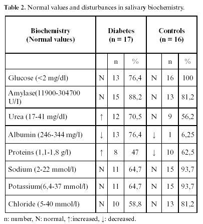
In the sample of diabetic patients it was found that the majority of the results for the salivary biochemistry were normal for: the glucose (13/17=76,4%), the amylase (15/17=88,2%), the sodium (11/17=64,7%), the potassium (11/17=64,7%) and the chloride (10/17). While the total proteins (8/17=47%) and the urea (12/17=70%) were increased, and only for the albumina the majority of patients had a decreased concentration (13/17=76,4%). In the control group it was found that for the majority of patients the salivary biochemical parameters studied were in normal concentrations. Except for the albumin (1/16=6,25%) and for the total proteins (10/16=62,5%) that someone values were decreased.
In the analyzed blood tests of the 17 diabetic patients, 13 (76,4%) had glucemias above the normal values (>126 mg/dl) of they 5 (29,4%) patients with glucose values between 126-180mg/dl and the other 8 patients had values more to 180mg/dl (47%); having 4 patients (35,5%) with normal glucose values and 7 (41,1%) patients had values of glycosilated hemoglobin greater than 8% (bad metabolic control), 3 (17,6%) patients had values between 7-8% and 7 (41,1%) with values less to 7%. In the control group the glucemia and glycosilated hemoglobin values was normal. Only, diabetic patients with glucemia more of 180 mg/dl and glycosylated more of 8% had salivary glucose increased (4 patients, 23,5%). While there are no significant differences in the amylase values, total proteins or salivary electrolytes, between type 2 diabetic patients and control group (table 2).
3. Oral parameters
In the xerostomia subjective test, 13 patients (76,4%) with type 2 diabetic related presented "dry mouth" sensation and 4 patients (23,5%) not haven xerostomia. In the control group 3 patients (18,7%) related haven xerostomia.
In the oral cavity disease, among of the diabetic patients the 100% haven symptoms of periodontal disease, 4 patients (23,5%) with mild, 6 patients (35,5%) with moderate and 7 patients (41,1%) with severe. While that in the control group, only 8 patients (50%) shown periodontal disease, 3 patients (18,7%) initial, 4 patients (25%) moderate and 1 patients (6,3%) severe disease.
The dental caries it was found in 13 (76,4%) of the diabetics patients, 4 patients (23,5%) with moderate and 9 patients (52,9%) with severe caries dental. Among the control group, 2 patients (12,5%) haven dental caries moderate and 6 patients (37,5%) with severe.
The loss tooth between of the diabetic patients, 5 patients (29,4%) presented less to 10, 8 patients (47%) more to 10 and 4 patients (23,5%) were edentulous. In the control group, 4 patients (25%) presented less to 10 loss tooth, 3 patients (18,7%) more to 10 and 1 patients (6,3%) was edentulous. The results are show in table 3.
Discussion
Several authors have covered the subject of the biochemical changes found in the saliva of diabetic patients, existing published articles about the salivary biochemistry where several disturbances are described in the glucose, total proteins, lisozymes, peroxidases, electrolytes (sodium, potassium, chloride, phosphorus, magnesium, calcium), amylase, A immunoglobulin, pH, and buffer capacity. The results in many cases differ from one study to another. These may be due to the diversity in selection criteria of the samples and the type of design of each study (24-26).
Previously we had studied the effect of the alcoholic sialosis in the salivary biochemistry (29) and in taste perception (30). It can be concluded: there was a significant difference between the total concentration of salivary proteins in consumer and non consumer groups. The highest mean concentration was found in the second group.
According to the results of this study there exist changes in the salivary biochemistry of diabetic patients with respect to the non-diabetics. The urea and the total proteins were increased, which matches the results of Ben-Aryeh et al. (22), the albumin was decreased while the amylase, sodium, potassium and chloride where found normal in the majority of the patients. The increase in the salivary glucose was related with a poor metabolic control, this matches the results of Reuterbin et al. (24). The increase of the blood glucose levels is not related with an increase in the salivary glucose concentration, which matches the results of Forbat et al. (25). No significant relationship was found between the metabolic status and the other altered salivary parameters in the diabetic sample, like the total proteins, urea and albumin.
Clinically, the age was relationship whith the more presence the oral signs and symptoms between diabetic and control group; dont encounter statistically significant differences among male and female. All of the diabetic patients presented some degree of periodontal disease, associated with caries and loss of teeth matching the previous results (26-28,31).
In the xerostomy subjective test, 13 diabetic patients related symptoms of "dry mouth", matching the results of Arrieta et al. (27) and Llamas et al. (28), whereas only 3 in the control group had these complaints, they had more to 68 years old.
Type 2 diabetes could, therefore, be considered a risk factor for suffering from xerostomia, and this may be due to the structural changes caused by diabetes mellitus in the salivary glands previously described acinar atrophy a adipose infiltration (21). In accordance with our results, we suggest that the principal cause of the increase in glandular size could be the notable adipose stroma infiltration. Neither was there evidence of inflammatory processes that could justify the parotid hypertrophy. And the biochemical disorders in the saliva of the type 2 diabetic patients would be related with the structural changes previously observed in parotid glands.
Dra. Carmen Carda
Dpto de Patología, Facultad de Medicina y Odontología
Universidad de Valencia.
Avda. Blasco Ibañez, 17,
46010 Valencia, Spain.
Email: carmen.carda@uv.es
Received: 5-11-2005
Accepted: 25-01-2006
References
1. Garcia-Pola MJ, Vallejo A, Alvarez I, Lapiedra R. Manifestaciones Bucales de la Diabetes Mellitus. http:/www.coem.org/revista/anterior/0198/articulo.html [ Links ]
2. Pelayo Antuña V. Manifestaciones Orales en la Diabetes Mellitus. http.//www.clinidiabet.com/es/infodiabetes/02-educacio/07-educando/01.html [ Links ]
3. Bermejo Fenoll A. Medicina Bucal. Vol I. Madrid: Síntesis SA Editores; 1998. p. 372-4. [ Links ]
4. Rose L, Kaye D eds. Medicina Interna en Odontología. Tomo II. Barcelona: Editorial Salvat; 1997. p. 1375-427. [ Links ]
5. Bagan JV, Alapont L, Sanz C, del Olmo JA, Morcillo E, Cortijo J et al. Alteraciones dentales y salivales en los pacientes con cirrosis hepática: estudio de 100 casos. Med Clin (Barcelona) 1998;111:125-8. [ Links ]
6. Moret Y, Muller A, Pernía Y. Manifestaciones bucales de la diabetes mellitus gestacional. Dirección electrónica: http://www.actaodontologica.com/40_2_2002/32.asp [ Links ]
7. Escovich L, Novelli JL, eds. Glándulas Salivales. Patología, diagnóstico y tratamiento. Rosario (Argentina): Editorial de la Universidad Nacional de Rosario; 2002. [ Links ]
8. Abelson D, Mandel ID, Karmiol M. Salivary studies in alcoholic cirrhosis. Oral Surg Oral Med Oral Pathol 1976;41:188-91. [ Links ]
9. Dutta SK, Dukehart M, Narang A, Latham PS. Functional and structural changes in parotid glands of alcoholic cirrhotic patients. Gastroenterology 1989;96:510-8. [ Links ]
10. Neville BW, Damm DD, Allen CM, Bouquet JE eds. Oral and maxillofacial pathology. Philadelpia: Saunders Company Ed.; 2002. p. 404-5. [ Links ]
11. Ceballos Salobreña A eds. Medicina Bucal. Granada: Gráficas Anel SA Editores; 1993. p. 234-8. [ Links ]
12. Coll Daroca J. Enfermedades localizadas de las glándulas salivales. En: Farreras P, Rozman C eds. Medicina Interna. Vol I. Barcelona: Doyma Editores; 1992. p. 37-8. [ Links ]
13. Ferraris ME, Carranza M, Ferraris R, Fili T. Variaciones estructurales en glándulas salivales de alcohólicos crónicos. Rev Fac Odont UNC 1995;20:59-68. [ Links ]
14. Ferraris ME, Arriaga A, Busso C, Carranza M. Histological study of parotid, submaxillary and von Ebner salivary glands in chronic alcoholics. Acta Odont Latinoamer 1999;12:97-102. [ Links ]
15. Smith PF, Esguep AS. Estudio histopatológico de glándulas salivales menores en alcohólicos. Rev Med Chile 1995;123:1387-93. [ Links ]
16. Scott J, Burns J, Flower EA. Histological analysis of parotid and submandibular glands in chronic alcohol abuse: a necropsy study. J Clin Pathol 1988;41:837-40. [ Links ]
17. Guggenheirmer J, Myers D, Weyant R. Insulin dependent diabetes mellitus and oral soft tissue pathologies. Oral Surg Oral Med Pathol Oral Radiol Endod 2000;89:563-9. [ Links ]
18. Rodrigo Gómez JM, Peydro Olaya A, Llombart Bosch A, Aparisi Quereda L, Serra Desfilis MA, Guix Garcia J, et al. Parotid hypertrophy in liver cirrhosis. II. Morphological, histochemical and ultrastructural study. Rev Esp Enferm Apar Dig 1973;41:751-70. [ Links ]
19. Marco J. Patología de la parótida. En: Symposium sobre patología de la parótida. Valencia: Fundación García Muñoz Editores; 1980. p. 82-6. [ Links ]
20. Carda C, Ferraris ME, Arriaga A, Carranza M, Peydró A. Sialosis Parotídea Alcohólica. Estudio Estructural y Ultraestructural. Med Oral 2004;9:24-32. [ Links ]
21. Carda C, Carranza M, Arriaga A, Díaz A, Peydró A, Ferraris ME. Structural differences between alcoholic and diabetic parotid sialosis. Med Oral Patol Oral Cir Bucal 2005;10:309-14. [ Links ]
22. Ben-Aryeh H, Cohen M, Kanter Y, Szargel R, Laufer D. Salivary composition in diabetic patients. J Diabet Complications 1988;2:96-9. [ Links ]
23. Dodds MW, Dodds AP. Effects of glycemic control on saliva flow rates and protein composition in non-insulin-dependent diabetes mellitus. Oral Surg Oral Med Oral Pathol Oral Radiol Endod 1997;83:465-70. [ Links ]
24. Reuterving CO, Reuterving G, Hagg E, Ericson T. Salivary flow rate and salivary glucose concentration in patients with diabetes mellitus influence of severity of diabetes. Diab Metab 1987;13:457-62. [ Links ]
25. Forbat L, Collins R, Maskell G, Sonksen P. Glucose concentrations in parotid fluid and venous blood of patients attending a diabetic clinic. J R Soc Med 1981;74:725-8. [ Links ]
26. Ben-Aryeh H, Serouya R, Kanter Y, Szargel R, Laufer D. Oral health and salivary composition in diabetic patients. J Diabetes Complications 1993;7:57-62. [ Links ]
27. Arrieta J, Bartolomé B, Jiménez E, Saavedra P, Arrieta F. Problemas bucodentales en pacientes con diabetes mellitus. Med Oral 2003;8:97-109. [ Links ]
28. Llamas Cadaval R, Sampedro Abascal C, Segura Egea JJ, Lapetra Peralta J. La diabetes como factor de riesgo de edentación en la población geriátrica. Centro de Salud San Pablo y Facultad de Odontología de Sevilla, 1995. [ Links ]
29. Actis AB, Simbrón A, Brunotto M, Gómez de Ferraris ME. Concentración de proteínas totales en saliva de jóvenes consumidores sociales de alcohol. Acta Odontológica Venezolana 2006, 44 (2) (in press). [ Links ]
30. Ferraris ME, Simbrón A, Actis AB, Basso B, Moretti E, Postiglion G. Estudios de percepción gustativa y determinaciones bioquímicas en saliva de jóvenes consumidores sociales de alcohol. Revista de la Facultad de Odontología de la Universidad de Chile 2003;21:59-68. [ Links ]
31. Miralles-Jorda L, Silvestre-Donat FJ, Grau Garcia-Moreno DM, Hernández-Mijares A. Buccodental pathology in patients with insulin-dependent diabetes mellitus: a clinical study. Med Oral 2002;7:298-302. [ Links ]

