


Mi SciELO
 Servicios personalizados
Servicios Personalizados
Revista
Articulo
 Inglés (pdf)
 Articulo en XML
 Referencias del artículo
 Enviar articulo por email
Indicadores
-
 Citado por SciELO
 -
 Accesos
Links relacionados
-
 Citado por Google
 -
 Similares en
SciELO
Similares en
SciELO -
 Similares en Google
Compartir

 Permalink
Medicina Oral, Patología Oral y Cirugía Bucal (Internet)
versión On-line ISSN 1698-6946
Med. oral patol. oral cir.bucal (Internet) vol.12 no.3 may. 2007
Osteocalcin in serum, saliva and gingival crevicular fluid: their relation with periodontal treatment outcome in postmenopausal women
Pedro Bullon1, Lucy Chandler1, Juan J. Segura Egea1, Ramón Perez Cano2, Angel Martinez Sahuquillo1
(1) Periodontics Department
(2) Medicine Department, School of Medicine, University of Sevilla
ABSTRACT
Background. Osteocalcin levels have been postulated as a marker of inhibition of bone formation. The aim of the present study was to assess plasma, saliva and GCF levels of osteocalcin and correlate them with periodontal treatment outcome in postmenopausal women.
Methods. Thirty-nine postmenopausal women (57.8 ±8.5 years old) were recruited for the study. Periodontal examination of all women was carried out and plaque, bleeding on probing, probing depth (PD), and clinical attachment level (CAL) were recorded. Serum, saliva and gingival crevicular fluid osteocalcin were measured. Then, periodontal treatment was carried out. Six months after the first appointment a second periodontal examination was carried out.
Results. Mean PD and mean CAL decreased significantly at second appointment in the group with serum osteocalcin concentration < 10 ng/ml (15.8 ±15.8% and 15.3 ± 21.2% respectively; p < 0.05). Mean PD decreased significantly at second appointment in the groups with saliva osteocalcin concentration < 3 ng/ml (17.1 ± 15.9%; p < 0.05) and 3 – 7 ng/ml (16.2 ± 18.1%; p < 0.05).
Conclusions. Low serum osteocalcin concentration is associated to a significantly higher percentage of decrease in PD and CAL after periodontal treatment in postmenopausal women. Low saliva osteocalcin concentrations are significantly associated to a higher percentage of decrease in PD.
Key words: Osteocalcin, osteoporosis, periodontitis.
RESUMEN
Antecedentes. Los niveles de osteocalcina se han propuesto como marcador de la inhibición de la formación ósea. El propósito de este trabajo es determinar las concentraciones de osteocalcina en plasma, saliva y fluido crevicular correlacionándolo con el resultado del tratamiento periodontal en mujeres postmenopáusicas.
Pacientes y métodos. El estudio se realizó en treinta y nueve mujeres postmenopáusicas (57.8 ±8.5 años de edad). El examen periodontal incluyó el control de placa, el sangrado al sondaje, la profundidad de sondaje (PS) y la pérdida de inserción (CAL). Se determinaron los niveles de osteocalcina en suero, saliva y fluido crevicular. A continuación se llevó a cabo el tratamiento periodontal. Pasados seis meses tras la primera cita se llevó a cabo un segundo examen periodontal.
Resultados. Las medias de la PS y del CAL disminuyeron significativamente en el segundo examen periodontal en el grupo de mujeres con osteocalcina en suero < 10 ng/ml (15.8± 15.8% y 15.3± 21.2%, respectivamente; p < 0.05). La PS media disminuyó significativamente en el segundo exámen en los grupos con concentraciones de osteocalcina en saliva < 3 ng/ml (17.1± 15.9%; p < 0.05) y 3 – 7 ng/ml (16.2 ± 18.1%; p < 0.05).
Conclusiones. Los niveles bajos de osteocalcina en suero se asocian significativamente a un mayor porcentaje de disminución de la PS y del CAL tras el tratamiento periodontal en mujeres postmenopáusicas. Las bajas concentraciones de osteocalcina en saliva se asociaron significativamente a un mayor porcentaje de disminución de la PS.
Palabras clave: Osteocalcina, osteoporosis, periodontitis.
Introduction
Periodontitis is an inflammatory disease usually leading to loss of bone. In periodontitis, there is an increased turnover of alveolar bone although there may be a dominance of bone resorption over bone formation, leading to alveolar bone loss and loss of attachment (1). Increase in bone resorption due to gonadal reasons, and accelerated loss of bone in the first decade after menopause appear to be the main pathogenic factors in women (2).
Serum osteocalcin is presently considered a valid marker of bone turnover when resorption and formation are coupled and a specific marker of bone formation when formation and resorption are uncoupled (3). Gingival crevicular fluid (GCF), which is an exudate that can be harvested from the sulcus or periodontal pocket, has been regarded as a promising medium for the detection of periodontal disease activity (4,5). So, the majority of new diagnostic tests for periodontal disease utilize GCF ( 6,7). Several investigations on osteocalcin levels in GCF from patients with periodontitis (8-12) have been reported, suggesting that osteocalcin levels in GCF may reflect inflammation at diseased sites and there has been recent interest in osteocalcin as a potential marker of bone turnover in periodontal disease (1,3). But the role of osteocalcin in periodontal disease progression and periodontal treatment outcome is still unclear.
The aim of the present study was to assess plasma, saliva and GCF levels of osteocalcin and correlate it with periodontal treatment outcome in postmenopausal periodontally diseased women.
Materials and methods
Subjects selection and clinical examination
For 2 years all the periodontally diseased women with more than one year after menopausal attended in our Dental School, were proposed to participate in the study. The initial sample consisted of 81 women but the final sample was reduced to 39 women because some did not follow the treatment in their totality and others did not return to revision. Our protocol and consent forms were approved by the Committee of Ethics and Research of the University Hospital "Virgen Macarena". Periodontal examination of all women was carried out and the following measurements were recorded: a) O´Leary plaque index (13), b) Van der Velden bleeding on probing index (14), c) probing depth (PD): measured from the gingival margin to the most apical penetration of the probe, and d) recession was measured, from the cemento-enamel junction to the gingival margin. Clinical attachment level (CAL) was calculated adding recession to PD. PD and CAL were assessed at six sites per tooth: mesial edge, midtooth, and distal edge on both facial and lingual sides. Women were classified as periodontally diseased according to the criteria established by Machtei (15): the clinical entity of periodontitis is suggested based on the presence of CAL ≥ 6 mm in two or more teeth and one or more sites with PD ≥ 5 mm. Moreover, all the women had to meet the following criteria: at least 12 months since the last menstrual period, at least three functional teeth, no taking hormone therapy, no receiving medical treatment influencing the bone metabolism, no suffering any metabolic diseases, and without densitometric diagnosis of osteoporosis.
In the first appointment some general data were recorded: age, age at menopause, years since menopause, and cigarette smoking. Samples for the determination of osteocalcin concentration in serum, saliva and gingival crevicular fluid were taken, and a densitometry was prescribed. Following baseline examination, all patients were instructed in oral hygiene and scaling and root planning was performed under local anaesthesia in 4 sessions.
Six months after the first appointment a second periodontal examination of all patients was carried out. Samples for the determination of osteocalcin concentration in serum, saliva and gingival crevicular fluid were also taken. Percentages of variation of PD and CAL between the first and the second appointments were calculated by subtracting both determinations and expressing this value as percentage of the first determination.
Sample collection and analytical determinations
Blood was centrifuged to obtained plasma. Gingival crevicular fluid (GCF) sampling was performed based on the previously method described by Nomura et al. (16,17). Each subject was monitored at 4 sites in the upper incisors teeth. The sites always were PD ≥ 5 mm. After isolation from saliva, the area around each site sampled was dried and supragingival plaque removed. GCF was collected for 2 minutes using four strips (20 x 4 mm) of Whatmann 1 paper (Whatmann Lab Sales Ltd, Maidstone, Kent, UK). Each strip was placed gently into the gingival crevice until slight resistance was felt, and GCF volume was collected. The GCF absorbed (µl) / wet area (mm2) in the strip paper was calculated by comparing it to the amount of a standard plasma containing a low concentration of 125I used as a tracer that the strip can absorb, similar to what has been described for blood specimens (18,20). A saliva sample was also collected from the sublingual zone with the same paper strips. The plasma, saliva and GCF samples were placed in a microcentrifuge tube and vortexed in 200 µL of extraction buffer consisting of 0.5 NaCl, 50mM Tris-HCL (ph 7.4), 5 mM CaCl2 and 0.05% Brij 35 (polyoxyethylene lauryl ether), following the method described by Lee et al. (20).
Osteocalcin assay
Osteocalcin measurements in serum, saliva and GCF were made using an electrochemiluminescence technique described previously (12).
Statistical analysis
Data are expressed as means ± standard deviation. Differences between groups in normally continuous variables were assessed by analysis of variance (ANOVA), using the Bonferroni test to establish differences between groups. Discrete variables were analysed by mean of the frequencies distribution.
Results
The mean age of the 39 women was 57.8 ± 8.5 years. Only four of them were smoker. Mean menopausal age was 48.3 ± 5.7. Mean number of years since menopause was 9.61 ± 8.1.
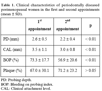
Mean PD, mean CAL, and mean BOP decreased significantly after periodontal treatment (p < 0.01) (Table 1). There were no significant differences in the percentage of sites with plaque between first and second appointment.
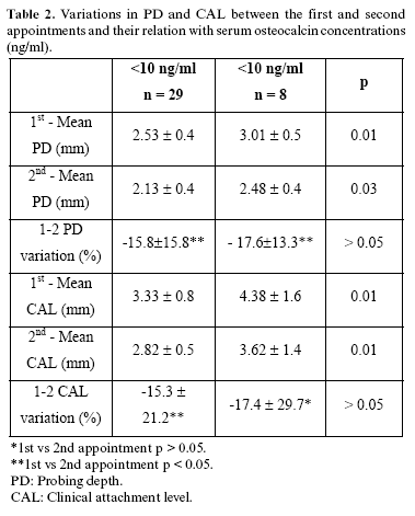
Variations in PD and CAL were analysed according to serum osteocalcin levels (Table 2). In two patients it was not possible to obtain serum samples. Patients were classified in two groups according to the osteocalcin level in serum: a) <10 ng/ml osteocalcin concentration (n = 29); and b) > 10 ng/ml osteocalcin concentration (n = 8). PD and CAL were significantly higher both in the first and second appointments in the group with serum osteocalcin concentration > 10 ng/ml (p < 0.05). Mean PD and mean CAL decreased significantly at second appointment, after periodontal treatment, in the group with serum osteocalcin concentration < 10 ng/ml (-15.8 ± 15.8% and -15.3 ± 21.2% respectively; p < 0.05). In the group with serum osteocalcin concentration > 10 ng/ml mean PD decreased significantly at second appointment (-17.6 ± 13.3 %; p < 0.05) but CAL did not differ (-17.4 ± 29.7%; p >0.05). There were not found significant differences between the two groups in PD and CAL percentages of variation between the first and second appointments.
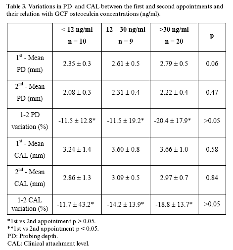
Variations in PD and CAL were analysed according to GCF osteocalcin levels (Table 3). Patients were classified in three groups according to the osteocalcin level in GCF: a) <12 ng/ml osteocalcin concentration (n = 10); b) 12 – 30 ng/ml (n = 9); and c) > 30 ng/ml osteocalcin concentration (n = 20). There were not significant differences between the three groups in PD and CAL neither in the first nor in the second appointments. Mean PD and mean CAL did not vary significantly at second appointment, after periodontal treatment, in the three groups. PD and CAL percentages of variation between the first and second appointment did not differ significantly in the three groups.
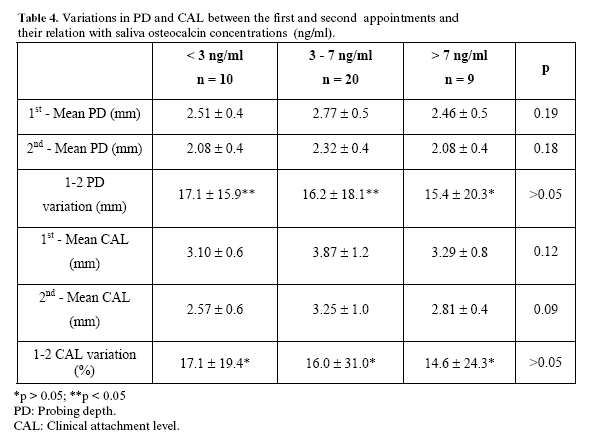
Variations in PD and CAL were analysed according to saliva osteocalcin levels (Table 4). Patients were classified in three groups according to the osteocalcin level in saliva: a) < 3 ng/ml osteocalcin concentration (n = 10); b) 3-7 ng/ml (n = 20); and c) > 7 ng/ml osteocalcin concentration (n = 9). There were not significant differences between the three groups in PD and CAL neither in the first nor in the second appointments. Mean PD decreased significantly at second appointment, after periodontal treatment, in the groups with saliva osteocalcin concentration < 3 ng/ml (-17.1 ± 15.9%; p < 0.05) and 3 – 7 ng/ml (-16.2 ± 18.1%; p < 0.05). PD and CAL percentages of variation between the first and second appointment did not differ significantly in the three groups.
Discussion
Using a longitudinal design, the periodontal treatment outcome in postmenopausal women and its correlation with GCF, saliva and serum osteocalcin concentrations has been studied. We showed that low serum osteocalcin concentration is associated to a significantly higher percentage of decrease in PD and CAL after periodontal treatment in postmenopausal women. We also showed that low saliva osteocalcin concentrations are significantly associated to a higher percentage of decrease in PD.
In a cross-sectional study carried out by Lee et al. (1) including 20 patients, no difference in GCF osteocalcin levels were observed between sites with and without periodontal pockets in the same patient. Recently, Wilson et al. (11) in a study including 14 patients without periodontal treatment, did not detect osteocalcin in GCF. However, most of these studies (1,10) have been carried out in periodontal patients and did not compare healthy and diseased subjects, but osteocalcin levels in healthy and periodontally affected sites in periodontal patients.
On the contrary, serum osteocalcin concentration correlated significantly with the periodontal treatment outcome. The use of serum osteocalcin as osteoporosis marker has been studied in 295 women over 67 years, without treatment with estrogens. The authors (21) conclude that high serum osteocalcin levels are associated with fast bone loss, but the individual value of this marker is limited. In some animal experimental models it has been possible to verify that serum osateocalcin levels are determinant of the bone formation (22-24) and it has been showed "in vitro" that has a role in the bone resorption process (25,26). Nevertheless, we must to be in mind that osteocalcin is produced by osteoblasts and only a small fraction is secreted directly in the circulation (27).
We can conclude that low serum osteocalcin concentration is associated to a significantly higher percentage of decrease in PD and CAL after periodontal treatment in postmenopausal women. Low saliva osteocalcin concentrations are significantly associated to a higher percentage of decrease in PD.
References
1. Lee AJ, Walsh TF, Hodges SJ, Rawlinson A. Gingival crevicular fluid osteocalcin in adult periodontitis. J Clin Periodontol 1999;26:252-6. [ Links ]
2. Riggs BL, Melton LJ. Evidence for two distinct syndromes of involutional osteoporosis. Am J Med 1986;75:899-901. [ Links ]
3. Giannobile WV, Al-Shammari KF, Sarment DP. Matrix molecules and growth factors as indicators of periodontal disease activity. Periodontol 2000 2003;31:125-34. [ Links ]
4. Curtis MA, Gillett IR, Griffiths GS, Maiden MF, Sterne JA, Wilson D et al. Detection of high-risk groups and individuals for periodontal diseases: laboratory markers from analysis of gingival crevicular fluid. J Clin Periodontol 1989;16:1–11. [ Links ]
5. Page RC. Host response tests for diagnosing periodontal diseases. J Periodontol 1992;63:356– 66. [ Links ]
6. Griffiths GS. Formation, collection and significance of gingival crevice fluid. Periodontol 2000 2003;31:32–42. [ Links ]
7. Uitto V-J, Overall CM, McCulloch C. Proteolytic host cell enzymes in ginigival crevice fluid. Periodontol 2000 2003;31:77–104. [ Links ]
8. Kunimatsu K, Mataki S, Tanaka H, Mine N, Kiyoki M, Hosoda K et al. A cross-sectional study on osteocalcin levels in gingival crevicular fluid from periodontal patients. J Periodontol 1993;64:865–9. [ Links ]
9. Nakashima K, Roehrich N, Cimasoni G. Osteocalcin, prostaglandin E2 and alkaline phosphatase in gingival crevicular fluid: their relations to periodontal status. J Clin Periodontol 1994;21:327–33. [ Links ]
10. Nakashima K, Giannopoulou C, Andersen E, Roehrich N, Brochut P, Dubrez B et al. A longitudinal study of various crevicular fluid components as markers of periodontal disease activity. J Clin Periodontol 1996;23:832–8. [ Links ]
11. Wilson AN, Schmid MJ, Marx DB, Reinhardt RA. Bone turnover markers in serum and periodontal microenvironments. J Period Res 2003; 38:355-61. [ Links ]
12. Bullón P, Goberna B, Guerrero JM, Segura JJ, Pérez-Cano R, Martinez-Sahuquillo A. Serum, saliva, and gingival crevicular fluid osteocalcin: their relation to periodontal status and bone mineral density in postmenopausal women. J Periodontol 2005;76:513-9. [ Links ]
13. O´Leary TJ, Drake RB, Naylos JE. The plaque control record. J Periodontol 1972;43:38-41. [ Links ]
14. Van der Velden U. Probing force and relationship of the probe tip to the periodontal tissues. J Clin Periodontol 1979;6:106-14 [ Links ]
15. Machtei EE, Christersson LA, Grossi SG, Dunford R, Zambon JJ, Genco RJ. Clinical criteria for the definition of es tablished periodontitis. J Periodontol 1992;63:207-15. [ Links ]
16. Nomura T, Ishii A, Shimizu H, Taguchi N, Yoshie H, Kusakari H et al. Tissue inhibitor of metalloproteinases-1, matrix metalloproteinases-1 and -8, and collagenase activity levels in peri-implant crevicular fluid after implantation. Clin Oral Implants Res 2000;11:430-40 [ Links ]
17. Oringer RJ, Al-Shammary KF, Aldredge WA, Iacono VJ, Eber RM, Wang H-L et al. Effect of locally delivered minocycline microspheres on markers of bone resorption. J Periodontol 2002;73:835-42. [ Links ]
18. Gonzalez C, Guerrero JM, Elorza FL, Molinero P, Goberna R. Evaluation of maternal serum alpha-foetoprotein assay using dry blood spot samples. J Clin Chem 1988;26:79-84 [ Links ]
19. Chamoles NA, Blanco MB, Gaggioli C. Hurther-like phenotype: enzymatic diagnosis in dried blood spots on filter paper. Clin Chem 2001; 47:2098-102 [ Links ]
20. Lee HJ, Auken S, Kulkarni G, Birek P, Overall CM, Sodek J et al. Collagenase activity in recurrent periodontics: relationship to disease progression and doxycyclin therapy. J Per Research 1991;26:479-85. [ Links ]
21. Bauer DC, Sklarin PM, Stone K, Black DM. Biochemical markers of bone turnover and prediction of hip bone loss in older women: the study of osteoporotic fractures. J Bone Miner Res 1999; 14: 1404-10. [ Links ]
22. Lopez-Marcos JF, Garcia-Valle S, Garcia-Iglesias AA. Periodontal aspects in menopausal women undergoing hormone replacement therapy. Med Oral Patol Oral Cir Bucal 2005;10:132-41. [ Links ]
23. Castro CE, Koss MA, Lopez ME. Biochemical markers of the periodontal ligament. Med Oral 2003;8:322-8. [ Links ]
24. Frutos R, Rodriguez S, Miralles-Jorda L, Machuca G. Oral manifestations and dental treatment in menopause. Med Oral 2002;7:26-30, 31-5. [ Links ]
25. Ducy P, Desbois C, Boyce B, Pinero G, Story B. Increased bone formation in osteocalcina deficient mice. Nature 1996;382:448-52. [ Links ]
26. Chenu C, Colucci S, Grano M, Zigrino P, Barattolo R. Osteocalcin induce chemotaxis, secretion of matriz proteins, and calcium-mediated intracellular signaling in human osteoclast-like cells. J Cell Biol 1994; 127:1149-58. [ Links ]
27. Lian JB, Gundberg M. Osteocalcin: biochemical consideration and clinical applications. Clin Orthop 1988;226:267-91. [ Links ]
![]() Correspondence:
Correspondence:
Prof. Pedro Bullón Fernández
C/ Luis Montoto 105 - 1º
41007 - Sevilla
E-mail: pbullon@us.es
Received: 20-02-2006
Accepted: 25-03-2007

