


Meu SciELO
 Serviços customizados
Serviços Personalizados
Journal
Artigo
 Inglês (pdf)
 Artigo em XML
 Referências do artigo
 Enviar este artigo por email
Indicadores
-
 Citado por SciELO
 -
 Acessos
Links relacionados
-
 Citado por Google
 -
 Similares em
SciELO
Similares em
SciELO -
 Similares em Google
Compartilhar

 Permalink
Medicina Oral, Patología Oral y Cirugía Bucal (Internet)
versão On-line ISSN 1698-6946
Med. oral patol. oral cir.bucal (Internet) vol.12 no.5 Set. 2007
A boy with oral hair: Case report
Farzaneh Agha-Hosseini1, Farideh Etesam2, Bita Rohani3
(1) D.D.S, M.S.C. Associated professor of Oral Medicine, Oral Medicine Department Faculty of Dentistry Tehran University of Medical Sciences (TUMS)
(2) D.D.S, PHD. Associated professor of Histology, Anatomy Department, Faculty of Medicine Tehran University of Medical Sciences (TUMS)
(3) D.D.S, M.S.C. Assistant Professor of Oral Medicine, Oral Medicine Department, Faculty of Dentistry Tehran University of Medical Sciences (TUMS), Tehran, Iran
ABSTRACT
In personal communication we have never seen or heard of hair being detected in the oral cavity. Even Julia Pastrana, the famous "Bearded Lady" of the 1800s, had no record of oral hair, although her entire body was covered with hair. Extensive records of her oral condition, including plaster models of her teeth have been preserved in the Odontological Museum of the Royal College of Surgeons in London city. She suffered from excessive gingival hyperplasia, but apparently no hair existed within the mouth. Some rodents have oral hair as a normal occurrence, but the condition is apparently limited in the animal kingdom.
A case of hair occurring naturally in the mouth has been reported only twice previously. A third case of this rare anomaly is reported here. In this case, multiple hairs were found at the gingival sulcus in the labial, buccal, lingual and palatal tooth surfaces in an 11-year-old boy.
Key words: Hair, oral cavity.
Introduction
A review of the literature relative to the existence of developmental hair in the oral cavity revealed only two incidences. One was reported by Miles (1), the other by Baughman (2).
The former was discovered by Miles at autopsy in a 57-year-old man. Miles stated that the hair was approximately 0.5cm in length, and located 0.5cm behind, and at the level of, the parotid papilla. In his extensive research reports on the subject of sebaceous gland distribution and involvement of oral mucosa, no other mention of the subject is made (3,4).
The other case was reported by Baughman et al. upon routine examination of the oral cavity of a 45-year-old white male presented at the Gainesville Veterans Administration Hospital Dental Clinic. A single dark brown hair measuring 3.5cm was seen extending from the base of the attached gingiva at the distal margin of the mandibular right cuspid (2).
Because of the extreme rarity of this phenomenon, this case with unique properties is presented.
Case Report
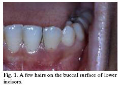
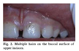
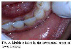
An 11-year-old boy presented at the Department of Oral Medicine, School of Dentistry, Tehran University of Medical Sciences, complaining of hairs eruption from his gingiva during the previous 5 days. Upon examination of his oral cavity, multiple thin black hairs measuring approximately 8-9mm were seen extending from the gingival sulcus in the labial, buccal, lingual and palatal surfaces of permanent teeth (Fig 1-2-3). In addition, the hairs were distributed on the dorsal surface of his tongue and oropharynx. He frequently felt nauseous because of this. The hairs fell into the oral cavity spontaneously, and had no adherence at the end to the attached gingiva. They also had a rapid growth rate – one hour after the mouth was completely cleared, the hairs reappeared. The problem caused suffering to the patient, and he wanted it treated.
His height and weight were normal. No problem was found during the examination of his systems. His sleep and appetite were completely normal. He was an athlete and played football, he was cheerful and was successful in his studies. Altogether he was healthy, had no systemic disease, and was not taking any drugs.
To assess the underlying tissue, after informed consent, an incisional biopsy was performed in the region of "4.2, 4.1, 3.1, 3.2" (as modified Widman flap).
Histologic features
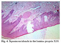
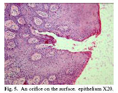
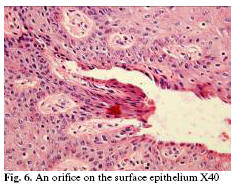
Histologic evaluation revealed parakeratinized squamous stratified epithelium of gingiva covering dense connective tissue, in some parts with chronic inflammatory infiltration. Epithelium was hyperplastic and showed marked acanthosis in some areas. In lamina propria a hypercellular connective tissue was observed, and also some epithelial nests similar to epithelial rests of Serres were evident. In some epithelial rests, clear cells (which seem to be melanocytes) were seen. In deep parts of lamina propria nests of ectopic squamous cells were seen (Fig 4). In different areas, deep notches appeared from the surface of epithelium to basal layer, and these notches were completely adjacent to ectopic squamous islands. Parakeratotic cells lined the wall of the notches, and the central part of these notches seemed empty. A hair-like structure was seen in this area on the surface of epithelium as a cell mass with well-defined eosinophilic cytoplasm and basophilic pyknotic nuclei (in contrast to normal hair structure) (Fig 5, 6).
The lining cells of the oral cavity are ectodermal derived, and can potentially produce hair, sebaceous gland and other skin apparatuses. The ducts of salivary and sweat glands form after canalization of the central part of the primitive epithelial solid cord following genetically programmed apoptosis . Thereafter, epithelial cords transform to a permanent canal. This phenomenon can help us to explain the mechanism of formation of these notches. Alternatively, these notches may be formed by the pressure of hair-like structures, which try to erupt from the epithelial surface. A few multinucleated cells were also seen in close proximity to the basement membrane. Extravasations of monocytes (probably for formation of these cells) were found in some areas. These cells may play a role in the elimination of apoptotic cells - the cells that occurred after notch formation in epithelium.
Discussion
The terms ectopia, heterotopia, or aberrance of tissues, are used for the development of tissues in situations where they are not normally found (4). For example, aberrant salivary glands have been reported in a variety of locations, including the middle-ear cleft, external auditory canal, neck, posterior and anterior mandible, pituitary, and cerebellopontine angle. These are usually incidental findings and do not require intervention (5).
The finding of naturally oral hair is certainly rare. It is puzzling why ectopic sebaceous glands are so common in the oral tissues (5), while their natural skin companion, hair, is so consistently absent. No explanation can be offered for this peculiarly common occurrence of one structure, with the almost universal absence of the other. Any attempted explanation would be sheer conjecture. In this report, our case is a young child, in contrast to the two previous reports.The hairs were multiple, and when an attempt was made to remove the hairs, they were found to be not attached to any structure and apparently growing from free gingival margin. These findings differ from the two citations.
In gingival connective tissue, copious remnants of dental lamina were seen. In parts of the gingivae where hair-like structures have been seen, those remnants transformed to prickle cells and gradually appeared as spindle cells with dense nuclides and eosinophilic cytoplasms adjacent to those hair-like structures. Which are factors, and which are causative interactional agents, is not accurately clear at the present time.
It seems that this ectopic phenomenon maybe a mutation in the gingival tissue. Whatever the cause, this phenomenon is extremely rare. Therefore its etiology is unclear, and we cannot treat this problem with our present knowledge.
Acknowledgements
The author very much appreciates the kind collaboration of Dr. Pouria Motahary.
References
1. Miles A.E.W. A Hair Follicle in Human Cheek Mucosa. Proc R Soc Med 1960;53:527-8. [ Links ]
2. Baughman R.A, Paul D, Heidrieh Jr, Gainesville and Winter Park, Fla. The oral hair: An extremely rare phenomenon. Oral Surg Oral Med Oral Pathol 1980;49:530-1. [ Links ]
3. Miles A.E.W. Sebaceous Glands in Oral and Lip Mucosa. Adv Biol skin 1963;4:1-32. [ Links ]
4. ORahilly R, Müller F. Human embryology & teratology. A John Wiley & Sons Editores; 1996. p. 123. [ Links ]
5. Greenberg M.S Glick M. Burkets oral medicine diagnosis and treatment.BC Decker Editor; 2003. p. 114. [ Links ]
![]() Correspondence:
Correspondence:
Dr. Farzaneh Agha-Hosseini
Oral Medicine Department,
Faculty of Dentistry Tehran
University of Medical Sciences (TUMS),
Tehran
Email: aghahose@sian.tums.ac.ir
Received: 24-07-2006
Accepted: 14-01-2007

