Mi SciELO
Servicios Personalizados
Revista
Articulo
Indicadores
-
 Citado por SciELO
 -
 Accesos
Links relacionados
-
 Citado por Google
 -
 Similares en
SciELO
 -
 Similares en Google
Compartir
Revista de Osteoporosis y Metabolismo Mineral
versión On-line ISSN 2173-2345versión impresa ISSN 1889-836X
Rev Osteoporos Metab Miner vol.5 no.1 Madrid ene./mar. 2013
https://dx.doi.org/10.4321/S1889-836X2013000100007
Zebra lines: Radiological repercussions of the action of bisphosphonates on the immature skeleton
Líneas cebra: Repercusión radiológica de la acción de los bifosfonatos en el esqueleto inmaduro
Etxebarria-Foronda I., Gorostiola-Vidaurrazaga L.
Servicio de Cirugía Ortopédica y Traumatología - Hospital Alto Deba - Mondragón - Gipuzkoa
SUMMARY
The bisphosphonates are used in the treatment of osteogenesis imperfecta, with a reduction in fractures in these patients observed with their use. However, the use of these drugs on the immature skeleton in these patients results in the formation of some radiologically visible hyperdense linear bands called zebra lines. We present the case of a patient with osteogenesis perfecta who started treatment with bisphosphonates at 10 years of age and after 2 years already showed these radiological images.
Key words: zebra lines, bisphosphonates, osteogenesis imperfecta.
RESUMEN
Los bifosfonatos son utilizados en el tratamiento de la osteogénesis imperfecta, observándose con ellos una reducción de las fracturas en estos pacientes. Sin embargo, el uso de dichos fármacos en el esqueleto inmaduro de estos pacientes da lugar a la formación de unas bandas lineales hiperdensas visibles radiológicamente, llamadas líneas cebra o zebra lines. Presentamos el caso de un paciente con osteogénesis imperfecta que inició tratamiento con bifosfonatos a los 10 años de edad y que al cabo de 2 años ya mostraba dichas imágenes radiológicas.
Palabras clave: líneas cebra, bifosfonatos, osteogénesis imperfecta.
Introduction
The use of bisphosphonates in the immature skeleton has been a great advance in the management of certain bone metabolic diseases, such as is the case with osteogenesis imperfecta, improving its clinical course and reducing the appearance of fractures. We describe the radiological repercussions which may be seen when these types of drugs are administered in growing bone, with the formation of a series of linear bands of increased density, known as zebra lines.
Clinical case
A male of 21 years of age diagnosed with osteogenesis imperfecta type IV-B. History of a tibial fracture and various femoral fractures between the ages of 6 months and 3 years (2 in the left femur and 2 in the right femur, treated conservatively. He received from the age of 10 to the age of 13 years a four-monthly infusion over three consecutive days of intravenous pamidronate (disodium pamidronate at a dose of 1 mg/kg/dose). From the age of 13 treatment with alendronate was initiated at a weekly dose of 40 mg, up to the age of 16, at which point the dose was reduced to 20 mg/week and maintained until he was 18 years of age. Analytical and densitometric controls were carried out. The last bone densitometry was carried out three years ago, with values within the normal range. The patient had not received any specific treatment in the last three years.
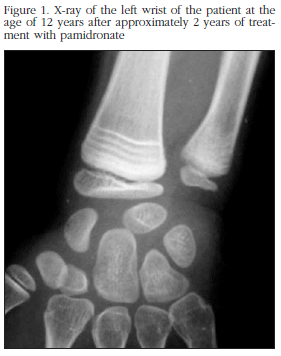
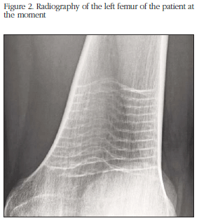
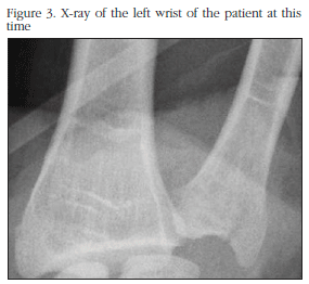
We show the radiological appearance of his left wrist at the age of 12 years, after approximately 2 years of treatment with pamidronate (Figure 1), and of his left femur at the current time (Figure 2). It is possible to see in both X-rays, in the metaphysary zone, some linear bands of increased density, called zebra lines, which correspond to the impact of the effect of the bisphosphonates on the growing skeleton. These lines shift towards the diaphysary zone, and may disappear or remain radiologically visible into adulthood, which may be seen in the X-ray of the same wrist carried out in the present, 9 years after the first (Figure 3).
Discussion
The use of bisphosphonates at a paediatric age has changed the clinical course of many bone metabolic diseases whose therapeutic management was limited to resolving its complications, as is the case with osteogenesis imperfecta [1]. These drugs increase bone mineral density, reduce the incidence of fractures and therefore secondary bone deformities, and improve functional parameters such as mobility and autonomy. They also diminish bone pain, above all dorso-lumbar pain, and definitely improve the quality of life of these patients [2]. In spite of there being questions to be answered, such as the age of initiation of treatment, its duration, the most appropriate type of bisphosphonates and their doses, the ideal form of administration and the long-term secondary effects, their use is already common in centres which treat this type of patient.
One of the consequences seen in patients with immature skeletons who receive treatment with bisphosphonates is the appearance of sclerotic lines, essentially metaphysary, parallel to the growth cartilage which have been called zebra lines [3]. In spite of their probable clinical insignificance, it is interesting to know of their presence at the time of radiologically evaluating these patients.
Their radiological pattern depends on the number of doses received: each cycle of pamidronate, for example, represents a dense band. If the doses are more frequent, the lines appear closer together. There are aligned circumferentially with the surface of the epiphysis, representing the progressive dose of the nuclei of secondary ossification, which means, on the other hand, that skeletal growth does not seem to be affected by the action of these drugs, which has also been corroborated clinically [3]. These finding are not found in bones of mature skeletons, being intimately related with metabolic activity: thus, in metaphysary regions with great growth potential, such as the distal section of the femur and the proximal section of the tibia, the lines are more prominent and marked. In zones of less activity, such as the pelvis or the calcaneum, the lines are finer and closer together, and less evident radiologically [4,5].
These bands have a great similarity with so-called Harris lines, described by Henry Albert Harris in 1931, who defined them as dense lines parallel to the physis, whose appearance is related to the temporary arresting of growth provoked by certain medical conditions, including diabetes mellitus, and which were called at the time growth arrest lines [6]. These lines also appear in the metaphysary-epiphysary zone of the immature skeleton, subsequently migrating towards the diaphysary zone, and which may disappear or remain visible into adulthood. They have been related with certain chronic diseases, above all, nutritional, and are considered to be the latent result of a situation of physiological stress. They also arouse interest in the archaeological analysis of bones of populations in which certain episodes of disease, epidemics or conflict situations have been historically documented which had a great impact on the health of the population of the time.
Although they are similar radiologically speaking, the differences between these two types of lines may be established first in their duration, the Harris lines being of greater duration and therefore more visible. Furthermore, the zebra lines are more extensive, appearing in all bones, while the others appear only in bone with high potential for growth, such as the distal section of the femur, the proximal and distal sections of the tibia and the proximal humerus. The Harris lines, may also appear in an isolated fashion in only one bone, for example in the reparative process of a fracture [3,7].
The fundamental etiopathogenic substrate is an interruption of osteoclast activity promoted by the bisphosphonates, there being a temporary disequilibrium in bone turnover in these zones of high metabolic activity, which brings with it an increase in osteoblast function, and consequently in bone mineralisation, which becomes, radiologically, a radiolucid line parallel to the growth cartilage and which subsequently, with the growth of the bone, will shift to the diaphysary zone [8].
The diagnosis of the image is clear if there is a history of administration of bisphosphonates. If this is not the case, it would be necessary to suggest other options in the differential diagnosis, which may be, principally, nutritional deficiencies, and more rarely, lead or bismuth poisoning, congenital syphilis, scurvy, hypothyroidism or certain leukemias which may also result in the appearance of metaphysary radio-opaque bands.
Declaration of interest
The authors declare no conflict of interest.
![]() Correspondence:
Correspondence:
Iñigo Etxebarria
Servicio de Cirugía Ortopédica y Traumatología
Hospital Alto Deba
Avda. Navarra, 16
20500 Mondragón - Gipuzkoa (Spain)
e-mail:
ietxe@yahoo.es
Date of receipt: 14/01/2013
Date of acceptance: 15/03/2013
Bibliography
1. Salom M, Vidal S, Miranda L. Aplicaciones de los bifosfonatos en la ortopedia infantil. Rev Esp Cir Ortop Traumatol 2011;55:302-11. [ Links ]
2. Glorieux FH. Experience with bisphosphonates in osteogenesis imperfecta. Pediatrics 2007;119:S163-5. [ Links ]
3. Al Muderis M, Azzopardi T, Cundy P. Zebra lines of pamidronate theraphy in children. J Bone Joint Surg Am 2007;89:1511-6. [ Links ]
4. Grissom LE, Harcke HT. Radiographic features of bisphosphonate therapy in pediatric patients. Pediatr Radiol 2003;33:226-9. [ Links ]
5. Sarraf KM. Radiographic zebra lines from cyclical pamidronate therapy. N Engl J Med 2011;365;3:e5. [ Links ]
6. Harris HA. Lines of arrested growth in the long bones of diabetic children. Br Med J 1931;1:700-1. [ Links ]
7. Ogden JA. Growth slowdown and arrest lines. J Pediatr Orthop 1984;4:409-15. [ Links ]
8. Rauch F, Travers R, Munns C, Glorieux FH. Sclerotic metaphyseal lines in a child treated with pamidronate: histomorphometric analysis. J Bone Miner Res 2004;19:1191-3. [ Links ]


 texto en


