Meu SciELO
Serviços Personalizados
Journal
Artigo
Indicadores
-
 Citado por SciELO
 -
 Acessos
Links relacionados
-
 Citado por Google
 -
 Similares em
SciELO
 -
 Similares em Google
Compartilhar
Revista de Osteoporosis y Metabolismo Mineral
versão On-line ISSN 2173-2345versão impressa ISSN 1889-836X
Rev Osteoporos Metab Miner vol.5 no.4 Madrid Nov./Dez. 2013
https://dx.doi.org/10.4321/S1889-836X2013000400004
ORIGINAL ARTICLES
The reality of osteoporosis in patients hospitalized in Internal Medicine
La realidad de la osteoporosis en el paciente hospitalizado en Medicina Interna
Neila Calvo S., Nan Nan D., García Ibarbia C., Olmos Martínez J.M., González Macías J., Hernández Hernández J.L.
Unidad de Metabolismo Óseo - Servicio de Medicina Interna - Hospital Marqués de Valdecilla-IFIMAV - Universidad de Cantabria - RETICEF - Santander
SUMMARY
Purpose: a) to know the prevalence of previous osteoporosis and vertebral fractures in patients admitted to an Internal Medicine department from a third-level hospital; b) to determinate the proportion of patients discharged with a diagnosis of osteoporosis, and the percentage of those receiving treatment; c) to quantify the risk of fracture by applying the FRAX calculation tool; and d) to know the serum levels of 25OHD in these patients.
Patients and methods: Retrospective study, based on the review of clinical charts of all the patients admitted to the Internal Medicine department of Marqués de Valdecilla University Hospital, during April 2012. The information was gathered by a standardized protocol, including demographic, clinical, radiological and laboratory variables.
Results: Three hundred patients were studied (mean age, 80 years). Thirty-four (11.3%) had a previous diagnosis of osteoporosis and 14 (4.8%) of them were, or had been, on treatment. A diagnosis of osteoporosis, in the hospital discharge report, was noted in 14 patients. No treatment was prescribed in one of them. According to the FRAX calculation tool, mean risk for major osteoporotic fracture was 10.5%, and mean risk for hip fracture was 5.4%. Mean serum 25OHD level was 16 ng/ml, and more than 80% of patients had values below 20 ng/ml.
Conclusion: Osteoporosis is an underdiagnosed and undertreated disease, in patients admitted to an Internal Medicine Department, whatever the reason. Moreover, we have observed a high prevalence of 25OHD deficiency among these patients. Hospitalization can represent an excellent opportunity for the internists and other clinicians, to pay attention to the presence of osteoporosis and its related complications.
Key words: vertebral fracture, osteoporosis, diagnosis, chest-X-ray, 25OHD, Internal Medicine.
RESUMEN
Objetivos: a) conocer la prevalencia de osteoporosis previa y de fracturas vertebrales en los pacientes ingresados en un Servicio de Medicina Interna de un hospital terciario; b) determinar la proporción de pacientes dados de alta con el diagnóstico de osteoporosis y el porcentaje de los tratados; c) cuantificar el riesgo de fractura mediante la herramienta FRAX® y d) conocer los niveles de 25-hidroxivitamina D (25OHD) en estos pacientes.
Material y método: Estudio retrospectivo mediante la revisión de los informes de alta y las historias clínicas de todos los pacientes ingresados en el Servicio de Medicina Interna del Hospital Marqués de Valdecilla durante abril de 2012, analizando variables demográficas, clínicas, radiológicas y de laboratorio.
Resultados: Se estudiaron 300 pacientes. Un total de 34 (11,3%) tenían diagnóstico previo de osteoporosis y 14 (4,8%) recibían o habían recibido tratamiento. Solamente 14 pacientes tenían un diagnóstico de osteoporosis en el informe de alta. En uno de ellos no se pautó ningún tratamiento. Según el índice FRAX®, el riesgo medio de fractura osteoporótica mayor fue de 10,5%, y el riesgo de fractura de cadera fue de 5,4%. El valor medio de 25OHD sérico, fue de 16 ng/ml y, en más del 80% de los pacientes, los valores fueron <20 ng/ml.
Conclusión: La osteoporosis es una enfermedad infradiagnosticada e infratratada en los pacientes ingresados en Medicina Interna por cualquier causa. Además, hemos observado una alta prevalencia de deficiencia de vitamina D en estos sujetos. La hospitalización puede ser una excelente oportunidad para que los internistas, y los clínicos en general, prestemos una mayor atención a la osteoporosis y a sus complicaciones.
Palabras clave: fractura vertebral, osteoporosis, diagnóstico, radiografía de tórax, 25OHD, Medicina Interna.
Introduction
Osteoporosis is a disease characterised by a reduction in bone mass and by alterations in the microarchitecture of bone tissue, which leads to an increase in its fragility and, consequently, a higher risk of suffering fractures [1]. The prevalence of osteoporosis increases with age, from 15% in women between 50 and 59 years of age to more than 80% in those over 80 [2].
The principal osteoporotic fractures are vertebral, hip, wrist, humerus and pelvis. These fractures bear significant health and social implications. Furthermore, in a high percentage of patients they lead to significant morbidity, such as loss of mobility, loss of ability to carry out independently basic activities of daily living, chronic pain and even depression [3,4]. Similarly, osteoporotic fractures, above all hip fractures, are associated with a significant increase in mortality [5], this fact is especially important, since it has been estimated that the annual incidence worldwide of hip fractures in women will have increased by 3.5 times between 1990 and 2050 [6].
In the last few decades, in hospitals in general, and in general internal medicine services in particular, an increase has been seen in the average age of patients being hospitalised who, in addition, usually have multiple comorbidities and are polymedicated. In fact, the prevalence osteoporosis, as a disease linked to aging, is increased in these individuals [7]. Admission to hospital represents on many occasions an opportunity to diagnose, initiate treatment and programme appropriate monitoring in those patients with osteoporosis, with the aim of attempting to prevent the complications of the disease, mainly fractures. We have carried out this work with the aims of: a) ascertaining the prevalence of previous osteoporosis and of vertebral fractures in patients admitted to the internal medicine service of a tertiary hospital; b) determining the proportion of patients discharged with a diagnosis of osteoporosis and the percentage of those who are treated; c) quantifying the risk of fracture by applying the FRAX® tool, and; d) finding out the blood levels of 25-hydroxyvitamin D (25OHD) in these patients.
Material and methods
A retrospective descriptive study was designed, based on a review of the clinical histories of all patients admitted to the internal medicine service of the Marqués de Valdecilla University Hospital during the month of April 2012. This centre serves as a health referral centre for a population of 350,000 inhabitants of Cantabria. The internal medicine service has approximately 130 hospital beds.
The variable data for the study were gathered using a standardised protocol in a computerised database management system. These variables were: risk factors for osteoporosis, comorbidities, drugs with an influence on bone metabolism, results of X-rays of thorax and/or dorso-lumbar spine, blood levels of 25OHD and prescribed treatments, differentiating between treatments at time of hospital admission and at discharge. Also recorded was the presence or not of family history of hip fracture and personal history of fractures.
Menopause was defined as being early if it had occurred before the age of 45. It was considered that a patient had clinical osteoporosis if they had suffered a typical osteoporotic fracture (hip, vertebral, humerus, forearm) earlier or had a vertebral fracture in the radiological study requested on admission, in the absence of other causes which may have accounted for it (high impact trauma, bone metastasis, myeloma or other bone diseases).
The comorbidities analysed were: chronic obstructive pulmonary disease -COPD- by means of a diagnosis based on tests of compatible respiratory function); ischemic cardiopathy or cardiac insufficiency, cerebrovascular accident, dementia, Parkinson's disease; active neoplasia, or in remission; venous thromboembolism; cataracts; rheumatoid arthritis; diabetes mellitus type 1 or 2 (HbA1c ≥6.5% or two tests for glycemia in fasting ≥126 mg/dl or a casual glycemia of 200 mg/dl or more than one patient with cardinal symptoms); arterial hypertension (through measurement and confirmation of levels of TAS > 140 mmHg and/or TAD >90 mmHg persistently); dyslipemia; hyperthyroidism and hypothyroidism; hyperparathyroidism; hypogonadism; intestinal malabsorption syndrome; malnutrition, urolithiasis; chronic hepatopathy; chronic renal insufficiency (defined as a glomerular filtrate <60 ml/min/m2 according to the four variable MDRD equation, maintained for at least 3 months). The drugs evaluated were: corticoids, beta-blockers, anticonvulsives, statins, proton pump inhibitors (PPIs), serotonin re-uptake inhibitors, oral anticoagulants, opiates, hypnotics and benzodiazepines. Treatments for osteoporosis recorded in the discharge reports included: denosumab, selective estrogen receptor modulators (SERMs), strontium ranelate, teriparatide, PTH 1-84, calcitonin or bisphosphonates.
Blood levels of 25OHD were determined using electrochemiluminescence (Elecsys 2010, Roche Diagnostics, GMBH, Mannheim, Germany).
A vertebral fracture was defined as a reduction in height of the vertebral body greater than or equal to 20%, assessed through a review of lateral X-rays of the thorax and/or thoracic-abdominal spine. The X-rays were reviewed independently by the two authors and disagreements (<5%) were resolved by consensus. All the patients diagnosed with osteoporosis had had at least one laboratory study which included biochemistry, haemogram and VG, thyroid hormone, proteinogram and 24 hour calciuria.
Results
300 patients were included in the study of whom 157 (52.3%) were women and 143 were men. The average age was 80 years. The average body mass index (BMI) was 29.3 kg/m2.
In terms of the risk factors related to osteoporosis or fragility fracture, the most frequent was the presence of cataracts (76 patients), followed by malnutrition (33), hypothyroidism (26), malabsorption syndrome (24) and chronic hepatopathy (24). Seven patients had history of hypogonadism and hyperparathyroidism, and it was only possible to confirm 4 women with history of early menopause.
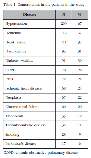
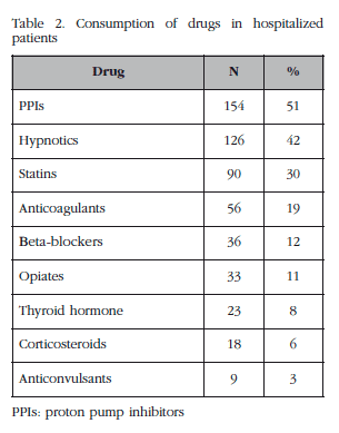
In tables 1 and 2, respectively, are shown the comorbidities of the patients included in the study and their consumption of drugs.
A total of 34 subjects (11.3%) had had a previous diagnosis of osteoporosis (5 men and 29 women), although only 14 of these (4.8%) were receiving or had received treatment for the disease (zoledronic acid in one patient, other bisphosphonates in eight patients, three patients were receiving denosumab and teriparatide and strontium ranelate had been subscribed in one case each). Of the 34 cases of osteoporosis, in 6 cases the patients were receiving oral corticoids, in one case for rheumatoid arthritis, and in five cases with chronic obstructive pulmonary disease (it was not possible to quantify the exact doses of the glucocorticoids administered). In one case there was history of early menopause, and in another long-term hyperthyroidism. Treatment had been prescribed in only two of these case.
On the other hand, of the 34 patients with diagnosis of osteoporosis, in 16 of them it was confirmed that they had suffered at least one vertebral fracture.
All the patients had an X-ray of the thorax taken during their admission and in 31 there was also an X-ray taken of the thoracic-lumbar spine. 50 patients were identified as having vertebral fractures. Taking into account the total of all the patients studied and given that only 16 had history of radiological vertebral fractures, in 12% of all patients the presence of vertebral fractures had passed unnoticed by the clinician responsible. On the other hand, 68% of all fractures had passed unnoticed. Of the 50 patients with vertebral fractures, in only one case was this attributed to multiple myeloma.
Overall, and in accord with earlier data, 74 cases of clinical osteoporosis (defined by vertebral, hip humerus or wrist fracture) were identified. Therefore, 40 subjects had not had a diagnosis of osteoporosis. At the time of discharge, in only 14 patients was the diagnosis of osteoporosis confirmed in the clinical record, and of these, in one case no treatment was programmed.
On the other hand, the average risk of major osteoporotic fracture (vertebral, hip, humerus or wrist) measured by application of the FRAX® scale was 10.5% (SD: 8.7) and the risk of hip fracture 5.4% (SD: 5.5). According to the FRAX® tool developed for Spain, there were 27 patients with a risk higher than 20% of major osteoporotic fracture, and 118 patients had a risk higher than 3% for fracture of the hip. The risk of fracture using the FRAX® tool could only be calculated in 174 patients, due to a lack of information in the clinical histories of the other subjects of the study, while those treated for osteoporosis were excluded.
The average blood value of 25OHD was 16 ng/ml, although this was only obtained from 45 patients, this being a test which was not included in the protocol of requests for laboratory tests on admission. More than 80% of the patients had values below 20 ng/ml, and more than 50% (26 patients) had serious hypovitaminosis (≤10 ng/ml). Only two patients with hypovitaminosis D did not receive oral supplements at discharge.
Discussion
Osteoporosis is a process which generally develops asymptomatically over a long period of time, a fracture being the first sign in the majority of cases. A predisposition to the development of fractures is, from a clinical point of view, the central phenomenon of the disease. Among these fractures, of greatest significance is fracture of the hip, which preferentially affects older people, is frequent in women and which varies greatly in its incidence, its mortality and in length of stay in hospital [8].
Vertebral fractures are another complication characteristic of osteoporosis, although more than 50% of them pass unnoticed [9,10]. It is important to remember that the presence of an osteoporotic fracture, independently of the densitometry value, increases even more the risk of subsequent fractures, which means that the diagnosis is as important as the monitoring of the disease. In a study published by Sosa et al. [11], it was concluded that the presence of vertebral fractures increased the risk of new fractures. Furthermore, the authors observed this type of fracture in 62.6% of patients hospitalised or treated for fracture of the hip. Although there is no universally accepted definition of vertebral fracture, the majority of authors are in agreement in considering that there must be a loss of height of the vertebral body of at least 20% [12]. We know that lateral X-rays of the thorax are a very useful tool in enabling the identification of vertebral fractures [9]. In our work 50 patients were identified with fractures of this type, mostly unnoticed by the clinician (in our service, thoracic X-rays are not analysed by a radiologist unless specifically requested) and as a consequence, are not reflected in the discharge notes.
On the other hand, according to the definition of clinical osteoporosis which we have used, 74 cases were identified, which shows that almost 50% were not recorded in the clinical record (there being 34 patients with a previous diagnosis of osteoporosis). Furthermore, only 13 patients in whom their discharge notes reflected a diagnosis of osteoporosis received treatment, which indicates once more that, although osteoporosis is a highly prevalent chronic disease, it continues to be underdiagnosed and undertreated.
In respect of the results obtained using the FRAX® tool, it is known that the Spanish version of FRAX® underestimates by 50% the number of major fractures [13]. So, we could take into account the recommendations of NOF, which is to treat patients if their risk of major fracture is ≥20% and of hip fracture ≥3%. Thus in our work, more than 30% of patients (for major fracture) and more than 80% (for hip fracture) had indications to receive treatment, although it should be borne in mind that this guide recommends starting treatment when there is a diagnosis of osteoporosis, whatever the FRAX® indicates. This makes us think that patients admitted to the internal medicine service usually have a high risk of osteoporotic fracture.
In terms of the determination of the level of 25OHD in the blood, it seems that, while this is not normally requested routinely during hospital admission, the staff do usually prescribe treatment when there is a deficit.
When dealing with a multifactorial disease in which are involved, among others, genetic and environmental factors, one of the keys to diagnosis is to identify its risk factors [2]. Hospitalisation may be an opportunity to diagnose osteoporosis and vertebral fracture. However, in line with the data provided in this work the risk factors for fracture are not generally evaluated during admission and osteoporosis continues to be forgotten by the clinician despite its known epidemiological, health and social significance. Furthermore, it is a highly prevalent disease in older patients, in whom, with increasing frequency, are associated malnutrition, comorbidities and polymedication (a notable finding in this work is the significant consumption of hypnotics) which themselves pose additional risk of falls and, therefore, of fracture [2].
However, there are highly efficacious and easy methods of treatment for osteoporosis, especially in polymedicated patients, which ensure therapeutic compliance, and which may be considered, when indicated, during admission to hospital [14].
Our study has various limitations. Firstly, those inherent in any retrospective study. In addition, not all the patients had X-rays of the lumbar spine, which means that it was not possible to determine the true prevalence of vertebral fractures in this section of the spine. Finally, the determination of 25OHD in the blood was carried out in a small sub-group of hospitalised patients, which means that we cannot generalise the results to all the patients studied. However, our work group has studied levels of 25OHD in a broad sample of patients hospitalised in our internal medicine service (approximately 400 individuals) and the results were similar (data not published).
In conclusion, osteoporosis is a disease rarely considered by the clinician in patients hospitalised due to other causes, in spite of its having a prevalence similar to other diseases such as diabetes mellitus, dyslipemia or dementia. As a consequence, it continues to be a condition which is underdiagnosed and secondarily undertreated, in spite of there being many therapeutic options. In the light of the findings of our work we suggest that the internist, and in general, any clinician, should maintain a high level of suspicion of this disease in hospitalised patients, in order to try to reduce associated complications, especially fractures.
![]() Correspondence:
Correspondence:
Sara Neila Calvo
Unidad de Metabolismo Óseo
Departamento de Medicina Interna
Hospital Universitario Marqués de Valdecilla
Avda. Valdecilla, s/n
39008 Santander (Spain)
e-mail: i491@humv.es
Date of receipt: 19/11/2013
Date of acceptance: 09/12/2013
Bibliography
1. Díaz Curiel M, García JJ, Carrasco JL, Honorato J, Pérez Cano R, Rapado A, et al. Prevalencia de osteoporosis determinada por densitometría en la población femenina española. Med Clin (Barc) 2001;116:86-8. [ Links ]
2. Rosen CJ. Postmenopausal osteoporosis. N Eng J Med 2005;353:595-603. [ Links ]
3. Ioannidis G, Papaioannou A, Hopman WM, Akhtar-Danesh N, Anastassiades T, Pickard L, et al. Relation between fractures and mortality: results from the Canadian Multicentre Osteoporosis Study. CMAJ 2009;181:265-71. [ Links ]
4. Poole KE, Compston JE. Osteoporosis and its management. BMJ 2006;333:1251-6. [ Links ]
5. Cauley JA, Thompson DE, Ensrud KC, Scott JC, Black D. Risk of mortality following clinical fractures. Osteoporos Int 2000;11:556-61. [ Links ]
6. Gullberg B, Johnell O, Kanis JA. World-wide projections for hip fracture. Osteoporosis Int 1997;7:407-13. [ Links ]
7. Cummings SR, Melton J. Epidemiology and outcome of osteoporotic fractures. Lancet 2002;359:1761-7. [ Links ]
8. Serra JA, Garrido G, Vidán M, Marañón E, Brañas F, Ortiz J. Epidemiología de la fractura de cadera en ancianos en España. An Med Interna (Madrid) 2002;19:389-95. [ Links ]
9. Hernández JL, Hidalgo I, López-Calderón M, Olmos JM, González Macías J. Diagnóstico de osteoporosis mediante radiografía lateral de tórax. Med Clin (Barc) 2001;117:734-6. [ Links ]
10. Becker C. Pathophysiology and clinical manifestations of osteoporosis. Clin Cornerstone 2006;8:19-27. [ Links ]
11. Sosa M, Saavedra P. Prevalencia de fracturas vertebrales en pacientes con fractura de cadera. Rev Clin Esp 2007;207:464-8. [ Links ]
12. Majumdar SR, Kim N, Colman I, Chahal AM, Raymond G, Jen H, et al. Incidental vertebral fractures discovered with chest radiography in the emergency department: prevalence, recognition, and osteoporosis management in a cohort of elderly patients. Arch Intern Med 2005;165:905-9. [ Links ]
13. González Macías J, Marin F, Vila J, Díez Pérez A. Probability of fractures predicted by FRAX® and observed incidence in the Spanish ECOSAP Study cohort. Bone 2012;50:373-7. [ Links ]
14. Lyles KW, Colón-Emeric CS, Magaziner JS, Adachi JD, Pieper CF, Mautalen C, et al. Zoledronic acid and clinical fractures and mortality after hip fracture. N Engl J Med 2007;18:1799-809. [ Links ]


 texto em


