


Mi SciELO
 Servicios personalizados
Servicios Personalizados
Revista
Articulo
 texto en
  Inglés (pdf)
 Articulo en XML
 Referencias del artículo
 Enviar articulo por email
Indicadores
-
 Citado por SciELO
 -
 Accesos
Links relacionados
-
 Citado por Google
 -
 Similares en
SciELO
Similares en
SciELO -
 Similares en Google
Compartir

 Permalink
Revista de Osteoporosis y Metabolismo Mineral
versión On-line ISSN 2173-2345versión impresa ISSN 1889-836X
Rev Osteoporos Metab Miner vol.13 no.1 Madrid ene./mar. 2021 Epub 17-Mayo-2021
https://dx.doi.org/10.4321/s1889-836x2021000100007
IMAGES IN OSTEOLOGY
Unusual case of bone proliferation: Nora's lesion
2Nuclear Medicine Clinical Management Unit. Virgen Macarena University Hospital. Sevilla (Spain)
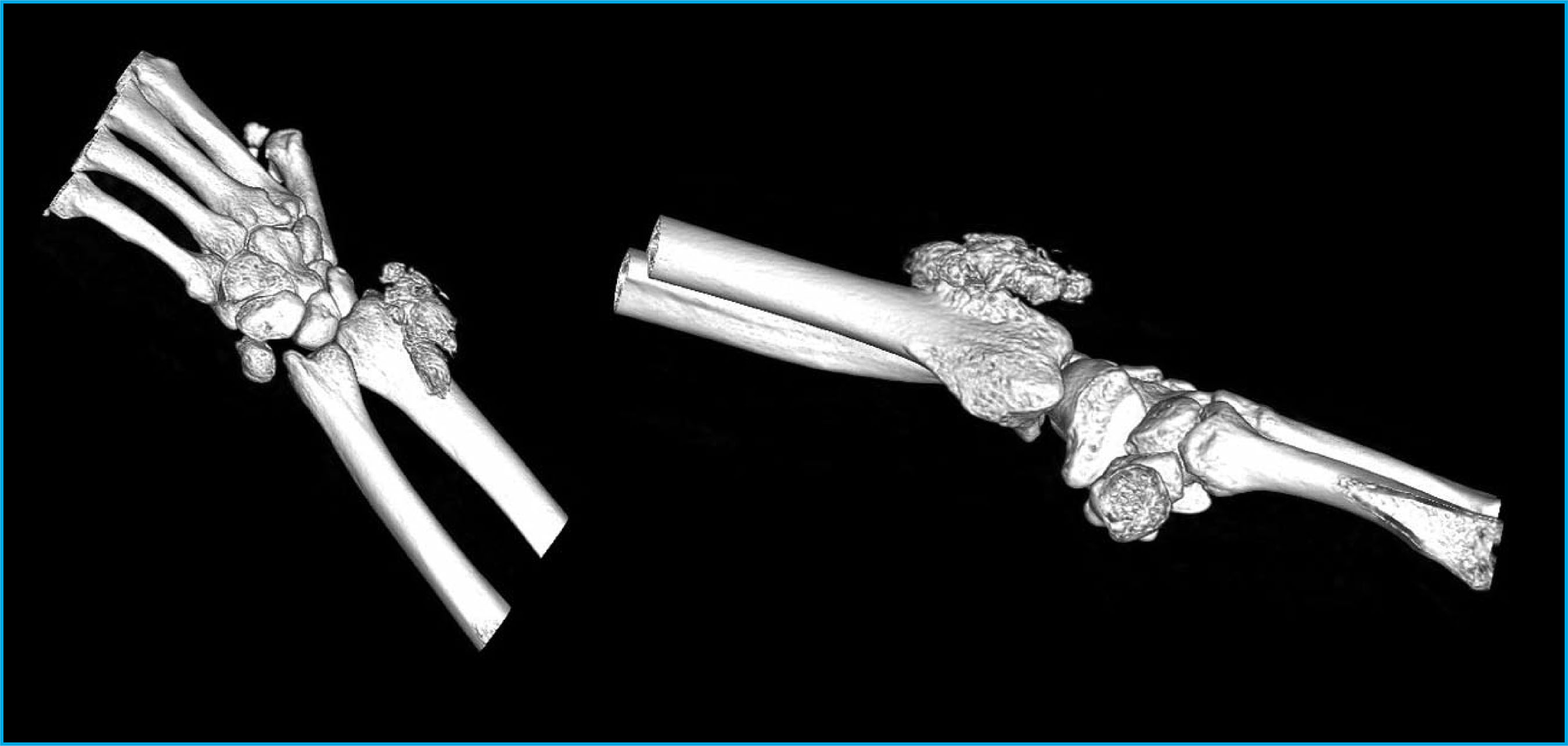
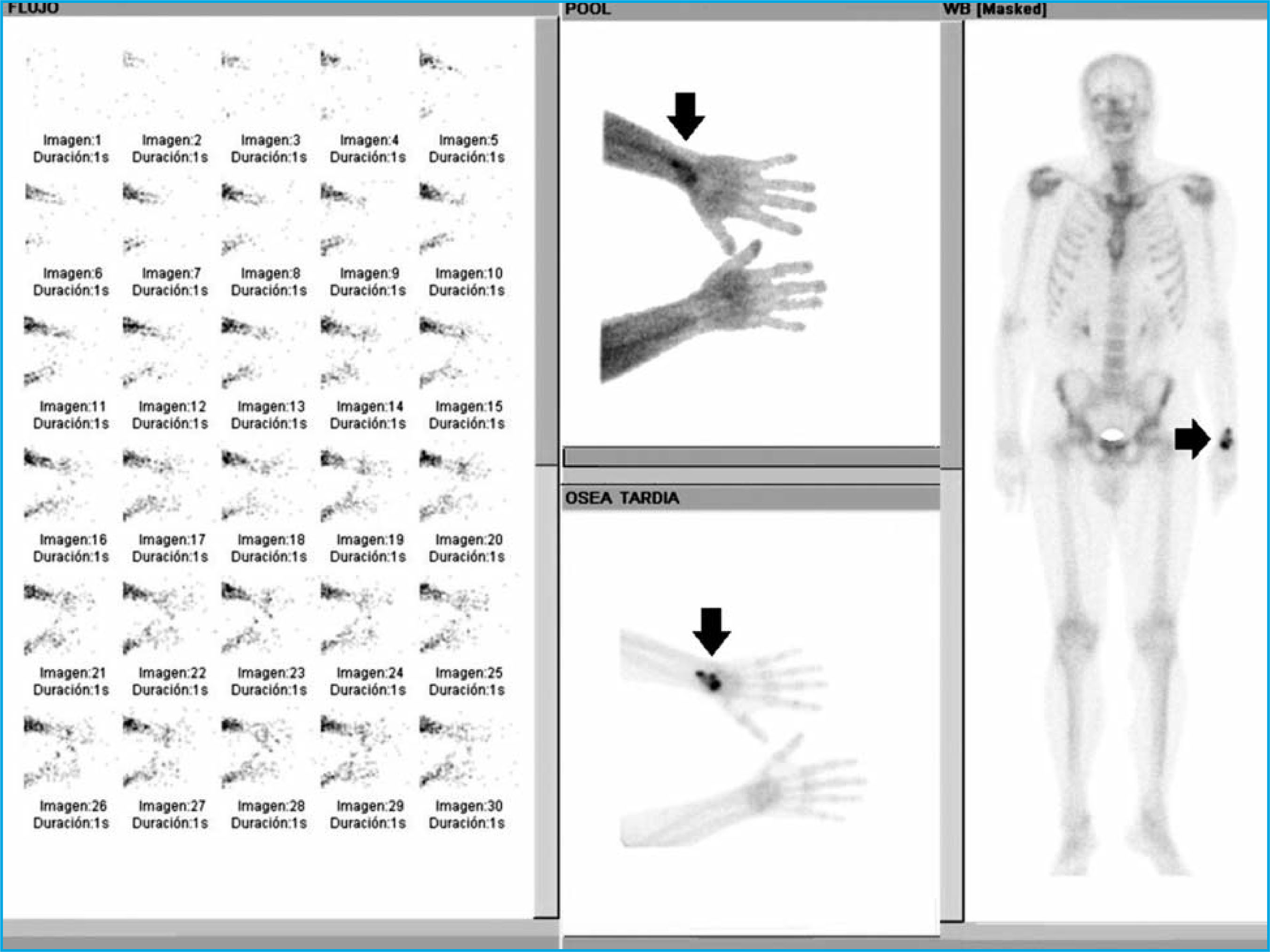
We present the case of a 43-year-old man who presents pain and functional impotence in the left wrist of one year of evolution. Upon examination, an indurated tumor adhered to deep planes is found in this location. Following findings on computerized axial tomography (CT) of images consistent with osteochondroma versus peripheral chondrosarcoma (Figure 1), a bone scan was requested. This bone scintigraphic study in three phases of the upper limbs and a subsequent full-body image (Figure 2), showed the early arrival of the tracer with an increase in the vascular pool of slight-moderate intensity in the distal portion of the left radius (arrow), which persisted with greater intensity in late images. No other diseased findings were observed in the rest of the skeleton. These findings revealed increased vascularity and osteoblastic activity at the distal end of the left radius.
A biopsy was carry out, with a pathological result of osteochondromatous proliferation compatible with Nora’s lesion, confirming this diagnosis after surgical resection. Nora's lesion occurs predominantly in the second or third decade of life1,2, without gender differences3, mainly affecting the extremities. Of uncertain etiology4,5, it consists of an excretory and exophytic lesion that originates in the bone cortex, formed by bone, cartilaginous and fibrous tissue, with nuclear atypia6,7. Bone scan allows us to know the metabolic characteristics of this lesion. Given its aggressive nature, a differential diagnosis should be made with malignant lesions such as osteosarcoma8.
Bibliografía
1 Nora FE, Dahlin DC, Beabout JW. Bizarre parosteal osteochondromatous proliferations of the hands and feet. Am J Surg Pathol. 1983;7:245-250. [ Links ]
2 Meneses MF, Unni KK, Swee RG. Bizarre parosteal osteochondromatous proliferation of bone (Nora’s lesion). Am J Surg Pathol. 1993;17:691-697. [ Links ]
3 Mahajan S, Chandra R, Mohan Lal Y. “Nora lesion” -Bizarre parosteal osteochondromatous proliferation. J Clin Orthop Trauma. 2012;3:119-121. [ Links ]
4 Matsui Y, Funakoshi T, Kobayashi H, Mitsuhashi T, Kamishima T, Iwasaki N. Bizarre parosteal osteochondromatous proliferation (Nora’s lesion) affecting the distal end of the ulna: a case report. BMC Musculoskelet Disord. 2016;17:130. [ Links ]
5 Joseph J, Ritchie D, MacDuff E, Mahendra A. Bizarre parosteal osteochondromatous proliferation. A locally aggressive benign tumor. Clin Orthop Relat Res. 2011. 469:2019-2027. [ Links ]
6 Gruber G, Giessauf C, Leithner A, Zacherl M, Clar H, Bodo K, et al. Bizarre parosteal osteochondromatous proliferation (Nora lesion): a report of 3 cases and a review of the literature. Can J Surg. 2008;51:486-489. [ Links ]
7 Cobo CE, Navarro R, Aracil EI, Velasco JA. Enfermedad de Nora: una entidad clínica infrecuente. Rev Pie Tobillo. 2018;32(1):43-46. [ Links ]
8 Berber O, Dawson-Bowling S, Jalgaonkar A, Miles J, Pollock RC, Skinner JA, et al. Bizarre parosteal osteochondromatous proliferation of bone.Clinical management of a series of 22 cases. J Bone Joint Surg. 2011;93-B:1118-1121 [ Links ]
Received: January 26, 2021; Accepted: February 27, 2021
 Este es un artículo publicado en acceso (Open Access) abierto bajo la licencia Creative Commons Attribution Non-Commercial, que permite su uso, distribución y reproducción en cualquier medio, sin restricciones siempre que sin fines comerciales y que el trabajo original sea debidamente citado.
