


 Custom services
 English (pdf)
 Article in xml format
 Article references
 Send this article by e-mail
 Cited by SciELO
  Cited by Google
  Similars in
SciELO
Similars in
SciELO  Similars in Google
 Permalink
Highlights
Aquasome is a self-assembling nanoparticulate carrier system with a polyhydroxy oligomer-coated nanocrystalline solid core. Recent decades have seen aquasomes become a promising innovative drug delivery technology as researchers investigate numerous approaches.
Aquasome formulation, including its structural properties, formulation methodologies, and pros and cons, is covered in this review.
Aquasomes protects biologically active compounds’ structural integrity and conformation, its crystalline ceramic core gives structural stability and carbohydrate coating maintains bioactive molecules’ structural integrity, suggesting aquasomes might carry peptide, protein, hormones, antigens, genes, and hydrophobic drugs to specified sites.
Introduction
The delivery of drug has always been an area of concern and a challenging aspect of drug discovery, formulation and delivery. A number of difficulties being faced by most drug delivery systems include poor bioavailability, physicochemical instability, poor solubility, poor intestinal absorption, potentially serious side effects and toxic effects, nonspecific drug delivery and poor therapeutic efficacy1. However, the past few decades have seen revolutionary progress in the approaches for drug delivery2. Researchers discovered a number of novel drug delivery systems which chiefly include vesicular, colloidal, liposomal, microparticulate, nanoparticulate, and lipid-based submicron system3.
Nanotechnology is one of the novel fields of research and innovation that has changed people’s lives in many ways and one of its main applications is in biomedical science, specifically the drug delivery system4. The human body has biological components in the size range similar to that of nanomaterials makes it easier for the nanocarriers to modulate the biological system and the intense research in this factor shows positive prospects for an efficient outcome. Nano-sized particles can pass through the bloodstream easily without any blockage or sedimentation in the blood vessel. Nanocarriers have the potential to shield the encapsulated drug from the first-pass metabolism, to provide site-specific drug delivery over a wide range of drugs in a sustained as well as controlled fashion5. Nanoparticles can be broadly classified as organic and inorganic nanoparticles. Organic particles can contain carbon nanoparticles (fullerenes) while magnetic nanoparticles, fine metal nanoparticles (such as gold and silver) and semiconductor nanoparticles (such as titanium dioxide and zinc oxide) are the inorganic ones6,7. The inorganic nanoparticles have grabbed more attention due to numerous characteristics such as high availability, better compatibility, rich functionality and their ability for site-specific drug delivery in a controlled manner8. Kossovsky et al.9) first proposed a new drug delivery system using an inorganic nanoparticle showing the possibilities of overcoming most of the challenges faced in a conventional drug delivery system. These drug delivery systems (also known as aquasomes) were surface modified crystalline ceramic carbohydrates of nano sizes.
The word ‘aquasome’ has been derived from two words i.e., ‘aqua’ meaning water and ‘somes’ meaning body and thus, ‘bodies of water’. Unlike other nanoparticles, aquasome are three-layered self-assembled spherical nanoparticulate carrier system comprising of an inner nanocrystalline solid core10. The solid is coated with polyhydroxy oligomers over which adsorption of the drug molecules or the biochemically active molecules takes place (Figure 1)11. Self-assemblance in this case is the spontaneous aggregation of molecules in organised, stable and noncovalently bonded arrangements. The chemistry involved is based on non-covalent interactions where most of the elements of the aquasome self-assemble12.
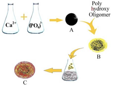
Figure 1: Schematic elucidation of the nanoparticulate three-layered aquasomal drug delivery system. The innermost central core (A) is a self-assembled ceramic core (calcium phosphate core) which is later coated with a polyhydroxy oligomeric film (B). The drug/ bio-actives / peptides are adsorbed on the surface of the oligomeric film to form the aquasome (C).
Mechanism of self-assembly
Kossovsky et al. who first proposed and developed the novel aquasomal drug delivery system described the principle of self-assembly in 19949. Self-assembly is the phenomenon in which the pre-existing components assume spontaneously determined structural orientations in a 2D or 3D space; traditionally studied under supramolecular chemistry or nanotechnology. The macromolecular self-assembly in the aqueous domain both for the purpose of building smart nanostructure materials as well as naturally occurring biochemistry are governed by three physicochemical processes:
Interactions between charged groups
The inherent chemical moieties or adsorbed ions from the biological environment charges most of the synthetic and biological surfaces. Interactions of charged groups, such as amino, sulphate, carboxyl and phosphate groups, easing up the long-range approach of the self-assembling subunits. Another pivotal role of the charged group is the stabilization of the tertiary structures of crimped proteins13..
Hydrogen bonding and dehydration effect
Hydrogen bonds potentially constitute the principal role of molecular interaction in self-assembly. Hydrophilic molecules provide greater stability and a notable degree of organization in the surrounding water molecules14. Hydrophobic molecules cannot form hydrogen bonds with the surrounding water. However, their tendency to repel water also provides greater stability to the surrounding environment. Organized water reduces the level of entropy of the environment. Since organized water is not thermodynamically favourable, molecules tend to ‘accept’ the opportunity to drain the surrounding water or dehydrate, favouring their assembly. This step is the basis of self-assembly.
Structural stability
Aquasome conserves the molecular structure and biological activity of active drugs. However, in the delivery of proteins by aquasome, protein molecules undergo many biophysical constraints like temperature, pH, solvent, salt, and protein are unstable in a liquid state, ultimately leading to irreversible denaturation of proteins. In such a scenario, carbohydrates play a vital role in overcoming barriers to keep the molecule intact. Van der Waals forces, usually within the molecule, also play a small but important role in the interaction of proteins with carbohydrates. As carbohydrate is a natural stabilizer, it functions as a dehydroprotectant and thus avert denaturation and maintains the stability of the protein structure(11, 15).
Strategies behind the chemical synthesis of the core
Aquasome as a drug delivery system is a three-layered self-assembled nanostructure. The basic strategies underlining the assembly of the nanostructures have been discussed below:
Sequential covalent synthesis
One of the basic strategies used in the synthesis of nanoparticles is the sequential covalent synthesis, which involves the binding of molecules through reversible interactions (H-bond). This strategy is applied to produce arrays of atoms that are covalently bonded with proper composition, shape and connectivity. For example, cyanocobalamin (Vit B12)16.
Covalent polymerization
This strategy is very essential in the formulation of aquasomes, where high molecular weight substances are produced by allowing the low molecular weight substances to react among themselves ultimately resulting in molecules with numerous covalently linked monomers.
Self-organizing synthesis
It is one of the most commonly used strategies which are dependent mostly on weak and fewer directional bonds, viz. hydrogen, ionic and Van der Waals interactions for the arrangements of the atoms, molecules or ions in the structure. Colloids, micelles, molecular crystals, ligand crystals, emulsions, self-assembled monolayer and phase-separated polymers are the varying types of structures formulated using this strategy. This unsolicited attribute of self-organization is used for the preparation of aquasomes, where the molecules revamp themselves to reach a minimum value of entropy and internal energy at equilibrium17.
Molecular self-assembly
Molecular self-assembly may be stated as the phenomenon in which an unorganized structure with pre-existing components rearranges itself in an organized form. The evolution of aquasomes has many interesting applications in nanoscience and nanotechnology. This method is used to make complex nanostructures based on weak noncovalent bonds such as hydrogen and ionic bonds or Van der Waals and hydrophobic interaction18,19.
Composition
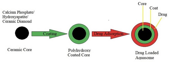
Aquasome is basically a simple three-layered structure whose composition or the principal layers have been discussed below along with their significant roles:
Centralised nanocrystalline core
The core material plays the pivotal role of the base or the foundation of the whole structure. The core should provide a high degree of stability, structural uniformity and greater surface energy for efficient binding of the coating material. The most popularly used substances for the preparation of the core are brushite (Calcium Phosphate), Ceramic Diamond (nano-crystalline carbon-ceramic), polymers (gelatin, albumin and acrylates) and Tin oxides16,20. The presence of Calcium Phosphate naturally in the body makes it a favourable candidate for the core material21,22. The biocompatibility and biodegradable nature of the ceramics, easy availability and cost-efficient manufacturing process of the ceramics make it another good choice of core material for the aquasome23.
Core coating material
The coating material generally used is the polyhydroxy oligomeric compounds. The coated layer of carbohydrate plays a number of crucial roles such as maintaining shape, chemical stability and confirmational integrity of both pharmaceutical active ingredients as well as the bioactive molecules. Some of the preferred coating materials are cellobiose, sucrose, lactose monohydrate, trehalose, citrate, chitosan and pyridoxal-5-phosphate, etc24.
API/ bioactive molecules
The bioactive molecules or the APIs are adsorbed on the surfaces of the carbohydrate coated core by various forces such as ionic and non-covalent interaction15.
Formulation techniques
The formulation techniques basically involve three steps:
Formulation of the inorganic core
The inorganic core can be manufactured by various methods. According to Nir Kossovsky15, the internal core can be prepared by colloidal precipitation technique, plasma condensation technique and inverted magnetron sputtering. Some of the most popularly employed techniques for this process have been discussed below:
a.// Co-precipitation technique by reflux.
Correia et al.25and Patil et al.26 used the co-precipitation technique for the preparation of hydroxyapatite core. This method involves the addition of 0.19 N diammonium hydrogen phosphate [(NH4)2HPO4] solution from a burette in a dropwise manner to calcium nitrate [Ca (NO3)2] solution of 0.32M at a regulated temperature of 50-75°C with uninterrupted stirring alongside maintaining the pH of Ca(NO3)2 between 8 to 10 with a concentrated solution of ammonia. Three neck round bottom flask can be used as described by Pavani et al. each bearing a thermometer, a reflux condenser attached with a CO2 trap and a charged funnel27.
The chemical reaction involved in this phenomenon is as follow:

The mixture formed in the flask is to be stirred in the similar pH and temperature condition. The precipitate formed is being dried for 24 hours at 100 °C after washing thoroughly with ultrapure water.
b.// Self-precipitation
Vengala et al.27 performed the self-precipitation method for the preparation of the core. In this method, the simulated body fluid (SBF) of pH 7.2 is prepared which contains Sodium Chloride (NaCl), Potassium Chloride (KCl), Magnesium Chloride (MgCl2), Calcium Chloride (CaCl2), sodium hydrogen carbonate (NaHCO3), disodium hydrogen phosphate (Na2HPO4), and disodium sulfate (Na2SO4). HCl is used every 24 hours to maintain a pH of 7.26 and the solution is stored in polystyrene bottles. 100 ml of this solution is poured into a series of 100 ml polystyrene bottles and firmly sealed and stored at 37±1°C for 7 days. The inner surface appears to develop precipitate, which is then filtered, washed multiple times with double or triple distilled water and finally dried overnight at 100°C.
c.// Co-precipitation technique by sonication
Jain S et. al.24 used this technique to prepare the core of the aquasome as discussed by Kossovsky. A solution of disodium hydrogen phosphate (Na2→HPO4, 0.75 M) is steadily added to a solution of calcium chloride (CaCl2, 0.25M) in sonication at 4°C for 2 hours (Figure 2).

The reaction taking place in this process is:

The precipitate so obtained is isolated by centrifugation (15,000 rpm, 2 hours) and then washed multiple times with bi-distilled water to remove the sodium chloride formed during the reaction. The precipitate is redispersed in bi-distilled water and filtered using a 0.2 µm Millipore filter to obtain core particles of sizes less than 0.2 µm.
d.// Poly (Amidoamine) (PAMAM):
This method of core formation was used by Khopade et al.28 They prepared SBF as reported by Kim et al.29 (except calcium, no other divalent salt was used). In the simulated body fluid (SBF), PAMAM is dissolved at a pH of 7.4 and stored for 1 week at 37°C to allow nucleation and the growth of crystals alongside maintaining the pH with the aid of NaOH solution. Deionized water is used to wash the precipitate so formed, which is later filtered and dried overnight.
e.// Inverted magnetron sputtering
Kossovsky et al.30 synthesized nanocrystalline tin oxide core using direct current reactive magnetron sputtering (inverted cathode). In this technique, extremely pure tin is sputtered in an argon and oxygen gas mixture at high pressure. The particles (ultrafine) so formed in the gaseous mixture are accumulated on cooled copper tubes at 77K with liquid nitrogen flowing continuously.
Coating of the core
The inorganic core so formed is coated with a polyhydroxy oligomer film. The process substantially requires the adsorption of the carbohydrate epitaxially on the core substrate. The cleaned preformed cores are dispersed in doubled distilled water. Generally, core particles obtained are dispersed in bi-distilled water and is subjected to sonication and finally lyophilization to enhance the irrevocable adsorption of the carbohydrate epitaxially on the ceramic surfaces. The coating materials that have been previously used are cellobiose, sucrose, trehalose, pyridoxal-5-phosphate and citrate and has been reported that variation is there in the drug loading based on the coating material31. Why Disaccharide Coating? In the past few decades, the application of disaccharide or other sugars has gained importance, especially in the field of nanotechnology. Delivery of proteins, hydrophobic molecules and other drugs is a very challenging task through oral routes due to various hindrances and poor bioavailability. Based on the reports in various studies, the abilities of trehalose and sucrose to withstand various stress condition have been exploited in the selection of coating material(32,33). In the process of desiccation, trehalose usually works by protecting proteins and membranes within the plant cell and thus maintaining cell structure, natural colours, taste and texture. The hydroxyl group in carbohydrates combines with the polar and charged groups of the proteins and maintains the structure when dehydrated. These disaccharides contain a large number of hydroxyl groups that help to replenish water around polar residues in proteins and preserves their integrity in a dearth of water. Thus, the carbohydrate layer plays a very vital role in the formulation of aquasome13,15.
Drug loading
The final step in the formulation of aquasome is the drug’s immobilization by physical adsorption on the surface-modified nano-crystalline cores. Drug loading on the coated core is a simple physical process of adsorption: a solution of known concentration of a drug of choice is to be prepared in a suitable solvent and added to a flask containing an accurate quantity of coated cores. The flask is stoppered and shaken continuously under controlled conditions overnight or lyophilized to procure drug-loaded aquasomes.
The three major steps involved in the formulation of an aquasomal drug delivery system have been summarized below (Figure 3).
Characterization of the aquasome
Several tests are being performed for the characterization of the aquasomes. It basically involves the evaluation of morphology of the particles, structural properties, size distribution and drug loading.
Evaluation of the nanocrystalline core
a.// Structure analysis
The structure of the nanocrystalline core formed is examined with the help of Fourier transform infrared spectroscopy (FTIR). FTIR spectrophotometer is used to record the infrared spectra in the range of 400-4000 cm−1 wave number34,35.
b.// Size distribution
The morphological characterization and the analysis of size distribution of the core particles can be performed using scanning electron microscopy (SEM) and transmission electron microscopy (TEM). Core, coated core, as well as drug-loaded aquasomes, are analysed by these techniques30,31.
c.// Crystallinity
The prepared ceramic core can be analysed for its crystalline/ amorphous nature in which the X-Ray diffraction pattern is interpreted on a comparative study with that of a standard diffractogram36.
Evaluation of sugar coating
a.// Colorimetric analysis of sugar coating on to the ceramic core
A number of methods are available such as concanavalin A-induced aggregation method is used for the determination of the quantity of sugar on the coated core while the anthrone method is utilized for quantification of the residual sugar remaining or residual sugar unbound after coating36. Anthrone method is the most preferred method. Anthrone gives green coloured product, which is formed after the hydrolysis of carbohydrate into simple sugars and eventually to hydroxyl methyl furfural. After the preparation of the calibration curve aliquots of samples are transferred to boiling tubes and diluted to an appropriate concentration. After the addition of anthrone reagent, the samples are heated in a boiling water bath and cooled rapidly. When a greenish solution is obtained, its absorbance is recorded using a UV-visible spectrophotometer, taking glucose as standard. For the analysis of the sample, the sugar-coated core is accurately weighed and dissolved in distilled water. The solution is treated with the anthrone reagent using the procedure as mentioned37.
Evaluation of aquasome
a.// Structural analysis
Structural analysis is assessed by Fourier-transform infrared spectroscopy (FT-IR) in the wave number range of 400-4000 cm−1. FTIR spectroscopy is used for the confirmation of the presence of ceramic core, presence of cellobiose on the ceramic core, and the presence of the drug on the cellobiose coated ceramic core38.
b.// Structural analysis
Structural analysis is assessed by Fourier-transform infrared spectroscopy (FT-IR) in the wave number range of 400-4000 cm−1. FTIR spectroscopy is used for the confirmation of the presence of ceramic core, presence of cellobiose on the ceramic core, and the presence of the drug on the cellobiose coated ceramic core38.
c.// Particle size analysis and morphology
The morphological examination of the drug-loaded aquasomes can be performed using a SEM. The mean particle size and size distribution are determined by SEM. The chemical composition and the crystalline structure of all samples are obtained through X-ray powder diffractometry38,39.
d.// Glass transition temperature
The effect of carbohydrate on the drug-loaded aquasomes can be analysed by differential scanning calorimetry (DSC). DSC studies are being performed extensively to record the transition from glass to rubber state with alteration in temperature for carbohydrates and proteins16.
e.// Estimation of percentage yield
After drying the formulated drug-loaded ceramic nanoparticles, free-flowing powdered nanoparticles may be obtained. These ceramic nanoparticles are to be collected carefully and accurately weighed. The percentage yield of the nanoparticles is calculated by the following equation40.

f.// Entrapment efficiency and drug loading
Entrapment efficiency is the percentage of the actual mass of drug entrapped in the carrier relative to the initial amount of loaded drug41. The % entrapment efficiency is calculated by:

g.// In-process stability studies
The impact of the process on the integrity and stability of the protein or bio-actives can be evaluated by performing SDS-PAGE (sodium dodecyl sulphate polyacrylamide gel)29,42.
h.// In vitro drug release studies
In vitro release study of the drug-loaded in the aquasomes can be performed using suitable phosphate buffer as dissolution media by dialysis bag method (12000-14000 Dalton). Accurately weighed aquasomal powder equivalent to a known amount of the API/drug is suspended in 10 ml of phosphate buffer. This suspension is poured into the activated dialysis bag and is sealed from both ends properly. The bag is then placed with 300 ml buffer and media must be stirred at a speed of 100 rpm at 37±0.5oC. Aliquots of 5ml samples are withdrawn at various time intervals and analysed for the drug content at a specific wavelength using a UV spectrophotometer. Sink condition is to be maintained with 5 ml of fresh dissolution medium7,43.
i// Drug release kinetics
The drug release kinetics is established by plotting several data obtained from the in vitro drug release studies in numerous kinetics models to understand the linear relationship, that is, kinetic principles. The data are processed for regression analysis using MS Excel statistical functions. To comprehend the release mechanisms, in vitro drug release data are analysed using Zero Order and First Order Kinetics, Higuchi’s model, Hixson-Crowell Cube root law and Korsmeyer-Peppas model.
The fate of the aquasome
Aquasome is a nanosized biodegradable drug delivery vehicle, they get accumulated in the liver and muscles. The fact that the drugs get adsorbed onto the surface of the aquasome without any alteration or modification helps to bring about immediate pharmacological effect as there is no hindrance in the recognition of the receptors at the site of action12. Overall, in a normal system, the ceramic core, that is, calcium phosphate is a biodegradable material. In vitro biodegradation of the ceramic is carried out by monocytes and osteoclasts (multicellular cells) because of their intervention initially at the site of biomaterial implantation during an inflammatory reaction.
Phagocytosis of two different types was reported to occur when cells interact with biomaterials; either crystals of calcium phosphate are segregated and eventually dissolved in the cytoplasm after the disappearance of the phagosome membrane or dissolution after the formation of heterophagosomes44. IFN-g (interferon-gamma) or dihydroxy cholecalciferol have an impact on the monocytic activities and can be synthesized with a number of soluble substances. Some cytokines can also develop an inflammatory process and may be involved in the biodegradation process45.
Applications
Aquasome has found numerous applications (Table 1) in the delivery of drugs and bio actives. Some of them has been discussed below.
Parenteral delivery of insulin
Cherian et al.46 formulated aquasomes for the delivery of insulin. They used calcium phosphate as the core material which was coated using numerous disaccharides (trehalose, cellobiose and pyridoxal-5-phosphate). The drug was later adsorbed onto this coated core using simple adsorption. in vitro studies revealed that the performance of the insulin loaded aquasome was far better than insulin solution.
Gene delivery
Studies have shown the possibility for delivery of a gene segment maintaining structural integrity. This drug carrier provides all the features of a viral vector without compromising its structure and activity15.
Aquasomes for antigen delivery
The delivery of antigen is always a challenging task as the adjuvant added for the enhancement of the immuno-property tend to alter or change the conformation of the antigen. Kossovsky et al.9 illustrated the efficiency of the aquasomes as a vehicle for antigen delivery. These aquasomes proffered conformational stabilization along with extensive surface exposure to antigen protein. Aquasomes were prepared by Goyal et al.47 by self-assembling of hydroxyapatite (using co-precipitation technique). In this process, trehalose and cellobiose were used as the coating material. Ultimately, model antigen, Bovine Serum Albumin (BSA) was adsorbed on the coated hydroxyapatite core. The spherical aquasomes so formed were reported to attain a better immunological response as compared to that of plain BSA, alongside maintaining the structural integrity and conformation of the protein48.
Oral delivery of lipophilic drug through aquasomes
Vengala et al.49 formulated aquasomes for the delivery of the drug pimozide having poor solubility. A number of techniques were employed for the core formation of which the coprecipitation technique by sonication was a productive one. Cellobiose was coated on the calcium phosphate [Ca3(PO4)2] core after which the drug pimozide was allowed to adsorb onto it. Studies revealed that the pimozide loaded aquasomes were spherical and uniform in shape and showed an enhanced release profile compared to pure drug. First-order release kinetics and Higuchi diffusion-controlled release mechanism were observed, thus making them potent for the enhancement of the solubility of poorly soluble drugs31. Vengala et al. also successfully formulated aquasomes loaded with piroxicam (2016) and lornoxicam (2017)38,39. Oviedo44 and his team also formulated aquasomes for the delivery of poorly soluble drug indomethacin adsorbed on a lactose coated calcium phosphate core.
Aquasomes as oxygen carrier
Khopade et al.28 and Patil et al.26 successfully prepared aquasomes for their functioning as oxygen carrier.
Khopade and his team developed haemoglobin aquasomes using spherical hydroxyapatite core. They formulated the core with carboxylic acid-terminated half-generation poly (amidoamine) dendrimer. Sugar-coating was performed on the hydroxyapatite cores after which the haemoglobin was adsorbed on the surfaces of the coated core. Aquasomes are characterized by size, loading of haemoglobin, oxygen-binding properties and storage capacity. They could successfully load 13.7mg haemoglobin per g of the nanosized aquasome and preserve oxygen concentration and consistency and stability for a minimum of 30 days. The efficacy of the formulation was tested in albino mice and indicated that it may be used as a blood transfusion.
After two years, Patil et al.26 again formulated nanocrystalline aquasomes as a vehicle for haemoglobin which in turn serves as a carrier for oxygen delivery. The formulated haemoglobin aquasome were suspended in phosphate buffer (having 0.01%w/v lecithin and 7.5%w/v albumin) and examined for its oxygen carrying capacity which was later found to be similar to the actual blood. In vivo study revealed that there was no notable variation in heart rate and blood pressure when the rats were transfused with the suspension of aquasome on 50% exchange transfusion.
Enzymes delivery through aquasomes
Aquasomes’ ability to protect the molecular conformation and structural integrity has been taken into account for the delivery of enzymes. Goyal et. al.50 successfully immobilized DNAase (therapeutic enzyme) on the surface of the aquasomes and was able to target the specific site of action and to bring about a prominent therapeutic effect as desired.
Table 1: Various applications of aquasomes.
| Sl No. | API/Bio-actives | Application | References |
|---|---|---|---|
| Dithranol | Treatment of psoriasis | 51 | |
| Insulin | Blood Sugar regulation | 46 | |
| Etoposide | Anticancer targeting | 52 | |
| Bromelain | multi-particulate drug carrier for oral delivery | 53 | |
| Hepatitis B Vaccine | Antigen for prevention of jaundice | 36 | |
| Haemoglobin | carrier for oxygen | 26 | |
| Merozoite Surface Protein-119 (MSP-119) | To improve immune adjuvant property for antimalarial antigen | 50 | |
| Polypeptide-k | Blood sugar regulation | 54 | |
| Pimozide Piroxicam Lornoxicam Indomethacin | To enhance the aqueous solubility on oral administration and improve their efficacy | 27,31,46,48 | |
| recombinant human interferon-α-2b (rhINF-α-2b) | To prolong release and enhance targeting in ovarian cancer | 45 | |
| Serratiopeptidase (STP) | Enhance proteolytic activity | 55 | |
| Interferon alpha (INF α) Cyclosporine A | To obtain sustained release | 56 | |
| Bovine Serum Albumin (BSA) | To preserve adjuvanticity and antigen properties | 47 |
Aquasomes as extract carrier
Banerjee and Sen58 formulated aquasomes of Chelidonium majus L extract and performed in vitro study to find out its activity on the liver. Characterization of the extract loaded nanoparticles indicated that the particles were stable and there was no interaction between the components and was found to have extract loading of 37.22 % on the carbohydrate coated core. In vitro histopathological analysis was performed (comparative histopathology results of toxic, silymarin treated, normal, extract-treated and formulation treated groups) which revealed the hepatoprotective activity of the extract loaded aquasome.
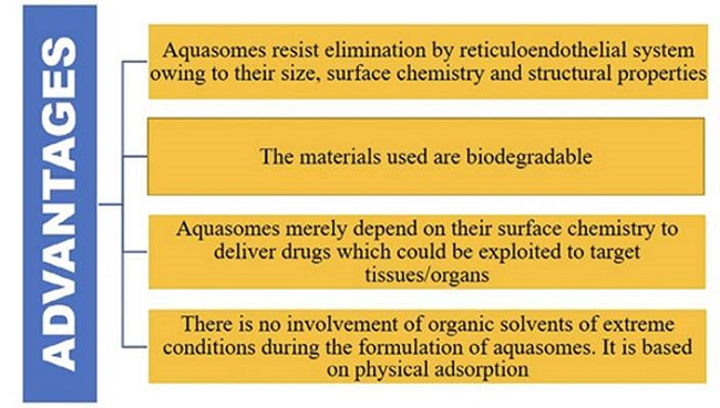
Advantages of aquasome
These systems act as a reservoir that can release the molecules either in a continuous or a pulsatile manner, avoiding a multiple-injection schedule. Nanoparticulate aquasomes have a huge surface area that enables the loading of a significant amount of bioactive molecules/drugs58.
This nanoparticulate system gives a suitable environment for protein delivery without having been denatured. This property is due to the presence of inorganic cores, which are coated with polyhydroxy compounds. The inorganic core in the aquasome prevents denaturation of the proteins and coats are responsible for the hydrophilic behaviour.
Aquasomes can increase the therapeutic efficacy of pharmaceutically active agents and also protect the active molecule from phagocytosis and environmental degradation59.
Multi-layered aquasomes, conjugated with antibiotics, nucleic acid, peptides etc can be used in various imaging tests.
Enzyme activity and sensitivity towards molecular conformation made aquasome a novel carrier for enzymes such as DNAaes and pigment/dyes.
Aquasomes-based vaccines offer many advantages as a vaccine delivery system due to the aquasomal potency to elicit both cellular and humoral mediated immune responses56,60.
Challenges
Aquasomes can be seen to have the potential to change the future of the delivery of proteins/ peptides and other pharmaceutically active ingredients. However, various challenges have come across the successful formulation and delivery. There are a number of sectors of aquasomes to be studied, such as their irreconcilable behaviour to several sterilization techniques and their shelf-life in accordance with the ICH guidelines. Other challenges include the practicability of the large-scale production and their ensuing commercialization potential. The overall high cost of the ingredients and the lengthy production process are yet to be made feasible61.
The understanding and knowledge of this technique are still in the growing stage. Reproducibility after characterization of vital factors, like the in vitro and in vivo studies and their safety parameters, are some of the crucial sections yet to be extensively studied. With due course of time and the intense research going on in this direction, aquasomal drug delivery will be one of the most preferred and popularly used novel nanoparticulate drug delivery systems.
Conclusions
Aquasome, a three-layered self-assembled molecule is a simple but novel drug delivery system and it seems to have optimistic and prospective carrier potentials to improve the solubility of poorly soluble drugs and enhance their bioavailability. It has been found to maintain and protect the structural integrity and conformation of the biologically active molecules. The crystalline ceramic core provides structural stability and overall integrity. The carbohydrate coating has the property of maintaining the conformational integrity of bioactive molecules which have led to the proposal that aquasomes have potential as a carrier system for delivery of peptide, protein, hormones, antigens, genes and hydrophobic drugs to specific sites. Although there are several challenges to be addressed including time consumption, complicated process, safety and high cost in the development, aquasome can emerge as an alternative vesicular carrier in the future. Still, considerable further study of aquasomes is necessary concerning pharmacokinetics, toxicology, and animal studies to confirm their efficiency as well as safety, to establish their clinical usefulness and to launch them commercially.

