Brief Report
Sympathovagal equilibrium analysis in patients with COVID-19
Análisis del equilibrio simpatovagal en pacientes con COVID-19
1Department of Internal Medicine, Hospital J. B. Iturraspe, Santa Fe, Argentina
Abstract
Introduction: With the increase of COVID-19 cases, an unusual manifestation for this type of virus began to appear anosmia and dysgeusia, which could indicate a neurologic alteration. In this context, it seems likely that subclinical manifestations of baroreflex involvement occur. The vegetative nervous system carries out the regulation of the baroreflex through the balance between sympathetic and parasympathetic activity. The objective of this study is to verify whether patients with COVID-19 present alteration of this equilibrium.
Material and methods: Patients included had a confirmed diagnosis of COVID-19 admitted to the Internal Medicine Department of JB Iturraspe Hospital. A Holter recording was performed at rest for 5 minutes, determining the variables in the frequency domain using Fourier transform analysis. We excluded patients with diabetes, medicated with drugs that modify heart rate or with a history of irradiation to the neck.
Results: 68 patients were studied. The mean age was 49±13 years. The median systolic blood pressure was 120 mmHg and the diastolic blood pressure 80 mmHg. The heart rate was 76±13 beats per minute and the median respiratory rate was 24 (16 to 40). Anosmia was observed in 22% and dysgeusia in 19% The variables in the frequency domain were: Low-frequency power (LF) 135.8ms2 (13.7-2861.7); High-frequency power (HF), 89.04ms2 (4.1-5234.4), LFnu 57.5±22.3, HFnu 43.1±22.6. LF:HF 2.1±2. 41.2% of the patients had a high LF:HF.
Conclusions: LF and HF components can be obtained through frequency analysis. The relationship between these two elements would thus represent the sympathovagal balance and is expressed as the LF/HF ratio. We observed that 41.2% of the studied patients showed elevated LF/HF ratio. The 41.2% of the patients presented an increased LF:HF ratio, which could be interpreted as an alteration in autonomic function.
Keywords Dysautonomia; COVID-19; Heart rate variability
Resumen
Introducción: Con el aumento de casos de COVID-19, unas manifestaciones inusuales para este tipo de virus como la anosmia y disgeusia comenzaron a aparecer, lo que podría indicar una alteración neurológica. En este contexto, parece probable que se produzcan manifestaciones subclínicas de afectación barorrefleja. El sistema nervioso vegetativo lleva a cabo la regulación del barorreflejo a través del equilibrio entre la actividad simpática y parasimpática. El objetivo de este estudio es verificar si los pacientes con COVID-19 presentan alteración de este equilibrio.
Material y métodos: Se evaluaron pacientes con diagnóstico confirmado de COVID-19 ingresados en el Servicio de Medicina Interna del Hospital JB Iturraspe. Se realizó un registro Holter en reposo durante 5 minutos, determinando las variables en el dominio de la frecuencia mediante análisis por transformada de Fourier. Se excluyeron pacientes con diabetes, medicados con fármacos que modifican la frecuencia cardiaca o con antecedentes de irradiación al cuello.
Resultados: Se estudiaron 68 pacientes. La edad media fue de 49±13 años. La mediana de la presión arterial sistólica fue de 120 mmHg y la diastólica de 80 mmHg. La frecuencia cardiaca fue de 76±13 latidos por minuto y la mediana de la frecuencia respiratoria fue de 24 (16 a 40). Se observó anosmia en 22% y disgeusia en 19% Las variables en el dominio frecuencial fueron: Potencia de baja frecuencia (LF) 135,8ms2 (13,7-2861,7); Potencia de alta frecuencia (HF), 89,04 ms2 (4,1-5234,4), LFnu 57,5±22,3, HFnu 43,1±22,6. LF:HF 2.1±2. El 41,2% de los pacientes tenían una relación LF:HF alta.
Conclusiones: Los componentes de LF y HF se pueden obtener a través del análisis de frecuencia. La relación entre estos dos elementos representaría así el equilibrio simpatovagal y se expresa como la relación LF/HF. Observamos que el 41,2% de los pacientes estudiados presentaban una relación LF/HF elevada. El 41,2% de los pacientes presentó una relación LF:HF aumentada, lo que podría interpretarse como una alteración en la función autonómica.
Palabras clave Disautonomía; COVID-19; Variabilidad frecuencia cardiaca
INTRODUCTION
At the end of 2019, a hitherto unknown coronavirus was identified in Wuhan (China), which showed unusual characteristics and spread to the entire planet. Due to the predominantly pulmonary involvement in its severe forms and its similarities with the microorganism that caused the pandemic in 2003, it was called SARS CoV-2 (severe acute respiratory syndrome coronavirus-2) and the disease caused by it was named COVID-19 [1].
Already in the early days of the pandemic, increasingly frequent reports of an unusual manifestation for this type of virus began to appear anosmia and dysgeusia [1]. Although the mechanism of production of both symptoms is not exactly known, it has been postulated that they are due to neurological involvement [2]. It has been suggested the possible relationship between the autonomic nervous system with numerous manifestations including postural tachycardia syndrome with dysautonomia (POTS) orthostatic hypotension, bradycardia and syncope [3, 4, 5].
In this context, it seems likely that subclinical manifestations of baroreflex involvement occur, a mechanism responsible for 90% of the short-term variability in blood pressure and heart rate [6]. If there is an alteration of the baroreflex with a decrease in its sensitivity, this could cause difficulties in the diagnosis and treatment of arterial hypertension. Moreover, if it persists over time, it could constitute a cardiovascular risk factor of unknown importance [7].
The vegetative nervous system carries out the regulation of the baroreflex through the balance between sympathetic and parasympathetic activity, which can be evaluated by analysis in the time domain: mean of RR intervals, standard deviation, RMSSD, etc [8]. Likewise, the analysis of heart rate variability in the frequency domain using the Fourier transform is of immense value. In this way, the low-frequency (LF) and high-frequency (HF) values are obtained, the quotient of which represents the sympathetic vagal equilibrium [8].
The calculation of the LF:HF (low frequency/high frequency) ratio in the spectral analysis of the heart rate variability is a simple method for estimating sympathovagal equilibrium [9]. The objective of this study is to verify whether patients with COVID-19 present alteration of this equilibrium by means of Holter recording.
MATERIAL AND METHODS
The study design was cross-sectional, observational, descriptive, prospective inclusion and the sampling was non-probabilistic of convenience.
Patients were included if they had a confirmed diagnosis of COVID-19 according to the clinical, epidemiological and laboratory criteria established by the Argentine Health Ministry [10], admitted to the Internal Medicine Department of JB Iturraspe Hospital in Santa Fe city, Argentina, in the period from May 10 to July 20, 2021. Patients with a diagnosis of diabetes mellitus, affected by a disease or medicated with drugs that affect heart rate or with a history of irradiation to the neck were excluded.
Verbal informed consent was obtained to avoid potential exposure to paper forms contaminated by the infected patient. Prior authorization was obtained from the hospital's Research Committee. At all times, our study was governed by the guidelines of good clinical practice, following the Declaration of Helsinki principles.
An ECG Holter recording was performed at rest for 5 minutes in the patient's room, on their bed. They were required to be in a quiet environment, with no other person in the room, in absence of another external stimuli. Variables in the frequency domain were determined using Fourier transform analysis through Kubios (v.3.0.0 [free version], HRV analysis, University of Eastern Finland).
Data are presented for low-frequency power (LF), and high-frequency power (HF) in both ms2 and normalized units, and the ratio of LF power to HF power (LF:HF). The normalized (or normalized unit) spectral indices are defined as LFnu = LF/(LF+HF) and HFnu = HF/(LF+HF).
The HF component can be influenced by the respiratory rate and thus overestimate LF:HF [11], but since COVID-19 is a disease that causes respiratory distress, we were not able to perform ECG with controlled breathing in the patients. Therefore, we conducted a logistic regression to evaluate the respiratory rate influence in LF:HF, controlling by age and the presence of anosmia/dysgeusia.
The results analysis was carried out with the statistical package IBM® SPSS® (23rd version). Normality in the distribution of continuous variables was determined using the Kolmogorov-Smirnov test. Quantitative variables of parametric distribution were described using mean and standard deviation and those non-parametric through median and range. Qualitative variables were expressed in percentages.
Reference values for normal population published in the literature were used, where a cut-off point greater than 2 was considered an alteration in the LF:HF ratio [12].
RESULTS
68 patients were studied. 62% were men and 38% women. The mean age was 49 ±13 years. The median systolic blood pressure (SBP) was 120 mmHg (range 95 to 170) and the diastolic blood pressure (DBP) 80 mmHg (range 60-110). The heart rate was 76±13 beats per minute and the respiratory rate was 24 (range 16 to 40). Anosmia was observed in 22% (n = 15) and dysgeusia in 19% (n = 13). 12 patients had both.
The variables of analysis in the frequency domain were: LF 135.8ms2 (range 13.7-2861.7); HF, 89ms2 (range 4.1-5234.4), LFnu 57.5 ±22.3, HFnu 43.1 ±22.6. LF:HF ratio 2.1±2. 41.2% (n = 28) of the patients had a high LF:HF ratio (> 2).
No association was found (p=NS) between the presence of anosmia or dysgeusia and a high LF:HF ratio (greater than 2) (Table 1).
Table 1. Low-frequency and high-frequency ratio (LH:HF) in patients affected or not of anosmia or dysgeusia
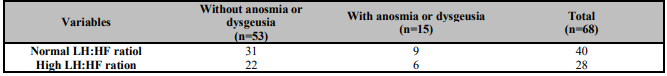
Logistic regression showed no influence of respiratory rate or the presence of anosmia/dysgeusia on LF:HF (p=0.13).
DISCUSSION
The theory that has been proposed to explain the development of neurological symptoms in SARS CoV-2 infection is that, due to the action of the virus, an internalization of angiotensin II receptors would occur in the solitary tract nucleus, altering sympathovagal equilibrium.
We observed that 41.2% of the studied patients showed elevated LF:HF ratio, so we interpret that this is due to an imbalance of autonomic function due to increased sympathetic activity. We do not know the clinical significance of this finding in the evolution of the disease.
We have not found any previous publications that demonstrate the presence of dysautonomia through an objective measurement such as that obtained by Holter in this type of patient.
Limitations of this work include the lack of knowledge of LF:HF values prior to infection and the absence of controls. In addition, no follow-up has been carried out to know the permanence of this alteration over time.
CONCLUSIONS
Through the analysis of a 5-minute Holter recording, it was found that 41.2% of the patients hospitalized with COVID-19 presented with an increase in the LF:HF ratio, and this result could be interpreted as an alteration in autonomic function due to an excess of sympathetic activity.
CONFLICT OF INTERESTS
The authors declare no conflict of interest.
REFERENCES
1. Lechien JR, Chiesa-Estomba CM, De Siati DR, Horoi M, Le Bon SD, Rodriguez A, et al. Olfactory and gustatory dysfunctions as a clinical presentation of mild-to-moderate forms of the coronavirus disease (COVID-19): a multicenter European study. Eur Arch Otorhinolaryngol. 2020;277(8):2251-61. doi: 10.1007/s00405-020-05965-1.
[ Links ]
2. Aghagoli G, Gallo Marin B, Katchur NJ, Chaves-Sell F, Asaad WF, Murphy SA. Neurological Involvement in COVID-19 and Potential Mechanisms: A Review. Neurocrit Care. 2021;34(3):1062-71. doi: 10.1007/s12028-020-01049-4.
[ Links ]
3. Ellul MA, Benjamin L, Singh B, Lant S, Michael BD, Easton A, et al. Neurological associations of COVID-19. Lancet Neurol. 2020;19(9):767-83. doi: 10.1016/S1474-4422(20)30221-0.
[ Links ]
4. Whittaker A, Anson M, Harky A. Neurological Manifestations of COVID-19: A systematic review and current update. Acta Neurol Scand. 2020;142(1):14-22. doi: 10.1111/ane.13266.
[ Links ]
5. Canetta C, Accordino S, Buscarini E, Benelli G, La Piana G, Scartabellati A, et al. Syncope at SARS-CoV-2 onset. Auton Neurosci. 2020;229:102734. doi: 10.1016/j.autneu.2020.102734.
[ Links ]
6. Parlow J, Viale JP, Annat G, Hughson R, Quintin L. Spontaneous cardiac baroreflex in humans. Comparison with drug-induced responses. Hypertension. 1995;25(5):1058-68. doi: 10.1161/01.hyp.25.5.1058.
[ Links ]
7. Balgobin B, Palma JA, Perez M, Norcliffe-Kaufmann L, Kaufmann H. Presentation, Causes, and Hemodynamic Features of Acquired Afferent Baroreflex Failure (4451). Neurology. 2020;94 (15 Supplement):4451.
[ Links ]
8. Agelink MW, Malessa R, Baumann B, Majewski T, Akila F, Zeit T, et al. Standardized tests of heart rate variability: normal ranges obtained from 309 healthy humans, and effects of age, gender, and heart rate. Clin Auton Res. 2001;11(2):99-108. doi: 10.1007/BF02322053.
[ Links ]
9. Eckberg DL. Sympathovagal balance: a critical appraisal. Circulation. 1997;96(9):3224-32. doi: 10.1161/01.cir.96.9.3224.
[ Links ]
10. Argentinean Health Minister. Definitions and classifications of case. Available from: https://www.argentina.gob.ar/salud/coronavirus/definicion-de-caso (accessed March 2022).
[ Links ]
11. Gerritsen J, TenVoorde BJ, Dekker JM, Kostense PJ, Bouter LM, Heethaar RM. Baroreflex sensitivity in the elderly: influence of age, breathing and spectral methods. Clin Sci (Lond). 2000;99(5):371-81.
[ Links ]
12. Nunan D, Sandercock GR, Brodie DA. A quantitative systematic review of normal values for short-term heart rate variability in healthy adults. Pacing Clin Electrophysiol. 2010;33(11):1407-17. doi: 10.1111/j.1540-8159.2010.02841.x.
[ Links ]
© 2022 The Authors. Published by Iberoamerican Journal of Medicine


 Servicios personalizados
 Inglés (pdf)
 Articulo en XML
 Referencias del artículo
 Enviar articulo por email
 Citado por SciELO
  Citado por Google
  Similares en
SciELO
Similares en
SciELO  Similares en Google
 Permalink
