<?xml version="1.0" encoding="ISO-8859-1"?><article xmlns:mml="http://www.w3.org/1998/Math/MathML" xmlns:xlink="http://www.w3.org/1999/xlink" xmlns:xsi="http://www.w3.org/2001/XMLSchema-instance">
<front>
<journal-meta>
<journal-id>1698-6946</journal-id>
<journal-title><![CDATA[Medicina Oral, Patología Oral y Cirugía Bucal (Internet)]]></journal-title>
<abbrev-journal-title><![CDATA[Med. oral patol. oral cir.bucal (Internet)]]></abbrev-journal-title>
<issn>1698-6946</issn>
<publisher>
<publisher-name><![CDATA[Sociedad Española de Medicina Oral]]></publisher-name>
</publisher>
</journal-meta>
<article-meta>
<article-id>S1698-69462006000500003</article-id>
<title-group>
<article-title xml:lang="en"><![CDATA[Spindle cell lipoma of the floor of the mouth: A case report]]></article-title>
</title-group>
<contrib-group>
<contrib contrib-type="author">
<name>
<surname><![CDATA[Coimbra]]></surname>
<given-names><![CDATA[Filipe]]></given-names>
</name>
<xref ref-type="aff" rid="A01"/>
</contrib>
<contrib contrib-type="author">
<name>
<surname><![CDATA[Lopes]]></surname>
<given-names><![CDATA[J.M.]]></given-names>
</name>
<xref ref-type="aff" rid="A02"/>
</contrib>
<contrib contrib-type="author">
<name>
<surname><![CDATA[Figueiral]]></surname>
<given-names><![CDATA[Helena]]></given-names>
</name>
<xref ref-type="aff" rid="A03"/>
</contrib>
<contrib contrib-type="author">
<name>
<surname><![CDATA[Scully]]></surname>
<given-names><![CDATA[Crispian]]></given-names>
</name>
<xref ref-type="aff" rid="A04"/>
</contrib>
</contrib-group>
<aff id="A01">
<institution><![CDATA[,University of Porto Faculty of Dental Medicine Department of Oral Medicine]]></institution>
<addr-line><![CDATA[ ]]></addr-line>
</aff>
<aff id="A02">
<institution><![CDATA[,University of Porto Faculty of Medicine Department of Pathology]]></institution>
<addr-line><![CDATA[ ]]></addr-line>
</aff>
<aff id="A03">
<institution><![CDATA[,University of Porto Faculty of Dental Medicine Department of Prosthodontics]]></institution>
<addr-line><![CDATA[ ]]></addr-line>
</aff>
<aff id="A04">
<institution><![CDATA[,University College London Eastman Dental Institute ]]></institution>
<addr-line><![CDATA[ ]]></addr-line>
<country>London</country>
</aff>
<pub-date pub-type="pub">
<day>00</day>
<month>09</month>
<year>2006</year>
</pub-date>
<pub-date pub-type="epub">
<day>00</day>
<month>09</month>
<year>2006</year>
</pub-date>
<volume>11</volume>
<numero>5</numero>
<fpage>401</fpage>
<lpage>403</lpage>
<copyright-statement/>
<copyright-year/>
<self-uri xlink:href="http://scielo.isciii.es/scielo.php?script=sci_arttext&amp;pid=S1698-69462006000500003&amp;lng=en&amp;nrm=iso"></self-uri><self-uri xlink:href="http://scielo.isciii.es/scielo.php?script=sci_abstract&amp;pid=S1698-69462006000500003&amp;lng=en&amp;nrm=iso"></self-uri><self-uri xlink:href="http://scielo.isciii.es/scielo.php?script=sci_pdf&amp;pid=S1698-69462006000500003&amp;lng=en&amp;nrm=iso"></self-uri><abstract abstract-type="short" xml:lang="en"><p><![CDATA[A sessile swelling in the floor of the mouth appeared three years ago in a 29-year old Caucasian female located laterally to the opening of the right duct of Wharton. The mass covered by normal looking mucosa exhibited slight growth since then. After excision, histological examination revealed the presence of a tumor formed by areas of mature adipose cells interspersed with extensions of tightly disposed fusiform fibroblasts immunoreactive for vimentin and CD-34. There were foci of concentric fibroblasts forming dense whorls. When large these conglomerates exhibited chondrocytes in the center. Mast-cells were not rare throughout the fibroblastic areas. No signs of malignancy occurred. These features led to the diagnosis of a spindle cell lipoma with chondrous metaplasia. The relative rarity of such a tumor in this location, especially in the chondrous variety, was considered worth of presentation, while the differential diagnosis with other intraoral tumors, namely mucoceles, dermoid cysts, mesenchimomas, fibromas and myxomas of the floor of the mouth, is discussed.]]></p></abstract>
<kwd-group>
<kwd lng="en"><![CDATA[Lipoma]]></kwd>
<kwd lng="en"><![CDATA[spindle cell lipoma]]></kwd>
<kwd lng="en"><![CDATA[intraoral tumor]]></kwd>
<kwd lng="en"><![CDATA[floor of the mouth]]></kwd>
</kwd-group>
</article-meta>
</front><body><![CDATA[ <p align="right"><font face="Verdana" size="2"><b>ORAL MEDICINE AND PATHOLOGY</b></font></p>     <p>&nbsp;</p>     <p><font face="Verdana" size="4"><b><a name="top"></a>Spindle cell lipoma of the floor of the mouth. A case report</b></font></p>     <p>&nbsp;</p>     <p>&nbsp;</p>     <p><font face="Verdana" size="2"><b>Filipe Coimbra<sup>1</sup>, J.M. Lopes<sup>2</sup>, Helena Figueiral<sup>3</sup>, Crispian Scully</b><sup>4</sup></font></p>     <p><font face="Verdana" size="2">(1) DDS, PhD, Auxiliary professor, Department of Oral Medicine, Faculty of Dental Medicine, University of Porto    <br> (2) MD, PhD, Associate professor, Department of Pathology, Faculty of Medicine, University of Porto    <br> (3) DDS, PhD, Auxiliary professor, Department of Prosthodontics, Faculty of Dental Medicine, University of Porto    <br> (4) CBE, MD, PhD, Dean and Director of Studies and Research: University College London, Eastman Dental Institute, 256 Gray’s Inn Road, London</font></p>     ]]></body>
<body><![CDATA[<p><font face="Verdana" size="2"><a href="#back">Address for correspondence</a></font></p>     <p>&nbsp;</p>     <p>&nbsp;</p><hr size="1">      <p><font face="Verdana" size="2"><b>ABSTRACT</b></font></p>     <p><font face="Verdana" size="2">A sessile swelling in the floor of the mouth appeared three years ago in a 29-year old Caucasian female located laterally to the opening of the right duct of Wharton. The mass covered by normal looking mucosa exhibited slight growth since then. After excision, histological examination revealed the presence of a tumor formed by areas of mature adipose cells interspersed with extensions of tightly disposed fusiform fibroblasts immunoreactive for vimentin and CD-34. There were foci of concentric fibroblasts forming dense whorls. When large these conglomerates exhibited chondrocytes in the center. Mast-cells were not rare throughout the fibroblastic areas. No signs of malignancy occurred. These features led to the diagnosis of a spindle cell lipoma with chondrous metaplasia. The relative rarity of such a tumor in this location, especially in the chondrous variety, was considered worth of presentation, while the differential diagnosis with other intraoral tumors, namely mucoceles, dermoid cysts, mesenchimomas, fibromas and myxomas of the floor of the mouth, is discussed.</font></p>     <p><font face="Verdana" size="2"><b>Key words:</b> Lipoma, spindle cell lipoma, intraoral tumor, floor of the mouth.</font></p>  <hr size="1">      <p>&nbsp;</p>     <p><font face="Verdana"><b>Introduction</b></font></p>     <p><font face="Verdana" size="2">Benign swellings in the floor of the mouth include mucoceles(1), dermoid cysts(2), mesenchymomas (3) and lipomas. In recent reviews of filed pathological cases Al-Naief et al (4) in the USA studied 164 oral lipomas, and Fregnani et al (5) in Brasil reported 46 tumors. The most common lesions were simple lipomas and fibrolipomas, and rarer variants included angiolipoma, intramuscular lipoma, pleomorphic lipoma and spindle cell lipoma. The spindle cell lipoma is a rather infrequent neoplasm, as shown by the fact that two recent surveys in the English language literature(5, 6) revealed only 25 cases. Of these only four were located in the floor of the mouth. Since spindle cell lipomas are composed of two tissues derived from embryonic mesenchyme, they bear some similarities to the also benign and rare mesenchymomas of which 21 cases have been reported so far in the oral cavity (3). However mesenchymomas usually contain more mesenchyme-derived tissues than the very cellular connective tissue and the mature adipose tissue occurring in spindle cell lipomas, and they lack the prominent fusiform fibroblasts of the latter.</font></p>     <p><font face="Verdana" size="2">We report a patient with a spindle cell lipoma in the floor of the mouth with conspicuous foci of chondrous metaplasia, which appears to be the fifth case described so far in this location.</font></p>     ]]></body>
<body><![CDATA[<p>&nbsp;</p>     <p><font face="Verdana"><b>Case report</b></font></p>     <p><font face="Verdana" size="2">
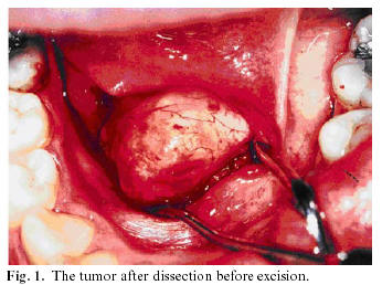
A 29-year-old Caucasian female complained of a right-sided swelling in the floor of the mouth. The lesion had appeared 3-years previously, growing very slowly and without any accompanying symptoms. Oral examination revealed a sessile mass, 1.5 cm in maximal diameter, in the floor of the mouth lateral to the opening of the duct of Wharton (<a href="#f1">Fig. 1</a>). It was firm and nonfluctuant on palpation, and not attached to the surrounding or deeper tissues. The overlying mucosa was normal and there were no salivary changes. There was no palpable cervical lymphadenopathy. The mass was dissected and excised. Upon formalin fixation and paraffin embedding, sections were treated with hematoxylin-eosine or the Giemsa stain diluted to 20 %. Other paraffin sections were immunostained with primary antibodies against vimentin (monoclonal antibody raised in mouse provided by Dakoppatts, Glostrup, Denmark), CD-34 or the S-100 protein (both polyclonal antibodies from Labvision, Fremont, USA). A kit of ABC, from Labvision, followed the secondary antibodies. The reactions were developed with diaminobenzidine (DAB) and hydrogen peroxide.</font></p>     <p align="center"><font face="Verdana" size="2"><a name="f1"><img border="0" src="/img/revistas/medicorpa/v11n5/03i.ht1.jpg" width="346" height="258"></a></font></p>      <p><font face="Verdana" size="2">    <br> 
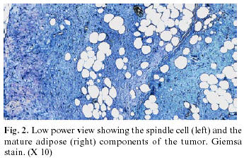
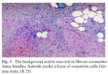
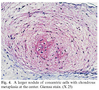
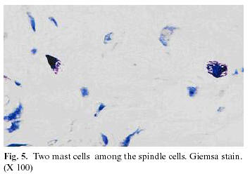
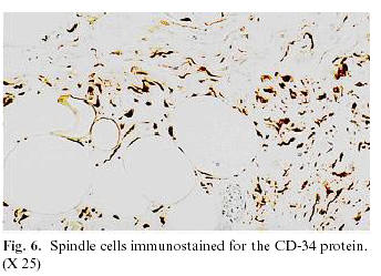
Histological examination revealed areas of mature adipose tissue interspersed with areas of spindle shaped fibroblasts, the extent occupied by both tissues being similar (<a href="#f2">Fig. 2</a>). Spindle cells were densely distributed in a fibrous matrix (<a href="#f3">Figs. 3</a> and <a href="#f5">5</a>) which also had some myxoid areas, as well as a few foci of concentric cells (<a href="#f3">Fig. 3</a>), often arranged in whorls. The larger foci showed cartilaginous cells at the center (<a href="#f4">Fig. 4</a>). Extensions of mature cartilage were rare. Occasionally the spindle shaped cells occurred in palisading arrangements. Mast cells were consistently present in the spindle cell areas (<a href="#f5">Fig. 5</a>). The mitotic index was low and there were no necrotic areas. Spindle cells were immunoreactive for vimentin and CD-34 (<a href="#f6">Fig. 6</a>). Immunoreactivity for the S-100 protein occurred in cartilaginous cells and the periphery of some adipocytes. There were no recurrences during a 3 year follow-up.</font></p>     <p align="center"><font face="Verdana" size="2"><a name="f2"><img border="0" src="/img/revistas/medicorpa/v11n5/03i.ht2.jpg" width="345" height="227"></a></font></p>     <p align="center"><font face="Verdana" size="2">    <br> <a name="f3"> <img border="0" src="/img/revistas/medicorpa/v11n5/03i.ht3.jpg" width="350" height="242"></a></font></p>     <p align="center"><font face="Verdana" size="2">    ]]></body>
<body><![CDATA[<br> <a name="f4"> <img border="0" src="/img/revistas/medicorpa/v11n5/03i.ht4.jpg" width="346" height="312"></a></font></p>     <p align="center"><font face="Verdana" size="2">    <br> <a name="f5"> <img border="0" src="/img/revistas/medicorpa/v11n5/03i.ht5.jpg" width="355" height="248"></a></font></p>     <p align="center"><font face="Verdana" size="2">    <br> <a name="f6"> <img border="0" src="/img/revistas/medicorpa/v11n5/03i.ht6.jpg" width="344" height="249"></a></font></p>      <p>&nbsp;</p>     <p><font face="Verdana"><b>Discussion</b></font></p>     <p><font face="Verdana" size="2">Swellings of the floor of the mouth are often benign tumors. The mucous extravasations or mucoceles of major salivary glands, also called blue ranulas, may occur in this location and they may plunge down sending one extension to the submandibular space in the neck(1). Dermoid cysts may occur in the floor of the mouth but they may also produce another mass in the neck from where they originally arose due to the sequestration of cutaneous tissue along cervical embryonic lines of closure, later extending to the mouth (2, 7). Fibromas and related entities caused by traumatic friction of prosthetic devices are common (8) and myxomas may arise from odontogenic rests (9). A small mass strictly circumscribed to the floor of the mouth with no signs of malignancy (no induration, adherence to deep plans, ulceration) is more likely to be an adenoma of the minor salivary glands if hard(8) or, if more tender, may be lipoma or a variant(5, 10) or a mesenchymoma (3).</font></p>     <p><font face="Verdana" size="2">The histopathology of our present case fits well with the prototype of the spindle cell lipoma, as described in the 38 (mostly cutaneous) cases reviewed by Fletcher and Martin-Bates(11). The distinction from the more common fibrolipoma was made by the greater density of fibroblasts in our tumor contrasting with the conspicuous collagen bundles intersecting the adipose tissue in fibrolipomas (5).</font></p>     <p><font face="Verdana" size="2">The occurrence of a spindle cell liposarcoma (11) or a spindle cell carcinoma (12, 13) was excluded by the lack of mitotic cells and lipoblasts. Although a rare case of lipomatous neurofibroma has recently been reported in the mouth(14), this possibility was ruled out in our case by the lack of specific immunostaining for S-100 in the palisading cell arrangements. Finally it should be stressed that the chondrous nodules described here were consistent with different steps of a metaplastic process occurring in cell foci distinct from the areas of stable cartilaginous tissue present in mesenchymomas<sup>3</sup>. In a case of the rare intramuscular variant of spindle cell lipoma, Fletcher and Martin-Bates (11)noted foci of chondrous and osseous metaplasia, while Nwaorgu et al (15)described striking chondrous metaplasia in a pharyngeal lipoma hence designated as chondrolipoma. However, to our best knowledge, no such data have been found in intraoral spindle cell lipomas (4, 6). Oral spindle cell lipomas appear not to differ from the cutaneous variety, which is much more common. In both recurrences are rare and histological signs of malignancy are lacking. Nevertheless, their very rarity demands reporting of every new cases.</font></p>     ]]></body>
<body><![CDATA[<p>&nbsp;</p>     <p><font face="Verdana"><b>References</b></font></p>     <!-- ref --><p><font face="Verdana" size="2">1. Anastassov GE, Haiavi J, Solodnik P, Lee H, Lumerman H: Submandibular gland mucocele, diagnosis and management. Oral Surg Oral Med Oral Pathol Oral Radiol Endod 2000; 89: 159-63.</font>&nbsp;&nbsp;&nbsp;&nbsp;&nbsp;&nbsp;&nbsp;&nbsp;[&#160;<a href="javascript:void(0);" onclick="javascript: window.open('/scielo.php?script=sci_nlinks&ref=2963296&pid=S1698-6946200600050000300001&lng=','','width=640,height=500,resizable=yes,scrollbars=1,menubar=yes,');">Links</a>&#160;]<!-- end-ref --><!-- ref --><p><font face="Verdana" size="2">2. Devine JC, Jones DC. Carcinomatous transformation a sublingual dermoid cyst, a case report. Int J Oral Maxillofac Surg 2000; 29: 126-7.</font>&nbsp;&nbsp;&nbsp;&nbsp;&nbsp;&nbsp;&nbsp;&nbsp;[&#160;<a href="javascript:void(0);" onclick="javascript: window.open('/scielo.php?script=sci_nlinks&ref=2963297&pid=S1698-6946200600050000300002&lng=','','width=640,height=500,resizable=yes,scrollbars=1,menubar=yes,');">Links</a>&#160;]<!-- end-ref --><!-- ref --><p><font face="Verdana" size="2">3. Jones CJ, Trochesset D, Freedman PD. Intraoral benign mesenchymoma: A report of 10 cases and review of the literature. Oral Surg Oral Med Oral Pathol Oral Radiol Endod 2003; 95: 67-76..</font>&nbsp;&nbsp;&nbsp;&nbsp;&nbsp;&nbsp;&nbsp;&nbsp;[&#160;<a href="javascript:void(0);" onclick="javascript: window.open('/scielo.php?script=sci_nlinks&ref=2963298&pid=S1698-6946200600050000300003&lng=','','width=640,height=500,resizable=yes,scrollbars=1,menubar=yes,');">Links</a>&#160;]<!-- end-ref --><!-- ref --><p><font face="Verdana" size="2">4. Al-Naief NS, Zahurullah FR, Sciubba JJ. Oral spindle cell lipoma. Ann Diagn Pathol 2001; 5:207-15.</font>&nbsp;&nbsp;&nbsp;&nbsp;&nbsp;&nbsp;&nbsp;&nbsp;[&#160;<a href="javascript:void(0);" onclick="javascript: window.open('/scielo.php?script=sci_nlinks&ref=2963299&pid=S1698-6946200600050000300004&lng=','','width=640,height=500,resizable=yes,scrollbars=1,menubar=yes,');">Links</a>&#160;]<!-- end-ref --><!-- ref --><p><font face="Verdana" size="2">5. Fregnani ER, Pires FR, Falzoni R, Lopes MA, Vargas PA. Lipomas of the oral cavity: clinical findings, histological classification and proliferative activity of 46 cases. Int J Oral Maxillofac Surg 2003; 32: 49-53.</font>&nbsp;&nbsp;&nbsp;&nbsp;&nbsp;&nbsp;&nbsp;&nbsp;[&#160;<a href="javascript:void(0);" onclick="javascript: window.open('/scielo.php?script=sci_nlinks&ref=2963300&pid=S1698-6946200600050000300005&lng=','','width=640,height=500,resizable=yes,scrollbars=1,menubar=yes,');">Links</a>&#160;]<!-- end-ref --><!-- ref --><p><font face="Verdana" size="2">6. Darling M, Thompson I, Schneider J. Spindle cell lipoma of the alveolar mucosa: a case report. Oral Surg Oral Med Oral Pathol Oral Radiol Endod 2002; 93: 171-3.</font>&nbsp;&nbsp;&nbsp;&nbsp;&nbsp;&nbsp;&nbsp;&nbsp;[&#160;<a href="javascript:void(0);" onclick="javascript: window.open('/scielo.php?script=sci_nlinks&ref=2963301&pid=S1698-6946200600050000300006&lng=','','width=640,height=500,resizable=yes,scrollbars=1,menubar=yes,');">Links</a>&#160;]<!-- end-ref --><!-- ref --><p><font face="Verdana" size="2">7. Godden DR, Boye T, Lloyd R. Sliding dermoid cyst, a case report. Int J Oral Maxillofac Surg 1999; 28: 459-60.</font>&nbsp;&nbsp;&nbsp;&nbsp;&nbsp;&nbsp;&nbsp;&nbsp;[&#160;<a href="javascript:void(0);" onclick="javascript: window.open('/scielo.php?script=sci_nlinks&ref=2963302&pid=S1698-6946200600050000300007&lng=','','width=640,height=500,resizable=yes,scrollbars=1,menubar=yes,');">Links</a>&#160;]<!-- end-ref --><!-- ref --><p><font face="Verdana" size="2">8. Eversole LR, Silverman S. Swellings and tumors of the oral cavity and face. In: Silverman S, Eversole LR, Truelove EL. editors. Essentials of Oral Medicine. Hamilton-London: BC Decker; 2001. p. 228-43.</font>&nbsp;&nbsp;&nbsp;&nbsp;&nbsp;&nbsp;&nbsp;&nbsp;[&#160;<a href="javascript:void(0);" onclick="javascript: window.open('/scielo.php?script=sci_nlinks&ref=2963303&pid=S1698-6946200600050000300008&lng=','','width=640,height=500,resizable=yes,scrollbars=1,menubar=yes,');">Links</a>&#160;]<!-- end-ref --><!-- ref --><p><font face="Verdana" size="2">9. Shimoyama T, Horie N, Kato T, Tojo T, Nasu D, Kaneko T, Ide F. Soft tissue myxoma of the gingiva: report of a case and review of the literature of soft tissue myxoma in the oral region. J Oral Sci 2000: 107-9.</font>&nbsp;&nbsp;&nbsp;&nbsp;&nbsp;&nbsp;&nbsp;&nbsp;[&#160;<a href="javascript:void(0);" onclick="javascript: window.open('/scielo.php?script=sci_nlinks&ref=2963304&pid=S1698-6946200600050000300009&lng=','','width=640,height=500,resizable=yes,scrollbars=1,menubar=yes,');">Links</a>&#160;]<!-- end-ref --><!-- ref --><p><font face="Verdana" size="2">10. Del Castillo Pardo de Vera JL, Cebrian Carretero JL, Gomez Garcia, E. Chronic lingual ulceration caused by lipoma of the oral cavity. Case report. Med Oral 2004 ; 9:163-7.</font>&nbsp;&nbsp;&nbsp;&nbsp;&nbsp;&nbsp;&nbsp;&nbsp;[&#160;<a href="javascript:void(0);" onclick="javascript: window.open('/scielo.php?script=sci_nlinks&ref=2963305&pid=S1698-6946200600050000300010&lng=','','width=640,height=500,resizable=yes,scrollbars=1,menubar=yes,');">Links</a>&#160;]<!-- end-ref --><!-- ref --><p><font face="Verdana" size="2">11. Fletcher CD, Martin-Bates E. Spindle cell lipoma: a clinicopathological study with some original observations. Histopathology 1987; 11:803-17.</font>&nbsp;&nbsp;&nbsp;&nbsp;&nbsp;&nbsp;&nbsp;&nbsp;[&#160;<a href="javascript:void(0);" onclick="javascript: window.open('/scielo.php?script=sci_nlinks&ref=2963306&pid=S1698-6946200600050000300011&lng=','','width=640,height=500,resizable=yes,scrollbars=1,menubar=yes,');">Links</a>&#160;]<!-- end-ref --><!-- ref --><p><font face="Verdana" size="2">12. Ferreras J, Junquera LM, Lopez JS, Gonzalez M, Villareal P, Cerrato E. Spindle cell carcinoma of the oral cavity. Report of a case. Med Oral 2000; 5: 47-53.</font>&nbsp;&nbsp;&nbsp;&nbsp;&nbsp;&nbsp;&nbsp;&nbsp;[&#160;<a href="javascript:void(0);" onclick="javascript: window.open('/scielo.php?script=sci_nlinks&ref=2963307&pid=S1698-6946200600050000300012&lng=','','width=640,height=500,resizable=yes,scrollbars=1,menubar=yes,');">Links</a>&#160;]<!-- end-ref --><!-- ref --><p><font face="Verdana" size="2">13. Sanchis JM. Spindle cell carcinoma. Med Oral Patol Oral Cir Bucal 2005; 10:280.</font>&nbsp;&nbsp;&nbsp;&nbsp;&nbsp;&nbsp;&nbsp;&nbsp;[&#160;<a href="javascript:void(0);" onclick="javascript: window.open('/scielo.php?script=sci_nlinks&ref=2963308&pid=S1698-6946200600050000300013&lng=','','width=640,height=500,resizable=yes,scrollbars=1,menubar=yes,');">Links</a>&#160;]<!-- end-ref --><!-- ref --><p><font face="Verdana" size="2">14. Shimoyama T, Kato T, Nasu D, Kaneko T, Horie N, Ide, F. Solitary neurofibroma of the oral mucosa: a previously undescribed variant of neurofibroma. J Oral Sci 2002; 44: 59-63.</font>&nbsp;&nbsp;&nbsp;&nbsp;&nbsp;&nbsp;&nbsp;&nbsp;[&#160;<a href="javascript:void(0);" onclick="javascript: window.open('/scielo.php?script=sci_nlinks&ref=2963309&pid=S1698-6946200600050000300014&lng=','','width=640,height=500,resizable=yes,scrollbars=1,menubar=yes,');">Links</a>&#160;]<!-- end-ref --><!-- ref --><p><font face="Verdana" size="2">15. Nwaorgu OG, Akang EE, Ahmad BM, Nwachokor FN, Odu-Eddo AN. Pharyngeal lipoma with cartilaginous metaplasia (chondrolipoma): a case report and literature review. J Laryngol Otol 1997; 111: 656-8.</font>&nbsp;&nbsp;&nbsp;&nbsp;&nbsp;&nbsp;&nbsp;&nbsp;[&#160;<a href="javascript:void(0);" onclick="javascript: window.open('/scielo.php?script=sci_nlinks&ref=2963310&pid=S1698-6946200600050000300015&lng=','','width=640,height=500,resizable=yes,scrollbars=1,menubar=yes,');">Links</a>&#160;]<!-- end-ref --><p>&nbsp;</p>     <p>&nbsp;</p>     <p><a href="#top"><img border="0" src="/img/revistas/medicorpa/v11n5/seta.gif" width="15" height="17"></a><font face="Verdana" size="2"><B><a name="back"></a> Address for correspondence:</B>    ]]></body>
<body><![CDATA[<br> Prof. Filipe Coimbra    <br> Faculdade de Medicina Dentária,     <br> Rua Doutor Manuel P. Silva 393,     <br> 4200-319, Porto, Portugal,     <br> E-mail:<a href="mailto:filipecoimbra@netcabo.pt">filipecoimbra@netcabo.pt</a></font></p>     <p><font face="Verdana" size="2">Received: 22-10-2005     <br> Accepted: 4-03-2006</font></p>       ]]></body><back>
<ref-list>
<ref id="B1">
<label>1</label><nlm-citation citation-type="journal">
<person-group person-group-type="author">
<name>
<surname><![CDATA[Anastassov]]></surname>
<given-names><![CDATA[GE]]></given-names>
</name>
<name>
<surname><![CDATA[Haiavi]]></surname>
<given-names><![CDATA[J]]></given-names>
</name>
<name>
<surname><![CDATA[Solodnik]]></surname>
<given-names><![CDATA[P]]></given-names>
</name>
<name>
<surname><![CDATA[Lee]]></surname>
<given-names><![CDATA[H]]></given-names>
</name>
<name>
<surname><![CDATA[Lumerman]]></surname>
<given-names><![CDATA[H]]></given-names>
</name>
</person-group>
<article-title xml:lang="en"><![CDATA[Submandibular gland mucocele, diagnosis and management]]></article-title>
<source><![CDATA[Oral Surg Oral Med Oral Pathol Oral Radiol Endod]]></source>
<year>2000</year>
<volume>89</volume>
<page-range>159-63</page-range></nlm-citation>
</ref>
<ref id="B2">
<label>2</label><nlm-citation citation-type="journal">
<person-group person-group-type="author">
<name>
<surname><![CDATA[Devine]]></surname>
<given-names><![CDATA[JC]]></given-names>
</name>
<name>
<surname><![CDATA[Jones]]></surname>
<given-names><![CDATA[DC]]></given-names>
</name>
</person-group>
<article-title xml:lang="en"><![CDATA[Carcinomatous transformation a sublingual dermoid cyst: a case report]]></article-title>
<source><![CDATA[IntJ Oral Maxillofac Surg]]></source>
<year>2000</year>
<volume>29</volume>
<page-range>126-7</page-range></nlm-citation>
</ref>
<ref id="B3">
<label>3</label><nlm-citation citation-type="journal">
<person-group person-group-type="author">
<name>
<surname><![CDATA[Jones]]></surname>
<given-names><![CDATA[CJ]]></given-names>
</name>
<name>
<surname><![CDATA[Trochesset]]></surname>
<given-names><![CDATA[D]]></given-names>
</name>
<name>
<surname><![CDATA[Freedman]]></surname>
<given-names><![CDATA[PD]]></given-names>
</name>
</person-group>
<article-title xml:lang="en"><![CDATA[Intraoral benign mesenchymoma: A report of 10 cases and review of the literature]]></article-title>
<source><![CDATA[Oral Surg Oral Med Oral Pathol Oral Radiol Endod]]></source>
<year>2003</year>
<volume>95</volume>
<page-range>67-76</page-range></nlm-citation>
</ref>
<ref id="B4">
<label>4</label><nlm-citation citation-type="journal">
<person-group person-group-type="author">
<name>
<surname><![CDATA[Al-Naief]]></surname>
<given-names><![CDATA[NS]]></given-names>
</name>
<name>
<surname><![CDATA[Zahurullah]]></surname>
<given-names><![CDATA[FR]]></given-names>
</name>
<name>
<surname><![CDATA[Sciubba]]></surname>
<given-names><![CDATA[JJ]]></given-names>
</name>
</person-group>
<article-title xml:lang="en"><![CDATA[Oral spindle cell lipoma]]></article-title>
<source><![CDATA[Ann Diagn Pathol]]></source>
<year>2001</year>
<volume>5</volume>
<page-range>207-15</page-range></nlm-citation>
</ref>
<ref id="B5">
<label>5</label><nlm-citation citation-type="journal">
<person-group person-group-type="author">
<name>
<surname><![CDATA[Fregnani]]></surname>
<given-names><![CDATA[ER]]></given-names>
</name>
<name>
<surname><![CDATA[Pires]]></surname>
<given-names><![CDATA[FR]]></given-names>
</name>
<name>
<surname><![CDATA[Falzoni]]></surname>
<given-names><![CDATA[R]]></given-names>
</name>
<name>
<surname><![CDATA[Lopes]]></surname>
<given-names><![CDATA[MA]]></given-names>
</name>
<name>
<surname><![CDATA[Vargas]]></surname>
<given-names><![CDATA[PA]]></given-names>
</name>
</person-group>
<article-title xml:lang="en"><![CDATA[Lipomas of the oral cavity: clinical findings, histological classification and proliferative activity of 46 cases]]></article-title>
<source><![CDATA[Int J Oral Maxillofac Surg]]></source>
<year>2003</year>
<volume>32</volume>
<page-range>49-53</page-range></nlm-citation>
</ref>
<ref id="B6">
<label>6</label><nlm-citation citation-type="journal">
<person-group person-group-type="author">
<name>
<surname><![CDATA[Darling]]></surname>
<given-names><![CDATA[M]]></given-names>
</name>
<name>
<surname><![CDATA[Thompson]]></surname>
<given-names><![CDATA[I]]></given-names>
</name>
<name>
<surname><![CDATA[Schneider]]></surname>
<given-names><![CDATA[J]]></given-names>
</name>
</person-group>
<article-title xml:lang="en"><![CDATA[Spindle cell lipoma of the alveolar mucosa: a case report]]></article-title>
<source><![CDATA[Oral Surg Oral Med Oral Pathol Oral Radiol Endod]]></source>
<year>2002</year>
<volume>93</volume>
<page-range>171-3</page-range></nlm-citation>
</ref>
<ref id="B7">
<label>7</label><nlm-citation citation-type="journal">
<person-group person-group-type="author">
<name>
<surname><![CDATA[Godden]]></surname>
<given-names><![CDATA[DR]]></given-names>
</name>
<name>
<surname><![CDATA[Boye]]></surname>
<given-names><![CDATA[T]]></given-names>
</name>
<name>
<surname><![CDATA[Lloyd]]></surname>
<given-names><![CDATA[R]]></given-names>
</name>
</person-group>
<article-title xml:lang="en"><![CDATA[Sliding dermoid cyst: a case report]]></article-title>
<source><![CDATA[Int J Oral Maxillofac Surg]]></source>
<year>1999</year>
<volume>28</volume>
<page-range>459-60</page-range></nlm-citation>
</ref>
<ref id="B8">
<label>8</label><nlm-citation citation-type="book">
<person-group person-group-type="author">
<name>
<surname><![CDATA[Eversole]]></surname>
<given-names><![CDATA[LR]]></given-names>
</name>
<name>
<surname><![CDATA[Silverman]]></surname>
<given-names><![CDATA[S]]></given-names>
</name>
</person-group>
<article-title xml:lang="en"><![CDATA[Swellings and tumors of the oral cavity and face]]></article-title>
<person-group person-group-type="editor">
<name>
<surname><![CDATA[Silverman]]></surname>
<given-names><![CDATA[S]]></given-names>
</name>
<name>
<surname><![CDATA[Eversole]]></surname>
<given-names><![CDATA[LR]]></given-names>
</name>
<name>
<surname><![CDATA[Truelove]]></surname>
<given-names><![CDATA[EL]]></given-names>
</name>
</person-group>
<source><![CDATA[Essentials of Oral Medicine]]></source>
<year>2001</year>
<page-range>228-43</page-range><publisher-loc><![CDATA[Hamilton-London ]]></publisher-loc>
<publisher-name><![CDATA[BC Decker]]></publisher-name>
</nlm-citation>
</ref>
<ref id="B9">
<label>9</label><nlm-citation citation-type="journal">
<person-group person-group-type="author">
<name>
<surname><![CDATA[Shimoyama]]></surname>
<given-names><![CDATA[T]]></given-names>
</name>
<name>
<surname><![CDATA[Horie]]></surname>
<given-names><![CDATA[N]]></given-names>
</name>
<name>
<surname><![CDATA[Kato]]></surname>
<given-names><![CDATA[T]]></given-names>
</name>
<name>
<surname><![CDATA[Tojo]]></surname>
<given-names><![CDATA[T]]></given-names>
</name>
<name>
<surname><![CDATA[Nasu]]></surname>
<given-names><![CDATA[D]]></given-names>
</name>
<name>
<surname><![CDATA[Kaneko]]></surname>
<given-names><![CDATA[T]]></given-names>
</name>
<name>
<surname><![CDATA[Ide]]></surname>
<given-names><![CDATA[F]]></given-names>
</name>
</person-group>
<article-title xml:lang="en"><![CDATA[Soft tissue myxoma of the gingiva: report of a case and review of the literature of soft tissue myxoma in the oral region]]></article-title>
<source><![CDATA[J Oral Sci]]></source>
<year>2000</year>
<page-range>107-9</page-range></nlm-citation>
</ref>
<ref id="B10">
<label>10</label><nlm-citation citation-type="journal">
<person-group person-group-type="author">
<name>
<surname><![CDATA[Del Castillo Pardo de Vera]]></surname>
<given-names><![CDATA[JL]]></given-names>
</name>
<name>
<surname><![CDATA[Cebrian Carretero]]></surname>
<given-names><![CDATA[JL]]></given-names>
</name>
<name>
<surname><![CDATA[Gomez Garcia]]></surname>
<given-names><![CDATA[E]]></given-names>
</name>
</person-group>
<article-title xml:lang="en"><![CDATA[Chronic lingual ulceration caused by lipoma of the oral cavity: Case report]]></article-title>
<source><![CDATA[Med Oral]]></source>
<year>2004</year>
<volume>9</volume>
<page-range>163-7</page-range></nlm-citation>
</ref>
<ref id="B11">
<label>11</label><nlm-citation citation-type="journal">
<person-group person-group-type="author">
<name>
<surname><![CDATA[Fletcher]]></surname>
<given-names><![CDATA[CD]]></given-names>
</name>
<name>
<surname><![CDATA[Martin-Bates]]></surname>
<given-names><![CDATA[E]]></given-names>
</name>
</person-group>
<article-title xml:lang="en"><![CDATA[Spindle cell lipoma: a clinicopathological study with some original observations]]></article-title>
<source><![CDATA[Histopathology]]></source>
<year>1987</year>
<volume>11</volume>
<page-range>803-17</page-range></nlm-citation>
</ref>
<ref id="B12">
<label>12</label><nlm-citation citation-type="journal">
<person-group person-group-type="author">
<name>
<surname><![CDATA[Ferreras]]></surname>
<given-names><![CDATA[J]]></given-names>
</name>
<name>
<surname><![CDATA[Junquera]]></surname>
<given-names><![CDATA[LM]]></given-names>
</name>
<name>
<surname><![CDATA[Lopez]]></surname>
<given-names><![CDATA[JS]]></given-names>
</name>
<name>
<surname><![CDATA[Gonzalez]]></surname>
<given-names><![CDATA[M]]></given-names>
</name>
<name>
<surname><![CDATA[Villareal]]></surname>
<given-names><![CDATA[P]]></given-names>
</name>
<name>
<surname><![CDATA[Cerrato]]></surname>
<given-names><![CDATA[E]]></given-names>
</name>
</person-group>
<article-title xml:lang="en"><![CDATA[Spindle cell carcinoma of the oral cavity: Report of a case]]></article-title>
<source><![CDATA[Med Oral]]></source>
<year>2000</year>
<volume>5</volume>
<page-range>47-53</page-range></nlm-citation>
</ref>
<ref id="B13">
<label>13</label><nlm-citation citation-type="journal">
<person-group person-group-type="author">
<name>
<surname><![CDATA[Sanchis]]></surname>
<given-names><![CDATA[JM]]></given-names>
</name>
</person-group>
<article-title xml:lang="en"><![CDATA[Spindle cell carcinoma]]></article-title>
<source><![CDATA[Med Oral Patol Oral Cir Bucal]]></source>
<year>2005</year>
<volume>10</volume>
<page-range>280</page-range></nlm-citation>
</ref>
<ref id="B14">
<label>14</label><nlm-citation citation-type="journal">
<person-group person-group-type="author">
<name>
<surname><![CDATA[Shimoyama]]></surname>
<given-names><![CDATA[T]]></given-names>
</name>
<name>
<surname><![CDATA[Kato]]></surname>
<given-names><![CDATA[T]]></given-names>
</name>
<name>
<surname><![CDATA[Nasu]]></surname>
<given-names><![CDATA[D]]></given-names>
</name>
<name>
<surname><![CDATA[Kaneko]]></surname>
<given-names><![CDATA[T]]></given-names>
</name>
<name>
<surname><![CDATA[Horie]]></surname>
<given-names><![CDATA[N]]></given-names>
</name>
<name>
<surname><![CDATA[Ide]]></surname>
<given-names><![CDATA[F]]></given-names>
</name>
</person-group>
<article-title xml:lang="en"><![CDATA[Solitary neurofibroma of the oral mucosa: a previously undescribed variant of neurofibroma]]></article-title>
<source><![CDATA[J Oral Sci]]></source>
<year>2002</year>
<volume>44</volume>
<page-range>59-63</page-range></nlm-citation>
</ref>
<ref id="B15">
<label>15</label><nlm-citation citation-type="journal">
<person-group person-group-type="author">
<name>
<surname><![CDATA[Nwaorgu]]></surname>
<given-names><![CDATA[OG]]></given-names>
</name>
<name>
<surname><![CDATA[Akang]]></surname>
<given-names><![CDATA[EE]]></given-names>
</name>
<name>
<surname><![CDATA[Ahmad]]></surname>
<given-names><![CDATA[BM]]></given-names>
</name>
<name>
<surname><![CDATA[Nwachokor]]></surname>
<given-names><![CDATA[FN]]></given-names>
</name>
<name>
<surname><![CDATA[Odu-Eddo]]></surname>
<given-names><![CDATA[AN]]></given-names>
</name>
</person-group>
<article-title xml:lang="en"><![CDATA[Pharyngeal lipoma with cartilaginous metaplasia (chondrolipoma): a case report and literature review]]></article-title>
<source><![CDATA[J Laryngol Otol]]></source>
<year>1997</year>
<volume>111</volume>
<page-range>656-8</page-range></nlm-citation>
</ref>
</ref-list>
</back>
</article>
