<?xml version="1.0" encoding="ISO-8859-1"?><article xmlns:mml="http://www.w3.org/1998/Math/MathML" xmlns:xlink="http://www.w3.org/1999/xlink" xmlns:xsi="http://www.w3.org/2001/XMLSchema-instance">
<front>
<journal-meta>
<journal-id>0004-0614</journal-id>
<journal-title><![CDATA[Archivos Españoles de Urología (Ed. impresa)]]></journal-title>
<abbrev-journal-title><![CDATA[Arch. Esp. Urol.]]></abbrev-journal-title>
<issn>0004-0614</issn>
<publisher>
<publisher-name><![CDATA[INIESTARES, S.A.]]></publisher-name>
</publisher>
</journal-meta>
<article-meta>
<article-id>S0004-06142006000700018</article-id>
<title-group>
<article-title xml:lang="en"><![CDATA[A new form of presentation of renal actinomycosis: renal tumor with retroperitoneal bleeding]]></article-title>
<article-title xml:lang="es"><![CDATA[Nueva forma de presentación de la actinomicosis renal: Tumor renal con sangrado retroperitoneal]]></article-title>
</title-group>
<contrib-group>
<contrib contrib-type="author">
<name>
<surname><![CDATA[Jalón Monzón]]></surname>
<given-names><![CDATA[Antonio]]></given-names>
</name>
<xref ref-type="aff" rid="A01"/>
</contrib>
<contrib contrib-type="author">
<name>
<surname><![CDATA[Álvarez Múgica]]></surname>
<given-names><![CDATA[Miguel]]></given-names>
</name>
<xref ref-type="aff" rid="A01"/>
</contrib>
<contrib contrib-type="author">
<name>
<surname><![CDATA[Seco Navedo]]></surname>
<given-names><![CDATA[Miguel Ángel]]></given-names>
</name>
<xref ref-type="aff" rid="A02"/>
</contrib>
<contrib contrib-type="author">
<name>
<surname><![CDATA[Fernández Gómez]]></surname>
<given-names><![CDATA[Jesús Maria]]></given-names>
</name>
<xref ref-type="aff" rid="A01"/>
</contrib>
<contrib contrib-type="author">
<name>
<surname><![CDATA[Bulnes Vázquez]]></surname>
<given-names><![CDATA[Verónica]]></given-names>
</name>
<xref ref-type="aff" rid="A03"/>
</contrib>
<contrib contrib-type="author">
<name>
<surname><![CDATA[González Álvarez]]></surname>
<given-names><![CDATA[Roberto Carlos]]></given-names>
</name>
<xref ref-type="aff" rid="A01"/>
</contrib>
<contrib contrib-type="author">
<name>
<surname><![CDATA[Martínez Gómez]]></surname>
<given-names><![CDATA[Francisco Javier]]></given-names>
</name>
<xref ref-type="aff" rid="A01"/>
</contrib>
<contrib contrib-type="author">
<name>
<surname><![CDATA[Regadera Sejas]]></surname>
<given-names><![CDATA[Francisco Javier]]></given-names>
</name>
<xref ref-type="aff" rid="A01"/>
</contrib>
</contrib-group>
<aff id="A01">
<institution><![CDATA[,Hospital Universitario Central de Asturias Urology Department ]]></institution>
<addr-line><![CDATA[Oviedo ]]></addr-line>
<country>Spain</country>
</aff>
<aff id="A02">
<institution><![CDATA[,Hospital Universitario Central de Asturias Anatomic Pathology Department ]]></institution>
<addr-line><![CDATA[Oviedo ]]></addr-line>
<country>Spain</country>
</aff>
<aff id="A03">
<institution><![CDATA[,Hospital Universitario Central de Asturias Radiology Department ]]></institution>
<addr-line><![CDATA[Oviedo ]]></addr-line>
<country>Spain</country>
</aff>
<pub-date pub-type="pub">
<day>00</day>
<month>09</month>
<year>2006</year>
</pub-date>
<pub-date pub-type="epub">
<day>00</day>
<month>09</month>
<year>2006</year>
</pub-date>
<volume>59</volume>
<numero>7</numero>
<fpage>756</fpage>
<lpage>759</lpage>
<copyright-statement/>
<copyright-year/>
<self-uri xlink:href="http://scielo.isciii.es/scielo.php?script=sci_arttext&amp;pid=S0004-06142006000700018&amp;lng=en&amp;nrm=iso"></self-uri><self-uri xlink:href="http://scielo.isciii.es/scielo.php?script=sci_abstract&amp;pid=S0004-06142006000700018&amp;lng=en&amp;nrm=iso"></self-uri><self-uri xlink:href="http://scielo.isciii.es/scielo.php?script=sci_pdf&amp;pid=S0004-06142006000700018&amp;lng=en&amp;nrm=iso"></self-uri><abstract abstract-type="short" xml:lang="en"><p><![CDATA[Objective: The aim of this case report is to add to the literature a new case of renal actinomycosis, but with a form of presentation that has never been reported: renal tumor with retroperitoneal bleeding. Methods/Results: We present the case of a 27 year old woman, with a 5-month history of general syndrome and right flank pain. Radiological findings showed a right renal tumor with suspicious of retroperitoneal bleeding. Right radical nephrectomy was performed and the pathological examination of the specimen found actinomyces colonies. The patient received 8 weeks of Penicillin after surgery and had none sequelae. Conclusions: Renal actinomycosis is an uncommon chronic infection caused by a gram-positive anaerobic actinomyces bacteria, usually actinomyces israelii, not easily diagnosed because of non-specific clinical and radiological findings. Early diagnosis of renal actinomycosis is important to avoid surgery since actinomyces respond well to high doses of Penicillin.]]></p></abstract>
<abstract abstract-type="short" xml:lang="es"><p><![CDATA[Objetivo: El objetivo de este artículo es añadir que en una forma de presentación nunca antes sido publicada: tumor renal con sangrado retroperitoneal. Métodos: Presentamos el caso de una mujer de 27 años con síndrome constitucional y dolor en flanco derecho de cinco meses de evolución. Las pruebas radiológicas mostraron un tumor renal derecho con sospecha de sangrado retroperitoneal. Se realizó nefrectomía radical derecha y el estudio patológico encontró colonias de actinomices. El paciente recibió penicilina durante ocho semanas después de la cirugía y no tuvo secuelas. Conclusiones: La actinomicosis renal es una infección crónica poco frecuente producida por actinomices, generalmente actinomices israelii, una bacteria gram positiva anaerobia de difícil diagnóstico debido a los hallazgos clínicos y radiológicos inespecíficos. Es importante diagnóstico precoz de la actinomicosis renal para evitar la cirugía, puesto que el actinomices responde bien a altas dosis de penicilina.]]></p></abstract>
<kwd-group>
<kwd lng="en"><![CDATA[Renal actinomycosis]]></kwd>
<kwd lng="en"><![CDATA[Chronic infection]]></kwd>
<kwd lng="en"><![CDATA[Penicillin]]></kwd>
<kwd lng="es"><![CDATA[Actinomicosis renal]]></kwd>
<kwd lng="es"><![CDATA[Infección crónica]]></kwd>
<kwd lng="es"><![CDATA[Penicilina]]></kwd>
</kwd-group>
</article-meta>
</front><body><![CDATA[ <p align="right"> <font face="Verdana" size="2"><b><a name="top"></a>INTERNATIONAL SECTION</b></font>     <p>&nbsp;</p>     <p><b><font face="Verdana" size="4">A new form of presentation of renal actinomycosis: renal tumor with retroperitoneal bleeding.</font></b></p>     <p><b><font face="Verdana" size="4">Nueva forma de presentación de la actinomicosis renal: Tumor renal con sangrado retroperitoneal.</font></b></p>     <p>&nbsp;</p>     <p>&nbsp;</p>     <p><b><font face="Verdana" size="2">Antonio Jalón Monzón<sup>1</sup>, Miguel Álvarez Múgica<sup>1</sup>, Miguel Ángel Seco Navedo<sup>2</sup>,&nbsp;Jesús Maria Fernández Gómez<sup>1</sup>,&nbsp;    <br>  Verónica Bulnes Vázquez<sup>3</sup>, Roberto Carlos González Álvarez<sup>1</sup>,&nbsp;Francisco Javier Martínez Gómez<sup>1</sup> &nbsp;    <br>  y Francisco Javier Regadera Sejas<sup>1</sup>.</font></b></p>     <p><font face="Verdana" size="2"><sup>1</sup>Urology Department. Hospital Universitario Central de Asturias. Oviedo. Spain.    ]]></body>
<body><![CDATA[<br> <sup>2</sup>Anatomic Pathology Department. Hospital Universtario Central de Asturias. Oviedo. Spain.    <br> <sup>3</sup>Radiology Department. Hospital Universitario Central de Asturias. Oviedo. Spain.</font></p>     <p><font face="Verdana" size="2"><a href="#bajo">Correspondence to</a></font></p>     <p>&nbsp;</p>     <p>&nbsp;</p> <hr size="1">     <p><font face="Verdana" size="2"><b>SUMMARY</b></font></p>     <p><font face="Verdana" size="2"><b>Objective</b>: The aim of this case report is to add to the literature a new case of renal actinomycosis, but with a form of presentation that has never been reported: renal tumor with retroperitoneal bleeding.    <br> <b>Methods/Results</b>: We present the case of a 27 year old woman, with a 5-month history of general syndrome and right flank pain. Radiological findings showed a right renal tumor with suspicious of retroperitoneal bleeding. Right radical nephrectomy was performed and the pathological examination of the specimen found actinomyces colonies. The patient received 8 weeks of Penicillin after surgery and had none sequelae.    <br> <b>Conclusions</b>: Renal actinomycosis is an uncommon chronic infection caused by a gram-positive anaerobic actinomyces bacteria, usually actinomyces israelii, not&nbsp;easily diagnosed because of non-specific clinical and radiological findings. Early diagnosis of renal actinomycosis is important to avoid surgery since actinomyces respond well to high doses of Penicillin.</font></p>     <p><font face="Verdana" size="2"><b>Key words</b>: Renal actinomycosis. Chronic infection. Penicillin.</font></p> <hr size="1">     ]]></body>
<body><![CDATA[<p><font face="Verdana" size="2"><b>RESUMEN</b></font></p>     <p><font face="Verdana" size="2"><b>Objetivo</b>: El objetivo de este artículo es añadir que en una forma de presentación nunca antes sido publicada: tumor renal con sangrado retroperitoneal.    <br> <b>Métodos</b>: Presentamos el caso de una mujer de 27 años con síndrome constitucional y dolor en flanco derecho de cinco meses de evolución. Las pruebas radiológicas mostraron un tumor renal derecho con sospecha de sangrado retroperitoneal. Se realizó nefrectomía radical derecha y el estudio patológico encontró colonias de actinomices. El paciente recibió penicilina durante ocho semanas después de la cirugía y no tuvo secuelas.    <br> <b>Conclusiones</b>: La actinomicosis renal es una infección crónica poco frecuente producida por actinomices, generalmente actinomices israelii, una bacteria gram positiva anaerobia de difícil diagnóstico debido a los hallazgos clínicos y radiológicos inespecíficos. Es importante diagnóstico precoz de la actinomicosis renal para evitar la cirugía, puesto que el actinomices responde bien a altas dosis de penicilina.</font></p>     <p><font face="Verdana" size="2"><b>Palabras clave</b>: Actinomicosis renal. Infección crónica. Penicilina.</font></p> <hr size="1">     <p>&nbsp;</p>     <p><b><font face="Verdana">Case Report</font></b></p>     <p><font face="Verdana" size="2">
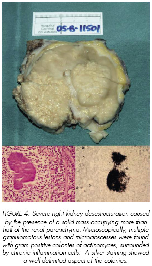
A 27 years old woman previously in good health, presented a 5-month history of general fatigue, a weight loss of 8-10 kg and right flank pain. On physical examination a palpable abdominal mass was detected. Laboratory tests showed severe anaemia (hemoglobine 6.1 g/dl). Abdominal ultrasonography and tomography yielded a right renal mass with retroperitoneal bleeding (<a href="#f1">Figure 1</a>) and multiple bilateral pulmonar nodules (<a href="#f2">Figures 2</a>). Neovascularization and chronic right renal vein thrombosis without evidence of active bleeding was demonstrated in selective angiography (<a href="#f3">Figure 3</a>). Blood, urine and sputum cultures were negative as well as tumor markers. Right nephrectomy was performed. The right kidney was firmly adherent to adjacent tissues and dissection was very difficult. Pathological examination showed fibrous tissue with mixed acute and chronic inflammatory cells. There were also multiple microabscesses. On further microscopic evaluation a branching, filamentous, gram-positive bacteria with a “sulfur granule” appearance consistent with actinomycosis was found (<a href="#f4">Figure 4</a>). In this patient the source and spread of actinomycosis can only be hypothesized. Since no intraabdominal etiology was identified, the origin was probably oral with thoracic and finally abdominal spread.</font></p>     <p align="center">&nbsp;</p>     <p align="center"><font face="Verdana" size="2"><a name="f1"><img border="0" src="/img/revistas/urol/v59n7/international1figura1.jpg" width="298" height="313"></a></font></p>     ]]></body>
<body><![CDATA[<p align="center"><font face="Verdana" size="2"><a name="f2"><img border="0" src="/img/revistas/urol/v59n7/international1figura2.jpg" width="298" height="313"></a></font></p>     <p align="center"><font face="Verdana" size="2"><a name="f3"><img border="0" src="/img/revistas/urol/v59n7/international1figura3.jpg" width="298" height="300"></a></font></p>     <p align="center"><font face="Verdana" size="2"><a name="f4"><img border="0" src="/img/revistas/urol/v59n7/international1figura4.jpg" width="299" height="521"></a></font></p>     <p>&nbsp;</p>     <p><font face="Verdana"><b>Discussion</b></font></p>     <p><font face="Verdana" size="2">Actinomycosis is a chronic granulomatous infection with a worldwide distribution (1). Actinomyces israelii, a gram-positive anaerobic microorganism, is the most common pathogenic agent involved. In the human, actinomyces is a harmless saprophyte in the mouth, tonsils and gastrointestinal tract, causing disease under special circumstances, during trauma, viscous perforation or surgery (2,3). Predisposing factors include diabetes mellitus, immunosupression, long term use of contraceptive intrauterine devices (pelvic disease), previous surgery (post-appendectomy abdominal actinomycosis), dental extraction, trauma and poor dental hygiene (cervico-facial disease) (4).</font></p>     <p><font face="Verdana" size="2"> Genitourinary actinomycosis is a very rare entity. Clinical findings are usually non specific and may include abdominal pain, urinary frequency or repetitive cystitis. Generally, there is a long term interval between the onset of symptoms and diagnosis. The diagnosis is primarily histological identification. Cultures are often unsuccessful in determining causative factors, being positive in less than 50 % of cases (1,5). In the genitourinary tract, actinomyces can cause primary renal infection (1,2,5,6), including a horseshoe kidney (7), reno-duodenal fistula (8) and reno-colic fistula (9). Other manifestations of actinomyces have been reported: retroperitoneal mass, ureteral stenosis, scrotal mass, prostatitis, penile pilonidal sinus and cystitis (10).</font></p>     <p><font face="Verdana" size="2"> The route of infection in renal actinomycosis is still controversial. In addition to direct spread from contiguous structures such as lungs or colon, (1,5,6) and aspiration of infected material, hematogeneous dissemination also occurs. By the time the kidney is involved the original lesions might have dissapeared completely. Four type of renal lesions have been&nbsp;described: chronic suppurative lesion (carbuncle), renal mass, pyelonephritis and pyonephritis (5,6).</font></p>     <p><font face="Verdana" size="2"> Renal actinomycosis is not easily diagnosed on the basis of clinical and radiological findings, because of its close resemblance to a variety of inflammatory processes and neplastic conditions (3,5). Clinical features are not specific and depends on the duration and site of involvement. Laboratory data often suggest infection but fever is not a frequent symptom (1).</font></p>     <p><font face="Verdana" size="2"> Computed Tomography (CT) is the method of choice for evaluation of characteristics and extent of the process. Its findings vary according to the stage of the disease and the region involved (1). There are two types of pattern in CT:</font></p>     ]]></body>
<body><![CDATA[<blockquote>     <p><font face="Verdana" size="2"> <b>1</b>. Predominantly solid masses with focal areas of low attenuation. Contrast enhancement is variable, although it is generally lower than that of muscles and solid viscera. If loculations are visible, contrast enhancement of the walls and septa may occur (3).</font></p>     <p><font face="Verdana" size="2"> <b>2</b>. Predominantly cystic masses with thick enhancing walls (1).</font></p> </blockquote>     <p><font face="Verdana" size="2"> Ultrasound image of the renal mass is non-specific, but different from the typically focal appearance of renal carcinoma.</font></p>     <p><font face="Verdana" size="2"> Magnetic Resonance Images (MRI) may be a useful tool for the diagnosis of renal actinomycosis. A low iso-intensity mass on T1 weighted MRI and a low intensity mass on T2 weighted MRI. These findings on MRI can help to distinguish actinomycosis from other entities such as malignant lymphoma. Differential diagnosis include inflammatory processes (tuberculosis), abscess and neoplasms (renal cell carcinoma, lymphoma, angiomyolipoma and oncocytoma). Although most cases of actinomycosis occur between 15 and 35 years of age, there are cases described in children under 10 years of age. In these situations the differential diagnoses has to be made with Wilms´ tumor, clear cell sarcoma, nephoblastomatosis, rhabdoid tumors and congenital mesoblastic nephroma (3,10).</font></p>     <p><font face="Verdana" size="2"> Pathological examination of the specimen shows fibroadipose tissue and a mix of acute and chronic inflammatory reaction with areas of fibrosis and granulation tissue. The presence of focal granulomatous lesions with giant cells can be noted as well as multiple irregularly shaped microabscessses with “sulfur granules”, oval basaloid bodies composed on of filamentous organisms that stained positively with Gram´s, periodic acid-Schiff and Gomori´s methenamine silver stains (1,3).</font></p>     <p><font face="Verdana" size="2"> The prognosis of this entity is well related with the progression at the time of diagnosis. Traditionally the treatment of renal actinomycosis has been surgical extirpation of the kidney followed by prolonged antibiotic therapy. However, in early stages, solitary renal actinomycosis, may be successfully treated using prolonged antibiotic therapy (8 weeks-1 year) with minimal or none sequelae (3,6).</font></p>     <p>&nbsp;</p>     <p><b><font face="Verdana">References and recomended reading (*of special interest, **of outstanding interest)</font></b></p>     <!-- ref --><p><font face="Verdana" size="2">*1. HORVÁTH, K.; PORKOLÁB, Z.; PALKÓ, A.: “Primary renal and retroperitonral Actinomycosis”. Eur. Radiol., 10: 287, 2000.</font>&nbsp;&nbsp;&nbsp;&nbsp;&nbsp;&nbsp;&nbsp;&nbsp;[&#160;<a href="javascript:void(0);" onclick="javascript: window.open('/scielo.php?script=sci_nlinks&ref=1081945&pid=S0004-0614200600070001800001&lng=','','width=640,height=500,resizable=yes,scrollbars=1,menubar=yes,');">Links</a>&#160;]<!-- end-ref --><!-- ref --><p><font face="Verdana" size="2">**2. DHANANI, N.N.; JONES, D.M.; GROSSMAN, H.B.: “Medical management of renal Actinomycosis”. J. Urol., 171: 2373, 2004.</font>&nbsp;&nbsp;&nbsp;&nbsp;&nbsp;&nbsp;&nbsp;&nbsp;[&#160;<a href="javascript:void(0);" onclick="javascript: window.open('/scielo.php?script=sci_nlinks&ref=1081946&pid=S0004-0614200600070001800002&lng=','','width=640,height=500,resizable=yes,scrollbars=1,menubar=yes,');">Links</a>&#160;]<!-- end-ref --><!-- ref --><p><font face="Verdana" size="2">*3. ALLEN, H.A.; SCATARIGE, J.C.; KIM, M.H.: “Actinomycosis: CT findings in six patients”. AJR, 149: 1255, 1987.</font>&nbsp;&nbsp;&nbsp;&nbsp;&nbsp;&nbsp;&nbsp;&nbsp;[&#160;<a href="javascript:void(0);" onclick="javascript: window.open('/scielo.php?script=sci_nlinks&ref=1081947&pid=S0004-0614200600070001800003&lng=','','width=640,height=500,resizable=yes,scrollbars=1,menubar=yes,');">Links</a>&#160;]<!-- end-ref --><!-- ref --><p><font face="Verdana" size="2">4. LEACH, T.D.; SADEK, S.A.; MASON, J.C.: “An unusual abdominal mass in a renal transplant recipient”. Transpl. Infect. Dis., 4: 218, 2002.</font>&nbsp;&nbsp;&nbsp;&nbsp;&nbsp;&nbsp;&nbsp;&nbsp;[&#160;<a href="javascript:void(0);" onclick="javascript: window.open('/scielo.php?script=sci_nlinks&ref=1081948&pid=S0004-0614200600070001800004&lng=','','width=640,height=500,resizable=yes,scrollbars=1,menubar=yes,');">Links</a>&#160;]<!-- end-ref --><!-- ref --><p><font face="Verdana" size="2">5. PATEL, B.J.; MOSKOWITZ, H.; HASHMAT, A.: “Unilateral renal Actinomycosis”. Urol., 21: 172, 1983.</font>&nbsp;&nbsp;&nbsp;&nbsp;&nbsp;&nbsp;&nbsp;&nbsp;[&#160;<a href="javascript:void(0);" onclick="javascript: window.open('/scielo.php?script=sci_nlinks&ref=1081949&pid=S0004-0614200600070001800005&lng=','','width=640,height=500,resizable=yes,scrollbars=1,menubar=yes,');">Links</a>&#160;]<!-- end-ref --><!-- ref --><p><font face="Verdana" size="2">**6. KHALAFF, H.; SRIGLEY, J.R.; KLOTZ, L.H.: “Recognition of renal Actinomycosis: Nephrectomy can be avoided. Report of a case”. Can. J. Surg., 38: 77, 1995.</font>&nbsp;&nbsp;&nbsp;&nbsp;&nbsp;&nbsp;&nbsp;&nbsp;[&#160;<a href="javascript:void(0);" onclick="javascript: window.open('/scielo.php?script=sci_nlinks&ref=1081950&pid=S0004-0614200600070001800006&lng=','','width=640,height=500,resizable=yes,scrollbars=1,menubar=yes,');">Links</a>&#160;]<!-- end-ref --><!-- ref --><p><font face="Verdana" size="2">*7. McGIBNEY, D.; CLARKE, P.B.: “Primary renal Actinomycosis in the presence of horseshoe kidney”. Br. J. Urol., 58: 566, 1986.</font>&nbsp;&nbsp;&nbsp;&nbsp;&nbsp;&nbsp;&nbsp;&nbsp;[&#160;<a href="javascript:void(0);" onclick="javascript: window.open('/scielo.php?script=sci_nlinks&ref=1081951&pid=S0004-0614200600070001800007&lng=','','width=640,height=500,resizable=yes,scrollbars=1,menubar=yes,');">Links</a>&#160;]<!-- end-ref --><!-- ref --><p><font face="Verdana" size="2">*8. CVETKOV, M.C.; ELENKOV, C.; GEORGIEV, M. y cols.: “Renal Actinomycosis complicated by renoduodenal fistula and diabetes mellitus”. Br. J. Urol., 75: 104, 1995.</font>&nbsp;&nbsp;&nbsp;&nbsp;&nbsp;&nbsp;&nbsp;&nbsp;[&#160;<a href="javascript:void(0);" onclick="javascript: window.open('/scielo.php?script=sci_nlinks&ref=1081952&pid=S0004-0614200600070001800008&lng=','','width=640,height=500,resizable=yes,scrollbars=1,menubar=yes,');">Links</a>&#160;]<!-- end-ref --><!-- ref --><p><font face="Verdana" size="2">9. YU, H.H.; YIM, C.M.; LEONG, C.H.: “Primary Actinomycosis of kidney presenting with reno-colic fistula”. Br. J. Urol., 50: 140, 1978.</font>&nbsp;&nbsp;&nbsp;&nbsp;&nbsp;&nbsp;&nbsp;&nbsp;[&#160;<a href="javascript:void(0);" onclick="javascript: window.open('/scielo.php?script=sci_nlinks&ref=1081953&pid=S0004-0614200600070001800009&lng=','','width=640,height=500,resizable=yes,scrollbars=1,menubar=yes,');">Links</a>&#160;]<!-- end-ref --><!-- ref --><p><font face="Verdana" size="2">10. MAKAR, A.P.; MICHIELSEN, J.P.; BOECKX, G.J.; VAN MARCK, E.A.: “Primary Actinomycosis of the urinary bladder”. Br. J. Urol., 70: 205, 1992.</font>&nbsp;&nbsp;&nbsp;&nbsp;&nbsp;&nbsp;&nbsp;&nbsp;[&#160;<a href="javascript:void(0);" onclick="javascript: window.open('/scielo.php?script=sci_nlinks&ref=1081954&pid=S0004-0614200600070001800010&lng=','','width=640,height=500,resizable=yes,scrollbars=1,menubar=yes,');">Links</a>&#160;]<!-- end-ref --><p>&nbsp;</p>     ]]></body>
<body><![CDATA[<p>&nbsp;</p>     <p><font face="Verdana" size="2"><b><a href="#top"><img border="0" src="/img/revistas/urol/v59n7/seta.gif" width="15" height="17"></a><a name="bajo"></a> Correspondence:    <br> </b>Antonio Jalón Monzón    <br> Servicio de Urología 1    <br> Hospital Universitario Central de Asturias    <br> C/ Celestino Villamil s/n    <br> 33006-Oviedo. Asturias. (Spain)    <br> <a href="mailto:ajalonm@hotmail.com"> ajalonm@hotmail.com</a></font></p>     <p><font face="Verdana" size="2"> Accepted for publication: 1st January, 2006</font></p>      ]]></body><back>
<ref-list>
<ref id="B1">
<label>1</label><nlm-citation citation-type="journal">
<person-group person-group-type="author">
<name>
<surname><![CDATA[HORVÁTH]]></surname>
<given-names><![CDATA[K]]></given-names>
</name>
<name>
<surname><![CDATA[PORKOLÁB]]></surname>
<given-names><![CDATA[Z]]></given-names>
</name>
<name>
<surname><![CDATA[PALKÓ]]></surname>
<given-names><![CDATA[A]]></given-names>
</name>
</person-group>
<article-title xml:lang="en"><![CDATA[Primary renal and retroperitonral Actinomycosis]]></article-title>
<source><![CDATA[Eur. Radiol]]></source>
<year>2000</year>
<volume>10</volume>
<page-range>287</page-range></nlm-citation>
</ref>
<ref id="B2">
<label>2</label><nlm-citation citation-type="journal">
<person-group person-group-type="author">
<name>
<surname><![CDATA[DHANANI]]></surname>
<given-names><![CDATA[N.N]]></given-names>
</name>
<name>
<surname><![CDATA[JONES]]></surname>
<given-names><![CDATA[D.M]]></given-names>
</name>
<name>
<surname><![CDATA[GROSSMAN]]></surname>
<given-names><![CDATA[H.B]]></given-names>
</name>
</person-group>
<article-title xml:lang="en"><![CDATA[Medical management of renal Actinomycosis]]></article-title>
<source><![CDATA[J. Urol]]></source>
<year>2004</year>
<volume>171</volume>
<page-range>2373</page-range></nlm-citation>
</ref>
<ref id="B3">
<label>3</label><nlm-citation citation-type="journal">
<person-group person-group-type="author">
<name>
<surname><![CDATA[ALLEN]]></surname>
<given-names><![CDATA[H.A]]></given-names>
</name>
<name>
<surname><![CDATA[SCATARIGE]]></surname>
<given-names><![CDATA[J.C]]></given-names>
</name>
<name>
<surname><![CDATA[KIM]]></surname>
<given-names><![CDATA[M.H]]></given-names>
</name>
</person-group>
<article-title xml:lang="en"><![CDATA[Actinomycosis: CT findings in six patients]]></article-title>
<source><![CDATA[AJR]]></source>
<year>1987</year>
<volume>149</volume>
<page-range>1255</page-range></nlm-citation>
</ref>
<ref id="B4">
<label>4</label><nlm-citation citation-type="journal">
<person-group person-group-type="author">
<name>
<surname><![CDATA[LEACH]]></surname>
<given-names><![CDATA[T.D]]></given-names>
</name>
<name>
<surname><![CDATA[SADEK]]></surname>
<given-names><![CDATA[S.A]]></given-names>
</name>
<name>
<surname><![CDATA[MASON]]></surname>
<given-names><![CDATA[J.C]]></given-names>
</name>
</person-group>
<article-title xml:lang="en"><![CDATA[An unusual abdominal mass in a renal transplant recipient]]></article-title>
<source><![CDATA[Transpl. Infect. Dis]]></source>
<year>2002</year>
<volume>4</volume>
<page-range>218</page-range></nlm-citation>
</ref>
<ref id="B5">
<label>5</label><nlm-citation citation-type="journal">
<person-group person-group-type="author">
<name>
<surname><![CDATA[PATEL]]></surname>
<given-names><![CDATA[B.J]]></given-names>
</name>
<name>
<surname><![CDATA[MOSKOWITZ]]></surname>
<given-names><![CDATA[H]]></given-names>
</name>
<name>
<surname><![CDATA[HASHMAT]]></surname>
<given-names><![CDATA[A]]></given-names>
</name>
</person-group>
<article-title xml:lang="en"><![CDATA[Unilateral renal Actinomycosis]]></article-title>
<source><![CDATA[Urol]]></source>
<year>1983</year>
<volume>21</volume>
<page-range>172</page-range></nlm-citation>
</ref>
<ref id="B6">
<label>6</label><nlm-citation citation-type="journal">
<person-group person-group-type="author">
<name>
<surname><![CDATA[KHALAFF]]></surname>
<given-names><![CDATA[H]]></given-names>
</name>
<name>
<surname><![CDATA[SRIGLEY]]></surname>
<given-names><![CDATA[J.R]]></given-names>
</name>
<name>
<surname><![CDATA[KLOTZ]]></surname>
<given-names><![CDATA[L.H]]></given-names>
</name>
</person-group>
<article-title xml:lang="en"><![CDATA[Recognition of renal Actinomycosis: Nephrectomy can be avoided. Report of a case]]></article-title>
<source><![CDATA[Can. J. Surg]]></source>
<year>1995</year>
<volume>38</volume>
<page-range>77</page-range></nlm-citation>
</ref>
<ref id="B7">
<label>7</label><nlm-citation citation-type="journal">
<person-group person-group-type="author">
<name>
<surname><![CDATA[McGIBNEY]]></surname>
<given-names><![CDATA[D]]></given-names>
</name>
<name>
<surname><![CDATA[CLARKE]]></surname>
<given-names><![CDATA[P.B]]></given-names>
</name>
</person-group>
<article-title xml:lang="en"><![CDATA[Primary renal Actinomycosis in the presence of horseshoe kidney]]></article-title>
<source><![CDATA[Br. J. Urol]]></source>
<year>1986</year>
<volume>58</volume>
<page-range>566</page-range></nlm-citation>
</ref>
<ref id="B8">
<label>8</label><nlm-citation citation-type="journal">
<person-group person-group-type="author">
<name>
<surname><![CDATA[CVETKOV]]></surname>
<given-names><![CDATA[M.C]]></given-names>
</name>
<name>
<surname><![CDATA[ELENKOV]]></surname>
<given-names><![CDATA[C]]></given-names>
</name>
<name>
<surname><![CDATA[GEORGIEV]]></surname>
<given-names><![CDATA[M]]></given-names>
</name>
</person-group>
<article-title xml:lang="en"><![CDATA[Renal Actinomycosis complicated by renoduodenal fistula and diabetes mellitus]]></article-title>
<source><![CDATA[Br. J. Urol]]></source>
<year>1995</year>
<volume>75</volume>
<page-range>104</page-range></nlm-citation>
</ref>
<ref id="B9">
<label>9</label><nlm-citation citation-type="journal">
<person-group person-group-type="author">
<name>
<surname><![CDATA[YU]]></surname>
<given-names><![CDATA[H.H]]></given-names>
</name>
<name>
<surname><![CDATA[YIM]]></surname>
<given-names><![CDATA[C.M]]></given-names>
</name>
<name>
<surname><![CDATA[LEONG]]></surname>
<given-names><![CDATA[C.H]]></given-names>
</name>
</person-group>
<article-title xml:lang="en"><![CDATA[Primary Actinomycosis of kidney presenting with reno-colic fistula]]></article-title>
<source><![CDATA[Br. J. Urol]]></source>
<year>1978</year>
<volume>50</volume>
<page-range>140</page-range></nlm-citation>
</ref>
<ref id="B10">
<label>10</label><nlm-citation citation-type="journal">
<person-group person-group-type="author">
<name>
<surname><![CDATA[MAKAR]]></surname>
<given-names><![CDATA[A.P]]></given-names>
</name>
<name>
<surname><![CDATA[MICHIELSEN]]></surname>
<given-names><![CDATA[J.P]]></given-names>
</name>
<name>
<surname><![CDATA[BOECKX]]></surname>
<given-names><![CDATA[G.J]]></given-names>
</name>
<name>
<surname><![CDATA[VAN MARCK]]></surname>
<given-names><![CDATA[E.A]]></given-names>
</name>
</person-group>
<article-title xml:lang="en"><![CDATA[Primary Actinomycosis of the urinary bladder]]></article-title>
<source><![CDATA[Br. J. Urol]]></source>
<year>1992</year>
<volume>70</volume>
<page-range>205</page-range></nlm-citation>
</ref>
</ref-list>
</back>
</article>
