<?xml version="1.0" encoding="ISO-8859-1"?><article xmlns:mml="http://www.w3.org/1998/Math/MathML" xmlns:xlink="http://www.w3.org/1999/xlink" xmlns:xsi="http://www.w3.org/2001/XMLSchema-instance">
<front>
<journal-meta>
<journal-id>0004-0614</journal-id>
<journal-title><![CDATA[Archivos Españoles de Urología (Ed. impresa)]]></journal-title>
<abbrev-journal-title><![CDATA[Arch. Esp. Urol.]]></abbrev-journal-title>
<issn>0004-0614</issn>
<publisher>
<publisher-name><![CDATA[INIESTARES, S.A.]]></publisher-name>
</publisher>
</journal-meta>
<article-meta>
<article-id>S0004-06142007000300020</article-id>
<title-group>
<article-title xml:lang="en"><![CDATA[Primary Ewing´s sarcoma/Primitive neuroectodermal tumor of the kidney: An infrequent finding]]></article-title>
<article-title xml:lang="es"><![CDATA[Sarcoma de Ewing primario/Tumor neuroectodérmico primitivo renal: Un hallazgo infrecuente]]></article-title>
</title-group>
<contrib-group>
<contrib contrib-type="author">
<name>
<surname><![CDATA[Parada]]></surname>
<given-names><![CDATA[David]]></given-names>
</name>
<xref ref-type="aff" rid="A01"/>
<xref ref-type="aff" rid="A03"/>
</contrib>
<contrib contrib-type="author">
<name>
<surname><![CDATA[Godoy]]></surname>
<given-names><![CDATA[Alí]]></given-names>
</name>
<xref ref-type="aff" rid="A02"/>
</contrib>
<contrib contrib-type="author">
<name>
<surname><![CDATA[Liuzzi]]></surname>
<given-names><![CDATA[Francisco]]></given-names>
</name>
<xref ref-type="aff" rid="A02"/>
</contrib>
<contrib contrib-type="author">
<name>
<surname><![CDATA[Peña]]></surname>
<given-names><![CDATA[Karla B.]]></given-names>
</name>
<xref ref-type="aff" rid="A01"/>
</contrib>
<contrib contrib-type="author">
<name>
<surname><![CDATA[Romero]]></surname>
<given-names><![CDATA[Aramis]]></given-names>
</name>
<xref ref-type="aff" rid="A01"/>
</contrib>
<contrib contrib-type="author">
<name>
<surname><![CDATA[Parada]]></surname>
<given-names><![CDATA[Arelí M.]]></given-names>
</name>
<xref ref-type="aff" rid="A03"/>
</contrib>
</contrib-group>
<aff id="A01">
<institution><![CDATA[,Vargas Hospital Department of Anatomic Pathology ]]></institution>
<addr-line><![CDATA[Caracas ]]></addr-line>
<country>Venezuela</country>
</aff>
<aff id="A02">
<institution><![CDATA[,Vargas Hospital Service of Surgery 1 ]]></institution>
<addr-line><![CDATA[Caracas ]]></addr-line>
<country>Venezuela</country>
</aff>
<aff id="A03">
<institution><![CDATA[,Central University of Venezuela School of Medicine, José María Vargas ]]></institution>
<addr-line><![CDATA[Caracas ]]></addr-line>
<country>Venezuela</country>
</aff>
<pub-date pub-type="pub">
<day>00</day>
<month>04</month>
<year>2007</year>
</pub-date>
<pub-date pub-type="epub">
<day>00</day>
<month>04</month>
<year>2007</year>
</pub-date>
<volume>60</volume>
<numero>3</numero>
<fpage>321</fpage>
<lpage>325</lpage>
<copyright-statement/>
<copyright-year/>
<self-uri xlink:href="http://scielo.isciii.es/scielo.php?script=sci_arttext&amp;pid=S0004-06142007000300020&amp;lng=en&amp;nrm=iso"></self-uri><self-uri xlink:href="http://scielo.isciii.es/scielo.php?script=sci_abstract&amp;pid=S0004-06142007000300020&amp;lng=en&amp;nrm=iso"></self-uri><self-uri xlink:href="http://scielo.isciii.es/scielo.php?script=sci_pdf&amp;pid=S0004-06142007000300020&amp;lng=en&amp;nrm=iso"></self-uri><abstract abstract-type="short" xml:lang="en"><p><![CDATA[Objetive: Ewing´s sarcoma/Primitive neuroectodermal tumor (ES/PNET) is an extraordinarily rare primary tumor in the kidney. We re-port herein the clinical, histological, and immunohisto-chemical features of a primary renal ES/PNET. Methods: A 19-year old male referred a two weeks history of constant, colic, left flank pain, and fever. A left radical nephrectomy was performed. Gross pathologic examination showed pink-tan, lobulated solid tumor, lo-calized at the superior pole. Results: Histologically, the tumor was solid with necrosis. The neoplastic cells showed a small amount of clear cytoplasm, and had vesicular nuclei with small nucleoli. Immunohistochemical studies showed strongly and diffusely positive staining for CD99 in a membranous pattern. Conclusions: This case represents a typical ES/PNET, affecting a young male patient. Adequate diagnosis is important because this neoplasm has an aggressive behaviour.]]></p></abstract>
<abstract abstract-type="short" xml:lang="es"><p><![CDATA[El sarcoma de Ewing/Tumor Neuroectodermico Primitivo (SE/TNEP) del riñón es una neoplasia extremadamente rara en el riñón. Presentamos los hallazgos clínicos, histológicos e inmunohistoquímicos de un SE/TNEP primario renal. Métodos: Un paciente varón de 19 años refirió historia de dos semanas de dolor tipo cólico, constante, en el flanco izquierdo y fiebre. Se hizo nefrectomía radical izquierda. El examen macroscópico mostró un tumor sólido, lobulado, pardo-rosado, localizado en el polo superior. Resultados: Histológicamente el tumor era sólido con necrosis. Las células neoplásicas mostraron citoplasma escaso claro y poseían un núcleo vesicular con nucléolo pequeño. Los estudios inmunohistoquímicos mostraron una fuerte y difusa positividad para el CD99 en un patrón membranoso. Conclusiones: Este caso representa un típico SE/TNEP, afectando a un varón joven. Es importante un diagnóstico adecuado debido a que esta neoplasia tiene una conducta agresiva.]]></p></abstract>
<kwd-group>
<kwd lng="en"><![CDATA[Kidney]]></kwd>
<kwd lng="en"><![CDATA[Ewing´s Sarcoma]]></kwd>
<kwd lng="en"><![CDATA[Primitive neuroectodermal tumor]]></kwd>
<kwd lng="en"><![CDATA[CD99]]></kwd>
<kwd lng="en"><![CDATA[Immunohistochemestry]]></kwd>
<kwd lng="es"><![CDATA[Riñón]]></kwd>
<kwd lng="es"><![CDATA[Sarcoma de Ewing]]></kwd>
<kwd lng="es"><![CDATA[Tumor neuroectodérmico primitivo]]></kwd>
<kwd lng="es"><![CDATA[CD99]]></kwd>
<kwd lng="es"><![CDATA[Inmunohistoquímica]]></kwd>
</kwd-group>
</article-meta>
</front><body><![CDATA[ <p align="right"><font face="Verdana" size="2"><B>INTERNATIONAL SECTION</B></font></p>     <p>&nbsp;</p>     <p><font face="Verdana" size="4"><B><a name="top"></a>Primary Ewing´s sarcoma/Primitive  neuroectodermal tumor of the kidney. An infrequent finding</B></font></p>     <p><b><font face="Verdana" size="4">Sarcoma de Ewing primario/Tumor  neuroectodérmico primitivo renal. Un hallazgo infrecuente</font></b></p>     <p>&nbsp;</p>     <p>&nbsp;</p>     <p><b><font face="Verdana" size="2">David Parada<sup>1,3</sup>, Alí Godoy<sup>2</sup>, Francisco Liuzzi<sup>2</sup>, Karla B. Peña<sup>1</sup>, Aramis Romero<sup>1</sup> and Arelí M. Parada<sup>3</sup></font></b></p>     <p><font face="Verdana" size="2"><sup>1</sup>Department of Anatomic Pathology, Vargas Hospital. Caracas. Venezuela.    <br> <sup>2</sup>Service of Surgery 1. Vargas Hospital. Caracas. Venezuela.    <br> <sup>3</sup>School of Medicine, “José María Vargas”. Central University of Venezuela. Caracas. Venezuela.</font></p>     ]]></body>
<body><![CDATA[<p><font face="Verdana" size="2"><a href="#back">Correspondence</a></font></p>     <p>&nbsp;</p>     <p>&nbsp;</p>  <hr size="1">      <p><font face="Verdana" size="2"><B>SUMMARY</B></font></p>     <p><font face="Verdana" size="2"><b>Objetive:</b> Ewing´s sarcoma/Primitive neuroectodermal tumor (ES/PNET) is an extraordinarily rare primary tumor in the kidney. We re-port herein the clinical, histological, and immunohisto-chemical features of a primary renal ES/PNET.    <br> <b>Methods:</b> A 19-year old male referred a two weeks history of constant, colic, left flank pain, and fever. A left radical nephrectomy was performed. Gross pathologic examination showed pink-tan, lobulated solid tumor, lo-calized at the superior pole.    <br> <b>Results:</b> Histologically, the tumor was solid with necrosis. The neoplastic cells showed a small amount of clear cytoplasm, and had vesicular nuclei with small nucleoli. Immunohistochemical studies showed strongly and diffusely positive staining for CD99 in a membranous pattern.    <br> <b>Conclusions:</b> This case represents a typical ES/PNET, affecting a young male patient. Adequate diagnosis is important because this neoplasm has an aggressive behaviour.</font></p>     <p><font face="Verdana" size="2"><B>Keywords:</B> Kidney. Ewing´s Sarcoma. Primitive neuroectodermal tumor. CD99. Immunohistochemestry.</font></p>  <hr size="1">      <p><b><font face="Verdana" size="2">RESUMEN</font></b></p>     ]]></body>
<body><![CDATA[<p><font face="Verdana" size="2">El sarcoma de Ewing/Tumor Neuroectodermico Primitivo (SE/TNEP) del riñón es una neoplasia extremadamente rara en el riñón. Presentamos los hallazgos clínicos, histológicos e inmunohistoquímicos de un SE/TNEP primario renal.    <br> <b>Métodos:</b> Un paciente varón de 19 años refirió historia de dos semanas de dolor tipo cólico, constante, en el flanco izquierdo y fiebre. Se hizo nefrectomía radical izquierda. El examen macroscópico mostró un tumor sólido, lobulado, pardo-rosado, localizado en el polo superior.    <br> <b>Resultados:</b> Histológicamente el tumor era sólido con necrosis. Las células neoplásicas mostraron citoplasma escaso claro y poseían un núcleo vesicular con nucléolo pequeño. Los estudios inmunohistoquímicos mostraron una fuerte y difusa positividad para el CD99 en un patrón membranoso.    <br> <b>Conclusiones:</b> Este caso representa un típico SE/TNEP, afectando a un varón joven. Es importante un diagnóstico adecuado debido a que esta neoplasia tiene una conducta agresiva.</font></p>     <p><font face="Verdana" size="2"><B>Palabras clave:</B> Riñón. Sarcoma de Ewing. Tumor neuroectodérmico primitivo. CD99. Inmunohistoquímica.</font></p>  <hr size="1">      <p>&nbsp;</p>     <p><font face="Verdana"><B>Introduction</B></font></p>     <p><font face="Verdana" size="2">The round cell tumors of the kidney include a wide range of unrelated neoplasms with overlapping morphologic features and different prognostic/therapeutic implications. In this group of tumors, Ewing´s sarcoma/primitive neuroectodermal tumor (ES/PNET) is an extremely rare primary tumor in the kidney (1). They ussually affect young adults, with slight male predominance, are large at presentation and tend to be highly aggressive tumors (1,2).</font></p>     <p><font face="Verdana" size="2">Immunohistochemical studies has proven to be valuable in the differencial diagnosis of renal round cell tumors, although some of these entities lack a characteristic immunophenotype (1). Herein we report the clinical, histological e immunohistochemical findings in a new case of primary Ewing´s sarcoma/Primitive neuroectodermal tumor of the kidney.</font></p>     <p>&nbsp;</p>     ]]></body>
<body><![CDATA[<p><font face="Verdana"><B>Material and methods</B></font></p>     <p><font face="Verdana" size="2"><I><B>Case Report</B></I></font></p>     <p><font face="Verdana" size="2">
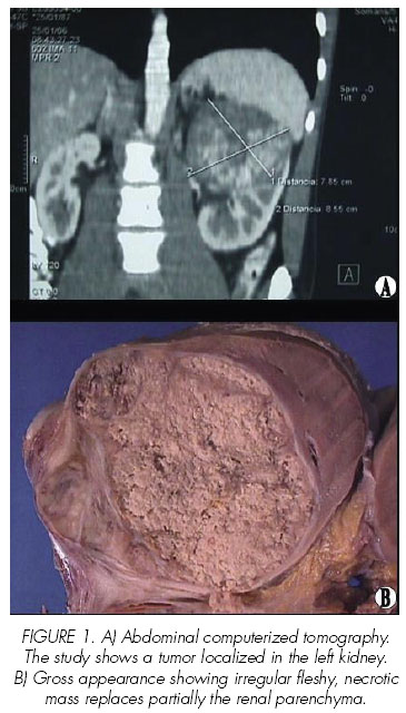
A 19-year-old male referred a two weeks history of constant, colic, left flank pain, and fever. His medical history was unremarkable. Abdominal physical examination demonstrated epigastric and left upper quadrant abdominal pain, at the superficial and profound palpation. The urinalysis was unremarkable. An abdominal ultrasonography and an abdominal computerized tomography were performed (<a href="#f1">Figure 1A</a>), followed by a left radical nephrectomy. The patient is alive after surgical intervention and he is receiving chemotherapy with vincristine, doxorubicin, cyclophosphamide, isofosphamide and ectoposide.</font></p>     <p align="center"><a name="f1"><img border="0" src="/img/revistas/urol/v60n3/19_f1.jpg" width="365" height="640"></a></p>      <p><font face="Verdana" size="2">The resected left kidney was fixed in buffer formalin pH 7.2, and tissue preparations were made by the routine procedure to hematoxylin-eosin stain. Paraffin-embedded preparation was stained immunohistochemically using the enzyme-conjugated polymer system (EnVision System, Dako,Glostrup, Denmark). For specific immunohistochemical details see <a target="_blank" href="/img/revistas/urol/v60n3/19_t1.jpg">Table I</a>.</font></p>     <p>&nbsp;</p>     <p><font face="Verdana"><B>Results</B></font></p>     <p><font face="Verdana" size="2"><I><B>Pathologics findings</B></I></font></p>     <p><font face="Verdana" size="2">Gross pathologic examination showed pink-tan, lobulated solid tumor (maximun diameter: 7.5 cm), localized at the superior pole, wich replaced the normal renal parenchima and had necrotic areas (<a href="#f1">Figure 1B</a>). No renal vein invasion was grossly identified.</font></p>     <p><font face="Verdana" size="2">Microscopically, the tumor was solid with necrosis and consisted of vaguely lobulated proliferation of round cells with high nuclear to cytoplasmic ratio (<a target="_blank" href="/img/revistas/urol/v60n3/19_f2.jpg">Figure 2, A-D</a>). The surrounding renal parenchyma showed infiltration by the malignant neoplasm, forming broad sheets. The tumor cells were present in groups separated by fibrovascular septae (<a target="_blank" href="/img/revistas/urol/v60n3/19_f2.jpg">Figure 2A</a>). The neoplastic cells showed a small amount of clear cytoplasm, and had vesicular nuclei with small nucleoli (Figure 2, B,C). Mitotic figures were commonly found (<a target="_blank" href="/img/revistas/urol/v60n3/19_f2.jpg">Figure 2, C,D</a>). Focal Homer-Wright type rosettes were seen (<a target="_blank" href="/img/revistas/urol/v60n3/19_f2.jpg">Figure 2, C</a>). No tubule formation, glomeruloid structures, pseudorosettes, cartilagenous, myogenous or spindle component were identified.</font></p>     ]]></body>
<body><![CDATA[<p><font face="Verdana" size="2">Immunohistochemically, the tumor cells failed to stain for cytokeratin AE1/AE3, cytokeratin 7, cytokeratin 20, epithelial membrane antigen, S-100 protein, alpha smooth muscle actin, Anti-Myo D1, Desmin, CD34 and CD31 (<a target="_blank" href="/img/revistas/urol/v60n3/19_f3.jpg">Figure 3, A</a>). The neoplastic cells were positive to vimentin and neuron specific enolase (<a target="_blank" href="/img/revistas/urol/v60n3/19_f3.jpg">Figure 3 B,C</a>), with strongly and diffusely positive in a membranous pattern for CD99 (<a target="_blank" href="/img/revistas/urol/v60n3/19_f3.jpg">Figure 3, D</a>).</font></p>     <p>&nbsp;</p>     <p><font face="Verdana"><B>Discussion</B></font></p>     <p><font face="Verdana" size="2">In the year 1994, Mor and coworkers (3) described a characteristic primary renal neoplasia consistent with the diagnosis of malignant peripheral primitive neuroectodermal tumor. Actually, most of the reported cases have occurred in young adults, with a mean age at presentation beetwen 28 and 34 years (range: 4-69 years) and a slight male predominance (1,2,4). Common symptoms at the presentation were flank pain and/or haematuia (1). In our case, a young male patient with left flank pain were consistent with previous reported findings of primary renal ES/PNET.</font></p>     <p><font face="Verdana" size="2">One possible source of renal ES/PNET is from neural ramifications that invest the kidney. The inervation of the kidney comes from adrenergic fibers originating in the celiac plexus and accompanying efferent arterioles and descending vasa recta (5). Another possibility is that embryonic neural crest cells migrate into the kidney and subsequently undergo tumorogenesis. An interplay between developing metanephros and neural differentiation factors such as c-ret (6) and neurotrophin-3 (7) is apparent in rodent kidneys and suggests that neural differentiation is essential to nephrogenesis.</font></p>     <p><font face="Verdana" size="2">Histologically, renal ES/PNET could be a diagnostic challenge because many tumors exhibited features of the so-called small round cell tumor. Malignant lymphoma, rhabdomiosarcoma, renal neuroblastoma, Wilms´ tumor, small cell osteosarcoma, desmoplastic small cell tumor and so, are disease for distinction (1). Therefore, immunohistochemestry has proven to be value in the differential diagnosis of these kidney tumors. The bases for diagnosis of the present case as ES/PNET were: a-) morphology of small round cell tumor was found histologically and b-) immunohistochemically, MIC2 showed strong membrane positivity. Similar features in renal ES/PNET has been previously reported (1,2,4).</font></p>     <p><font face="Verdana" size="2">Althought histological and immunohistochemical studies are important to reach a definitive diagnosis the chromosomal and moleular analyses could be required. The ES family have chromosomal translocations, t(11;22)(q24;q12), t(21;22) and chimera genes, EWS-FL1, EWS-ERG, EWS-ETV1 or EWS-EIAF as common abnormalities (8,9). Over 85% of ES/PNET are characterized by the translocation t(11;22)(q24;q22) that results in the fusion of the ews gene on chromosome 22 to the fli-1 gene on chromosome 11 (10-13). The chimeric EWS/FLI-1 fusion protein localizes to the nucleus, is a more powerful transcription activator than is normal fli-1 (14).</font></p>     <p><font face="Verdana" size="2">Primary renal ES/PNET is an aggressive neoplasm with poor prognosis. Metastases are usually absent at the time of first observation, but metastases affect the prognosis little (4). In previous reported cases, the patients died 10.3 months on average after their first diagnosis of the tumor. Radiotherapy or chemotherapy is not effective. Jimenez et al., (1) informed that chemotherapeutic treatment with ifosphamide and/or cyclophosphamide, could be included in treatment protocols for primary kidney ES/PNET. Obviously, further studies will be necessary to determine the precise impact of these drugs on survival in renal ES/PNET.</font></p>     <p><font face="Verdana" size="2">In summary, we report an additional case of primary renal ES/PNET. Our case confirmed that ES/PNET is a rare neoplasm, affecting young adults with aggressive behavior. It is important to distinguish primary renal ES/PNET from other round cell tumors, given his aggressive behavior. The immunohistochemical study may be valuable in the differential diagnosis of renal round cell tumors.</font></p>     <p>&nbsp;</p>     ]]></body>
<body><![CDATA[<p><font face="Verdana"><B>References and recomended readings (*of special interest, **of outstanding interest)</B></font></p>     <!-- ref --><p><font face="Verdana" size="2">**1. JIMENEZ, R.; FOLPE, A.; LAPHAM, R. y cols.: “Primary Ewing´s Sarcoma/Primitive Neuroectodermal Tumor of the kidney. A clinicopathologic and immnunohistochemical analysis of 11 cases”. Am. J. Surg. Pathol., 26: 320, 2002.</font>&nbsp;&nbsp;&nbsp;&nbsp;&nbsp;&nbsp;&nbsp;&nbsp;[&#160;<a href="javascript:void(0);" onclick="javascript: window.open('/scielo.php?script=sci_nlinks&ref=1100321&pid=S0004-0614200700030002000001&lng=','','width=640,height=500,resizable=yes,scrollbars=1,menubar=yes,');">Links</a>&#160;]<!-- end-ref --><!-- ref --><p><font face="Verdana" size="2">**2. PARHAM, D.; ROLOSON, G.; FEELY, M. y cols.: “Primary malignant neuroepithelial tumors of the Kidney: a clinicopathologic analysis of 146 adult and pediatric cases from the National Wilms´ Tumor Study Group Pathology Center”. Am. J. Surg. Pathol., 25: 133, 2001.</font>&nbsp;&nbsp;&nbsp;&nbsp;&nbsp;&nbsp;&nbsp;&nbsp;[&#160;<a href="javascript:void(0);" onclick="javascript: window.open('/scielo.php?script=sci_nlinks&ref=1100322&pid=S0004-0614200700030002000002&lng=','','width=640,height=500,resizable=yes,scrollbars=1,menubar=yes,');">Links</a>&#160;]<!-- end-ref --><!-- ref --><p><font face="Verdana" size="2">*3. MOR, Y.; NASS D.; RAVIV, G. y cols.: “Malignant peripheral primitive neuroectodermal tumro (PNET) of the kidney”. Med. Ped. Oncol., 23: 437, 1994.</font>&nbsp;&nbsp;&nbsp;&nbsp;&nbsp;&nbsp;&nbsp;&nbsp;[&#160;<a href="javascript:void(0);" onclick="javascript: window.open('/scielo.php?script=sci_nlinks&ref=1100323&pid=S0004-0614200700030002000003&lng=','','width=640,height=500,resizable=yes,scrollbars=1,menubar=yes,');">Links</a>&#160;]<!-- end-ref --><!-- ref --><p><font face="Verdana" size="2">4. KURODA, M.; URANO M.; ABE M. y cols.: “Primary primitive neuroectodermal tumor of the kidney”. Pathol. Int., 50: 967, 2000.</font>&nbsp;&nbsp;&nbsp;&nbsp;&nbsp;&nbsp;&nbsp;&nbsp;[&#160;<a href="javascript:void(0);" onclick="javascript: window.open('/scielo.php?script=sci_nlinks&ref=1100324&pid=S0004-0614200700030002000004&lng=','','width=640,height=500,resizable=yes,scrollbars=1,menubar=yes,');">Links</a>&#160;]<!-- end-ref --><!-- ref --><p><font face="Verdana" size="2">5. CLAPP, W.; CROKER, B.: “Adult kidney”. In: Histology for pathologists. 2nd ed. Sternberg S. (ed). Raven Press. New York. pp 799, 1997.</font>&nbsp;&nbsp;&nbsp;&nbsp;&nbsp;&nbsp;&nbsp;&nbsp;[&#160;<a href="javascript:void(0);" onclick="javascript: window.open('/scielo.php?script=sci_nlinks&ref=1100325&pid=S0004-0614200700030002000005&lng=','','width=640,height=500,resizable=yes,scrollbars=1,menubar=yes,');">Links</a>&#160;]<!-- end-ref --><!-- ref --><p><font face="Verdana" size="2">6. SCHUCHARDT, A.; D´AGATI, V.; PACHNIS, V. y cols.: “Renal agenesis and hypodyspalsia in ret-k-mutant mice result from defects in ureteric bud development”. Development., 122: 1919, 1996.</font>&nbsp;&nbsp;&nbsp;&nbsp;&nbsp;&nbsp;&nbsp;&nbsp;[&#160;<a href="javascript:void(0);" onclick="javascript: window.open('/scielo.php?script=sci_nlinks&ref=1100326&pid=S0004-0614200700030002000006&lng=','','width=640,height=500,resizable=yes,scrollbars=1,menubar=yes,');">Links</a>&#160;]<!-- end-ref --><!-- ref --><p><font face="Verdana" size="2">7. KARAVANOV, A.; SAINIO, K.; PALGI, J. y cols.: “Neurotrophin 3 rescues neuronal precursors from apoptosis and promotes neuronal differentiation in the embryonic metanephric kidney”. Proc. Natl. Acad. Sci. USA., 92: 11279, 1995.</font>&nbsp;&nbsp;&nbsp;&nbsp;&nbsp;&nbsp;&nbsp;&nbsp;[&#160;<a href="javascript:void(0);" onclick="javascript: window.open('/scielo.php?script=sci_nlinks&ref=1100327&pid=S0004-0614200700030002000007&lng=','','width=640,height=500,resizable=yes,scrollbars=1,menubar=yes,');">Links</a>&#160;]<!-- end-ref --><!-- ref --><p><font face="Verdana" size="2">*8. BIGGS, C.; ROWLAND, J.; CRABBE, D. y cols.: “MIC-2 expression in small round cell tumors of childhood”. Lab. Invest., 68: 126, 1993.</font>&nbsp;&nbsp;&nbsp;&nbsp;&nbsp;&nbsp;&nbsp;&nbsp;[&#160;<a href="javascript:void(0);" onclick="javascript: window.open('/scielo.php?script=sci_nlinks&ref=1100328&pid=S0004-0614200700030002000008&lng=','','width=640,height=500,resizable=yes,scrollbars=1,menubar=yes,');">Links</a>&#160;]<!-- end-ref --><!-- ref --><p><font face="Verdana" size="2">9. HESS, E.; COHEN, C; DEROSE, P.: “Nonspecificity of p30/32 MIC2 inmunolocalization with O13 monoclonal antibody in the diagnosis of Ewing´s sarcoma”. Appl. Immunohistochem., 5: 94, 1997.</font>&nbsp;&nbsp;&nbsp;&nbsp;&nbsp;&nbsp;&nbsp;&nbsp;[&#160;<a href="javascript:void(0);" onclick="javascript: window.open('/scielo.php?script=sci_nlinks&ref=1100329&pid=S0004-0614200700030002000009&lng=','','width=640,height=500,resizable=yes,scrollbars=1,menubar=yes,');">Links</a>&#160;]<!-- end-ref --><!-- ref --><p><font face="Verdana" size="2">10. TURC-CAREL, C. ; PHILIP, I. ; BERGER, M. y cols.: “Chromosome study of Ewing´s sarcoma (ES) cell lines. Consistency of a reciprocal translocation t(11;22)(q24;q12)”. Cancer Genet. Cytogenet., 12: 1, 1984.</font>&nbsp;&nbsp;&nbsp;&nbsp;&nbsp;&nbsp;&nbsp;&nbsp;[&#160;<a href="javascript:void(0);" onclick="javascript: window.open('/scielo.php?script=sci_nlinks&ref=1100330&pid=S0004-0614200700030002000010&lng=','','width=640,height=500,resizable=yes,scrollbars=1,menubar=yes,');">Links</a>&#160;]<!-- end-ref --><!-- ref --><p><font face="Verdana" size="2">**11. TURC-CAREL, C.; AURIAS, A.; MUGNERET, F. y cols.: “Chromosome in Ewing´s sarcoma I: an evaluation of 85 cases of remarkable consistency of t(11;22)(q24;q12)”. Cancer Genet. Cytogenet., 32: 229, 1988.</font>&nbsp;&nbsp;&nbsp;&nbsp;&nbsp;&nbsp;&nbsp;&nbsp;[&#160;<a href="javascript:void(0);" onclick="javascript: window.open('/scielo.php?script=sci_nlinks&ref=1100331&pid=S0004-0614200700030002000011&lng=','','width=640,height=500,resizable=yes,scrollbars=1,menubar=yes,');">Links</a>&#160;]<!-- end-ref --><!-- ref --><p><font face="Verdana" size="2">*12. ZUCMAN, J.; DELATTRE, O.; DESMAZE, C. y cols.: “Cloning and chracterization of Ewing´s sarcoma and peripheral neuroepitelioma t(11;22) translocation breakpoints”. Genes Chromosome., 5: 271, 1992.</font>&nbsp;&nbsp;&nbsp;&nbsp;&nbsp;&nbsp;&nbsp;&nbsp;[&#160;<a href="javascript:void(0);" onclick="javascript: window.open('/scielo.php?script=sci_nlinks&ref=1100332&pid=S0004-0614200700030002000012&lng=','','width=640,height=500,resizable=yes,scrollbars=1,menubar=yes,');">Links</a>&#160;]<!-- end-ref --><!-- ref --><p><font face="Verdana" size="2">**13. DE ALAVA, E; GERALD, W.: “Molecular biology of the Ewing´s sarcoma/primitive neuroectodermal tumor family”. J. Clin. Oncol., 18: 204, 2000.</font>&nbsp;&nbsp;&nbsp;&nbsp;&nbsp;&nbsp;&nbsp;&nbsp;[&#160;<a href="javascript:void(0);" onclick="javascript: window.open('/scielo.php?script=sci_nlinks&ref=1100333&pid=S0004-0614200700030002000013&lng=','','width=640,height=500,resizable=yes,scrollbars=1,menubar=yes,');">Links</a>&#160;]<!-- end-ref --><!-- ref --><p><font face="Verdana" size="2">14. MAY, W.; LESSNICK, S.; BRAUN, B. y cols.: “The Ewing´s sarcoma EWS/FLI-1 fusion gene encodes a more potent transcriptional activator and is more powerful transforming gene than FLI-1”. Mol. Cell Biol., 13: 7393, 1993.</font>&nbsp;&nbsp;&nbsp;&nbsp;&nbsp;&nbsp;&nbsp;&nbsp;[&#160;<a href="javascript:void(0);" onclick="javascript: window.open('/scielo.php?script=sci_nlinks&ref=1100334&pid=S0004-0614200700030002000014&lng=','','width=640,height=500,resizable=yes,scrollbars=1,menubar=yes,');">Links</a>&#160;]<!-- end-ref --><p>&nbsp;</p>     <p>&nbsp;</p>     <p><font face="Verdana" size="2"><b><a href="#top"><img border="0" src="/img/revistas/urol/v60n3/seta.gif" width="15" height="17"></a><a name="back"></a>Correspondence:</b>    <br> David Parada    <br> Department of Anatomic Pathology    ]]></body>
<body><![CDATA[<br> Vargas Hospital    <br> San Francisquito a Monte Carmelo    <br> Esquina El Recodo. San José    <br> Apdo. 1010, Caracas. (Venezuela).    <br> <a href="mailto:parada@cantv.net">parada@cantv.net</a></font></p>     <p><font face="Verdana" size="2">Acepted for publication: June 7th, 2006</font></p>       ]]></body><back>
<ref-list>
<ref id="B1">
<label>1</label><nlm-citation citation-type="journal">
<person-group person-group-type="author">
<name>
<surname><![CDATA[JIMENEZ]]></surname>
<given-names><![CDATA[R.]]></given-names>
</name>
<name>
<surname><![CDATA[FOLPE]]></surname>
<given-names><![CDATA[A.]]></given-names>
</name>
<name>
<surname><![CDATA[LAPHAM]]></surname>
<given-names><![CDATA[R.]]></given-names>
</name>
</person-group>
<article-title xml:lang="en"><![CDATA[Primary Ewing´s Sarcoma/Primitive Neuroectodermal Tumor of the kidney: A clinicopathologic and immnunohistochemical analysis of 11 cases]]></article-title>
<source><![CDATA[Am. J. Surg. Pathol.]]></source>
<year>2002</year>
<volume>26</volume>
<page-range>320</page-range></nlm-citation>
</ref>
<ref id="B2">
<label>2</label><nlm-citation citation-type="journal">
<person-group person-group-type="author">
<name>
<surname><![CDATA[PARHAM]]></surname>
<given-names><![CDATA[D.]]></given-names>
</name>
<name>
<surname><![CDATA[ROLOSON]]></surname>
<given-names><![CDATA[G.]]></given-names>
</name>
<name>
<surname><![CDATA[FEELY]]></surname>
<given-names><![CDATA[M.]]></given-names>
</name>
</person-group>
<article-title xml:lang="en"><![CDATA[Primary malignant neuroepithelial tumors of the Kidney: a clinicopathologic analysis of 146 adult and pediatric cases from the National Wilms´ Tumor Study Group Pathology Center]]></article-title>
<source><![CDATA[Am. J. Surg. Pathol.]]></source>
<year>2001</year>
<volume>25</volume>
<page-range>133</page-range></nlm-citation>
</ref>
<ref id="B3">
<label>3</label><nlm-citation citation-type="journal">
<person-group person-group-type="author">
<name>
<surname><![CDATA[MOR]]></surname>
<given-names><![CDATA[Y.]]></given-names>
</name>
<name>
<surname><![CDATA[NASS]]></surname>
<given-names><![CDATA[D.]]></given-names>
</name>
<name>
<surname><![CDATA[RAVIV]]></surname>
<given-names><![CDATA[G.]]></given-names>
</name>
</person-group>
<article-title xml:lang="en"><![CDATA[Malignant peripheral primitive neuroectodermal tumro (PNET) of the kidney]]></article-title>
<source><![CDATA[Med. Ped. Oncol.]]></source>
<year>1994</year>
<volume>23</volume>
<page-range>437</page-range></nlm-citation>
</ref>
<ref id="B4">
<label>4</label><nlm-citation citation-type="journal">
<person-group person-group-type="author">
<name>
<surname><![CDATA[KURODA]]></surname>
<given-names><![CDATA[M.]]></given-names>
</name>
<name>
<surname><![CDATA[URANO]]></surname>
<given-names><![CDATA[M.]]></given-names>
</name>
<name>
<surname><![CDATA[ABE]]></surname>
<given-names><![CDATA[M.]]></given-names>
</name>
</person-group>
<article-title xml:lang="en"><![CDATA[Primary primitive neuroectodermal tumor of the kidney]]></article-title>
<source><![CDATA[Pathol. Int.]]></source>
<year>2000</year>
<volume>50</volume>
<page-range>967</page-range></nlm-citation>
</ref>
<ref id="B5">
<label>5</label><nlm-citation citation-type="book">
<person-group person-group-type="author">
<name>
<surname><![CDATA[CLAPP]]></surname>
<given-names><![CDATA[W.]]></given-names>
</name>
<name>
<surname><![CDATA[CROKER]]></surname>
<given-names><![CDATA[B.]]></given-names>
</name>
</person-group>
<article-title xml:lang="en"><![CDATA[Adult kidney]]></article-title>
<person-group person-group-type="editor">
<name>
<surname><![CDATA[Sternberg]]></surname>
<given-names><![CDATA[S.]]></given-names>
</name>
</person-group>
<source><![CDATA[Histology for pathologists]]></source>
<year>1997</year>
<edition>2</edition>
<page-range>799</page-range><publisher-loc><![CDATA[New York ]]></publisher-loc>
<publisher-name><![CDATA[Raven Press]]></publisher-name>
</nlm-citation>
</ref>
<ref id="B6">
<label>6</label><nlm-citation citation-type="journal">
<person-group person-group-type="author">
<name>
<surname><![CDATA[SCHUCHARDT]]></surname>
<given-names><![CDATA[A.]]></given-names>
</name>
<name>
<surname><![CDATA[D´AGATI]]></surname>
<given-names><![CDATA[V.]]></given-names>
</name>
<name>
<surname><![CDATA[PACHNIS]]></surname>
<given-names><![CDATA[V.]]></given-names>
</name>
</person-group>
<article-title xml:lang="en"><![CDATA[Renal agenesis and hypodyspalsia in ret-k-mutant mice result from defects in ureteric bud development]]></article-title>
<source><![CDATA[Development.]]></source>
<year>1996</year>
<volume>122</volume>
<page-range>1919</page-range></nlm-citation>
</ref>
<ref id="B7">
<label>7</label><nlm-citation citation-type="journal">
<person-group person-group-type="author">
<name>
<surname><![CDATA[KARAVANOV]]></surname>
<given-names><![CDATA[A.]]></given-names>
</name>
<name>
<surname><![CDATA[SAINIO]]></surname>
<given-names><![CDATA[K.]]></given-names>
</name>
<name>
<surname><![CDATA[PALGI]]></surname>
<given-names><![CDATA[J.]]></given-names>
</name>
</person-group>
<article-title xml:lang="en"><![CDATA[Neurotrophin 3 rescues neuronal precursors from apoptosis and promotes neuronal differentiation in the embryonic metanephric kidney]]></article-title>
<source><![CDATA[Proc. Natl. Acad. Sci. USA.]]></source>
<year>1995</year>
<volume>92</volume>
<page-range>11279</page-range></nlm-citation>
</ref>
<ref id="B8">
<label>8</label><nlm-citation citation-type="journal">
<person-group person-group-type="author">
<name>
<surname><![CDATA[BIGGS]]></surname>
<given-names><![CDATA[C.]]></given-names>
</name>
<name>
<surname><![CDATA[ROWLAND]]></surname>
<given-names><![CDATA[J.]]></given-names>
</name>
<name>
<surname><![CDATA[CRABBE]]></surname>
<given-names><![CDATA[D.]]></given-names>
</name>
</person-group>
<article-title xml:lang="en"><![CDATA[MIC-2 expression in small round cell tumors of childhood]]></article-title>
<source><![CDATA[Lab. Invest.]]></source>
<year>1993</year>
<volume>68</volume>
<page-range>126</page-range></nlm-citation>
</ref>
<ref id="B9">
<label>9</label><nlm-citation citation-type="journal">
<person-group person-group-type="author">
<name>
<surname><![CDATA[HESS]]></surname>
<given-names><![CDATA[E.]]></given-names>
</name>
<name>
<surname><![CDATA[COHEN]]></surname>
<given-names><![CDATA[C]]></given-names>
</name>
<name>
<surname><![CDATA[DEROSE]]></surname>
<given-names><![CDATA[P.]]></given-names>
</name>
</person-group>
<article-title xml:lang="en"><![CDATA[Nonspecificity of p30/32 MIC2 inmunolocalization with O13 monoclonal antibody in the diagnosis of Ewing´s sarcoma]]></article-title>
<source><![CDATA[Appl. Immunohistochem.]]></source>
<year>1997</year>
<volume>5</volume>
<page-range>94</page-range></nlm-citation>
</ref>
<ref id="B10">
<label>10</label><nlm-citation citation-type="journal">
<person-group person-group-type="author">
<name>
<surname><![CDATA[TURC-CAREL]]></surname>
<given-names><![CDATA[C.]]></given-names>
</name>
<name>
<surname><![CDATA[PHILIP]]></surname>
<given-names><![CDATA[I.]]></given-names>
</name>
<name>
<surname><![CDATA[BERGER]]></surname>
<given-names><![CDATA[M.]]></given-names>
</name>
</person-group>
<article-title xml:lang="en"><![CDATA[Chromosome study of Ewing´s sarcoma (ES) cell lines: Consistency of a reciprocal translocation t(11;22)(q24;q12)]]></article-title>
<source><![CDATA[Cancer Genet. Cytogenet.]]></source>
<year>1984</year>
<volume>12</volume>
<page-range>1</page-range></nlm-citation>
</ref>
<ref id="B11">
<label>11</label><nlm-citation citation-type="journal">
<person-group person-group-type="author">
<name>
<surname><![CDATA[TURC-CAREL]]></surname>
<given-names><![CDATA[C.]]></given-names>
</name>
<name>
<surname><![CDATA[AURIAS]]></surname>
<given-names><![CDATA[A.]]></given-names>
</name>
<name>
<surname><![CDATA[MUGNERET]]></surname>
<given-names><![CDATA[F.]]></given-names>
</name>
</person-group>
<article-title xml:lang="en"><![CDATA[Chromosome in Ewing´s sarcoma I: an evaluation of 85 cases of remarkable consistency of t(11;22)(q24;q12)]]></article-title>
<source><![CDATA[Cancer Genet. Cytogenet.]]></source>
<year>1988</year>
<volume>32</volume>
<page-range>229</page-range></nlm-citation>
</ref>
<ref id="B12">
<label>12</label><nlm-citation citation-type="journal">
<person-group person-group-type="author">
<name>
<surname><![CDATA[ZUCMAN]]></surname>
<given-names><![CDATA[J.]]></given-names>
</name>
<name>
<surname><![CDATA[DELATTRE]]></surname>
<given-names><![CDATA[O.]]></given-names>
</name>
<name>
<surname><![CDATA[DESMAZE]]></surname>
<given-names><![CDATA[C.]]></given-names>
</name>
</person-group>
<article-title xml:lang="en"><![CDATA[Cloning and chracterization of Ewing´s sarcoma and peripheral neuroepitelioma t(11;22) translocation breakpoints]]></article-title>
<source><![CDATA[Genes Chromosome.]]></source>
<year>1992</year>
<volume>5</volume>
<page-range>271</page-range></nlm-citation>
</ref>
<ref id="B13">
<label>13</label><nlm-citation citation-type="journal">
<person-group person-group-type="author">
<name>
<surname><![CDATA[DE ALAVA]]></surname>
<given-names><![CDATA[E]]></given-names>
</name>
<name>
<surname><![CDATA[GERALD]]></surname>
<given-names><![CDATA[W.]]></given-names>
</name>
</person-group>
<article-title xml:lang="en"><![CDATA[Molecular biology of the Ewing´s sarcoma/primitive neuroectodermal tumor family]]></article-title>
<source><![CDATA[J. Clin. Oncol.]]></source>
<year>2000</year>
<volume>18</volume>
<page-range>204</page-range></nlm-citation>
</ref>
<ref id="B14">
<label>14</label><nlm-citation citation-type="journal">
<person-group person-group-type="author">
<name>
<surname><![CDATA[MAY]]></surname>
<given-names><![CDATA[W.]]></given-names>
</name>
<name>
<surname><![CDATA[LESSNICK]]></surname>
<given-names><![CDATA[S.]]></given-names>
</name>
<name>
<surname><![CDATA[BRAUN]]></surname>
<given-names><![CDATA[B.]]></given-names>
</name>
</person-group>
<article-title xml:lang="en"><![CDATA[The Ewing´s sarcoma EWS/FLI-1 fusion gene encodes a more potent transcriptional activator and is more powerful transforming gene than FLI-1]]></article-title>
<source><![CDATA[Mol. Cell Biol.]]></source>
<year>1993</year>
<volume>13</volume>
<page-range>7393</page-range></nlm-citation>
</ref>
</ref-list>
</back>
</article>
