Segmental testicular infarction vs testicular tumour: the usefulness of the excisional frozen biopsy
Infarto testicular segmentario vs. tumor testicular: utilidad de la biopsia fría
Jorge Hidalgo, Antonio Rodríguez, Joan Canalias1, Mª. Jesús Muntané2, Mª Victoria Huerta2, Nicolau Carrasco and Joaquim Vesa.
Department of Urology, Department of Radiology1, Department of Pathology2. Hospital de Figueres. Figueres. Girona. Spain.
]]>
SUMMARY
Objective: To report a case of a Segmental testicular infarction (STI) simulating a testicular tumour and to discuss the importance of the excisional frozen biopsy.
Methods: We present the case of a patient with STI mimicking a testicular tumour.
Results: The patient was treated with partial orchiectomy after excisional frozen biopsy.
Conclusions: The excisional frozen biopsy in testicular masses is a diagnostic maneuver to be considered in order to perform a testis-sparing surgery.
Key words: Segmental testicular infarction. Excisional. Biopsy. Testicular tumour.
RESUMEN
Objetivo: Presentar un caso de infarto testicular segmentario que simulaba ser un tumor testicular y la importancia de la realización de la biopsia perioperatoria.
Métodos: Se presenta el caso de un paciente con un infarto testicular segmentario simulando un tumor testicular. ]]>
Resultados: El paciente fue tratado con una orquiectomía parcial gracias a la biopsia perioperatoria.
Conclusiones: La biopsia perioperatoria testicular es una herramienta diagnostica a ser considerada para ofrecer una cirugía testicular conservadora.
Palabras clave: Infarto testicular segmentario. Tumor testicular. Biopsia perioperatoria.
Introduction
Segmental testicular infarction (STI) is a rare entity, usually of idiopathic etiology (1) and is commonly not considered in the preoperative differential diagnosis of scrotal masses (2). The STI normally presents as an acute or subacute testicular pain.
The diagnosis includes clinical presentation, colour Doppler ultrasound and nuclear magnetic resonance (MRI) (3-6).
Notwithstanding these diagnostic tools, it is not uncommon to find STI in the orchiectomy specimen due to the suspicion of testicular tumour (7).
Case report
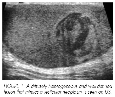
]]> An 18 year old man was brought to the emergency department with a severe and sudden episode of right testicular pain. Accordingly to the patient, there was not previous testicular trauma. Physical examination of the scrotum demonstrated tenderness and pain of the inferior pole of the right testis. Blood cell count, urine culture and serum biochemistry were normal. Ultrasound and color Doppler showed a well defined avascular lesion located at the testicular mediastinum (Figure 1). The possibility of an acute area of segmental ischaemia or infarction rather than a tumour was raised because of the absence of colour Doppler signal in the abnormal area. Nevertheless an ingui-nal exploration of the right testis was programmed. Tumour markers (human corionic gonadotropin and alpha-fetoprotein) were negative.
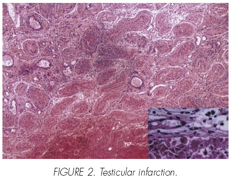
The right testis was explored via inguinal incision. An intraoperative biopsy was carried out and the pathological evaluation demonstrated testicular infarction (Figure 2). The patient recovered successfully and was discharged 24 h after surgery.
As possible causes, haematological disorders like leukaemia, sickle-cell anaemia, compression of blood vessels by hernia or tumour, thromboembolism and vasculitis were dismissed after appropriate studies.
Discussion
Segmental testicular infarction is rare, with 2 cases reported and is usually diagnosed following orchiectomy (6). Some haematologic disorders are considered as causative factors, although in most cases seems to be idiopathic (1).
]]> Clinical findings in most cases include pain and tenderness of the affected testis. Ultrasonographically the STI is characterized by hypo or isoechoic lesions and with the Doppler ultrasound is possible to observe an avascular pattern (2). MRI could be helpful in the diagnostic workup, but cannot rule out neoplasia (6).The problem is that it is almost impossible to di-fferentiate STI from neoplasia preoperatively. Final diagnosis is only established after surgery or during surgical exploration if we consider the possibility of a performing an excisional frozen biopsy. In most reported series radical orchiectomy was chosen (1). We propose inguinal surgical expiration of the testis clamping the spermatic cord and to ule out a neoplasia by excisional frozen biopsy in the cases of suspicion of STI.
References and recomended readings (*of special interest, **of outstanding interest)
*1. RUIBAL, M.; QUINTANA, J.; FERNANDEZ, G. y cols.: Segmental Testicular Infarction. J. Urol., 170: 187, 2003. [ Links ]
2. SRIPRASAD, S.; KOOIMAN, G.; MUIR,G. y cols.: Acute segmental testicular infarction: Differentiation from tumour using high frequency colour Doppler ultrasound. British Journal of Radiology, 74: 965, 2001. [ Links ]
3. PELLICE, C.; CASTELLA, J.; ALERT, E.: Focal infarction of the testis. Report of a case simulating a gonadal mass. Actas Urol. Esp., 19: 716, 1996. [ Links ]
*4. FERNANDEZ, J.; MARTIN, A.; RABADE, J. y cols.: Testicular infarction as a cause of benign intrascrotaltumor. Arch. Esp. Urol., 49: 72, 1996. [ Links ]
5. NAYAK, S.; PURANIK, S.; HOLLA, V.: Testicular infarction mimicking a neoplasm. Indian Journal of Surgery, 65: 284, 2003. [ Links ]
6. KODAMA, K.; YOTSUYANAGI, S.; FUSE, H. Y cols.: Magnetic resonante imaging to diagnose segmental testicular infarction. J. Urol., 163: 910, 2000. [ Links ]
**7. RIPA SALDIAS, R.; GUARCH TROYAS, R.; HUALDE ALFARO, A. y cols.: Infarto segmentario de testículo. Actas Urol. Esp., 30: 227, 2006. [ Links ]
![]() Correspondence:
Correspondence:
Jorge Hidalgo Arroyo
Hospital de Figueres
Ronda Rector Arolas, s/n
17600 Figueres. Girona. (Spain)
hidarro@hotmail.com
Accepted for publication: March 30th, 2007.
]]>