<?xml version="1.0" encoding="ISO-8859-1"?><article xmlns:mml="http://www.w3.org/1998/Math/MathML" xmlns:xlink="http://www.w3.org/1999/xlink" xmlns:xsi="http://www.w3.org/2001/XMLSchema-instance">
<front>
<journal-meta>
<journal-id>0210-5691</journal-id>
<journal-title><![CDATA[Medicina Intensiva]]></journal-title>
<abbrev-journal-title><![CDATA[Med. Intensiva]]></abbrev-journal-title>
<issn>0210-5691</issn>
<publisher>
<publisher-name><![CDATA[Elsevier España, S.L.]]></publisher-name>
</publisher>
</journal-meta>
<article-meta>
<article-id>S0210-56912008000300003</article-id>
<title-group>
<article-title xml:lang="en"><![CDATA[Comparison of bronchoscopic bronchoalveolar lavage vs blind lavage with a modified nasogastric tube in the etiologic diagnosis of ventilator-associated pneumonia]]></article-title>
<article-title xml:lang="es"><![CDATA[Comparación de lavado broncoalveolar broncoscópico frente a lavado ciego con sonda nasogástrico modificada en el diagnóstico etiológico de neumonía asociada a ventilador]]></article-title>
</title-group>
<contrib-group>
<contrib contrib-type="author">
<name>
<surname><![CDATA[Leo]]></surname>
<given-names><![CDATA[A.]]></given-names>
</name>
<xref ref-type="aff" rid="A01"/>
</contrib>
<contrib contrib-type="author">
<name>
<surname><![CDATA[Galindo-Galindo]]></surname>
<given-names><![CDATA[J.]]></given-names>
</name>
<xref ref-type="aff" rid="A01"/>
</contrib>
<contrib contrib-type="author">
<name>
<surname><![CDATA[Folch]]></surname>
<given-names><![CDATA[E.]]></given-names>
</name>
<xref ref-type="aff" rid="A02"/>
</contrib>
<contrib contrib-type="author">
<name>
<surname><![CDATA[Guerrero]]></surname>
<given-names><![CDATA[A.]]></given-names>
</name>
<xref ref-type="aff" rid="A01"/>
</contrib>
<contrib contrib-type="author">
<name>
<surname><![CDATA[Bosques]]></surname>
<given-names><![CDATA[F.]]></given-names>
</name>
<xref ref-type="aff" rid="A01"/>
</contrib>
<contrib contrib-type="author">
<name>
<surname><![CDATA[Mercado]]></surname>
<given-names><![CDATA[R.]]></given-names>
</name>
<xref ref-type="aff" rid="A01"/>
</contrib>
<contrib contrib-type="author">
<name>
<surname><![CDATA[Arroliga]]></surname>
<given-names><![CDATA[A.C.]]></given-names>
</name>
<xref ref-type="aff" rid="A03"/>
</contrib>
</contrib-group>
<aff id="A01">
<institution><![CDATA[,Hospital Universitario Dr. José Eleuterio González  ]]></institution>
<addr-line><![CDATA[Monterrey ]]></addr-line>
<country>México</country>
</aff>
<aff id="A02">
<institution><![CDATA[,The Cleveland Clinic Department of Pulmonary, Allergy and Critical Care Medicine ]]></institution>
<addr-line><![CDATA[Cleveland Ohio]]></addr-line>
<country>USA</country>
</aff>
<aff id="A03">
<institution><![CDATA[,Scott & White Hospital Division of Pulmonary and Critical Care Medicine ]]></institution>
<addr-line><![CDATA[Temple Texas]]></addr-line>
<country>USA</country>
</aff>
<pub-date pub-type="pub">
<day>00</day>
<month>04</month>
<year>2008</year>
</pub-date>
<pub-date pub-type="epub">
<day>00</day>
<month>04</month>
<year>2008</year>
</pub-date>
<volume>32</volume>
<numero>3</numero>
<fpage>115</fpage>
<lpage>120</lpage>
<copyright-statement/>
<copyright-year/>
<self-uri xlink:href="http://scielo.isciii.es/scielo.php?script=sci_arttext&amp;pid=S0210-56912008000300003&amp;lng=en&amp;nrm=iso"></self-uri><self-uri xlink:href="http://scielo.isciii.es/scielo.php?script=sci_abstract&amp;pid=S0210-56912008000300003&amp;lng=en&amp;nrm=iso"></self-uri><self-uri xlink:href="http://scielo.isciii.es/scielo.php?script=sci_pdf&amp;pid=S0210-56912008000300003&amp;lng=en&amp;nrm=iso"></self-uri><abstract abstract-type="short" xml:lang="en"><p><![CDATA[Objective. Our objective was to compare the results of a blind lavage vs a bronchoscopic-guided bronchoalveolar lavage for the etiologic diagnosis of ventilator-associated pneumonia (VAP). Design. Prospective study in consecutive patients with high probability of VAP. Every patient underwent both procedures, in a formally randomized fashion. The interpretation of quantitative cultures was done in a blind fashion. Setting. Single center study, with a 20 bed medical and surgical Intensive Care Unit of the University Hospital in Monterrey, Mexico. Patients. Twenty-five patients with high probability of VAP. Interventions. Every patient underwent blind bronchoalveolar lavage with a modified nasogastric tube, and a bronchospic-guided bronchoalveolar lavage. Results. Twenty-one patients underwent both procedures. Four patients were excluded due to contamination of the cultures. The quantitative cultures were compared in a paired fashion. Only two patients had discordant cultures. The correlation coefficient between the number of colonies was very high, r = 0.90 (95% confidence interval [CI], 0.77-0.96; p = 0.0001). Conclusions. The blind bronchoalveolar lavage with a modified nasogastric tube is a valuable tool for the identification of etiologic agent in VAP, particularly when trained bronchoscopists or the necessary resources for bronchoscopic-guided bronchoalveolar lavage are not readily available.]]></p></abstract>
<abstract abstract-type="short" xml:lang="es"><p><![CDATA[Objetivo. Nuestro objetivo fue el de comparar los resultados de un lavado ciego frente a un lavado broncoalveolar guiado con broncoscopio para el diagnóstico etiológico de neumonía asociada a ventilador (NAV). Diseño. Estudio prospectivo en pacientes consecutivos con alta probabilidad de NAV. En todos los pacientes se llevaron a cabo ambos procedimientos de manera aleatorizada. La interpretación de los cultivos cuantitativos fue hecha a ciegas. Ámbito. Estudio en un único centro, en una Unidad de Cuidados Intensivos Quirúrgicos con 20 camas del Hospital Universitario de Monterrey, en México. Pacientes. Veinticinco pacientes con alta probabilidad de NAV. Intervenciones. A cada paciente se le realizó un lavado broncoalveolar ciego con una sonda nasogástrica modificada y un lavado broncoalveolar guiado con broncoscopio. Resultados. Se realizaron ambos procedimientos en 21 pacientes. Cuatro fueron excluidos debido a contaminación de los cultivos. Los cultivos cuantitativos fueron comparados en pares. Solo dos pacientes tenían cultivos discordantes. El coeficiente de correlación entre el número de colonias fue muy alto, r = 0,90 (intervalo de confianza [IC] del 95% 0,77-0,96; p = 0,0001). Conclusiones. El lavado broncoalveolar ciego con sonda nasogástrica modificada es una herramienta de mucho valor para la identificación del agente etiológico en NAV, especialmente cuando un broncoscopista experto o los recursos necesarios para lavado broncoalveolar guiado con broncoscopio no están fácilmente disponibles.]]></p></abstract>
<kwd-group>
<kwd lng="en"><![CDATA[ventilator-associated pneumonia]]></kwd>
<kwd lng="en"><![CDATA[bronchoalveolar lavage]]></kwd>
<kwd lng="en"><![CDATA[nasogastric tube]]></kwd>
<kwd lng="en"><![CDATA[blind bronchoalveolar lavage]]></kwd>
<kwd lng="es"><![CDATA[neumonía asociada a ventilador]]></kwd>
<kwd lng="es"><![CDATA[lavado broncoalveolar]]></kwd>
<kwd lng="es"><![CDATA[sonda nasogástrica]]></kwd>
<kwd lng="es"><![CDATA[lavado broncoalveolar ciego]]></kwd>
</kwd-group>
</article-meta>
</front><body><![CDATA[ <p align="right"><font face="Verdana" size="2"><B>ORIGINALES</B></font></p>     <p>&nbsp;</p>     <p><font face="Verdana" size="4"><B><a name="top"></a>Comparison of bronchoscopic bronchoalveolar lavage vs blind lavage with a modified nasogastric tube in the etiologic diagnosis of ventilator-associated pneumonia</B></font></p> <font FACE="Verdana" SIZE="4"><b>     <p>Comparación de lavado broncoalveolar broncoscópico frente a lavado ciego con sonda nasogástrico modificada en el diagnóstico etiológico de neumonía asociada a ventilador</p> </b></font>     <p>&nbsp;</p>     <p>&nbsp;</p>     <p><font face="Verdana" size="2"><B>A. Leo<sup>a</sup>; J. Galindo-Galindo<sup>a</sup>; E. Folch<sup>b</sup>; A. Guerrero<sup>a</sup>; F. Bosques<sup>a</sup>; R. Mercado<sup>a</sup>; A.C. Arroliga<sup>c</sup></B></font></p> <font FACE="Verdana" SIZE="2">     <p><sup>a</sup>Hospital Universitario Dr. José Eleuterio González. Monterrey. México.    <br> <sup>b</sup>Department of Pulmonary, Allergy and Critical Care Medicine. The Cleveland Clinic. Cleveland. Ohio. USA.    <br> <sup>c</sup>Division of Pulmonary and Critical Care Medicine. Scott &amp; White Hospital. Temple. Texas. USA.</p>     ]]></body>
<body><![CDATA[<p><a href="#back">Correspondence</a></p>     <p>&nbsp;</p>     <p></font></p> <hr size="1">      <P><font face="Verdana" size="2"><B>ABSTRACT</B></font>    <P>     <p><font face="Verdana" size="2"><B>Objective.</B> Our objective was to compare the results of a blind lavage vs a bronchoscopic-guided bronchoalveolar lavage for the etiologic diagnosis of ventilator-associated pneumonia (VAP).    <BR><B>Design.</B> Prospective study in consecutive patients with high probability of VAP. Every patient underwent both procedures, in a formally randomized fashion. The interpretation of quantitative cultures was done in a blind fashion.    <BR><B>Setting.</B> Single center study, with a 20 bed medical and surgical Intensive Care Unit of the University Hospital in Monterrey, Mexico.    <BR><B>Patients.</B> Twenty-five patients with high probability of VAP. Interventions. Every patient underwent blind bronchoalveolar lavage with a modified nasogastric tube, and a bronchospic-guided bronchoalveolar lavage.    <BR><B>Results.</B> Twenty-one patients underwent both procedures. Four patients were excluded due to contamination of the cultures. The quantitative cultures were compared in a paired fashion. Only two patients had discordant cultures. The correlation coefficient between the number of colonies was very high, r = 0.90 (95% confidence interval &#091;CI&#093;, 0.77-0.96; p = 0.0001).    ]]></body>
<body><![CDATA[<BR><B>Conclusions.</B> The blind bronchoalveolar lavage with a modified nasogastric tube is a valuable tool for the identification of etiologic agent in VAP, particularly when trained bronchoscopists or the necessary resources for bronchoscopic-guided bronchoalveolar lavage are not readily available.</font></P>     <P><font face="Verdana" size="2"><B>Key words:</B> ventilator-associated pneumonia, bronchoalveolar lavage, nasogastric tube, blind bronchoalveolar lavage.</font></p>  <hr size="1">      <P><font face="Verdana" size="2"><B>RESUMEN</B></font>     <p><font face="Verdana" size="2"><B>Objetivo.</B> Nuestro objetivo fue el de comparar los resultados de un lavado ciego frente a un lavado broncoalveolar guiado con broncoscopio para el diagnóstico etiológico de neumonía asociada a ventilador (NAV).    <BR><B>Diseño.</B> Estudio prospectivo en pacientes consecutivos con alta probabilidad de NAV. En todos los pacientes se llevaron a cabo ambos procedimientos de manera aleatorizada. La interpretación de los cultivos cuantitativos fue hecha a ciegas.    <BR><B>Ámbito.</B> Estudio en un único centro, en una Unidad de Cuidados Intensivos Quirúrgicos con 20 camas del Hospital Universitario de Monterrey, en México.    <BR><B>Pacientes.</B> Veinticinco pacientes con alta probabilidad de NAV.    <BR><B>Intervenciones.</B> A cada paciente se le realizó un lavado broncoalveolar ciego con una sonda nasogástrica modificada y un lavado broncoalveolar guiado con broncoscopio.    <BR><B>Resultados.</B> Se realizaron ambos procedimientos en 21 pacientes. Cuatro fueron excluidos debido a contaminación de los cultivos. Los cultivos cuantitativos fueron comparados en pares. Solo dos pacientes tenían cultivos discordantes. El coeficiente de correlación entre el número de colonias fue muy alto, r = 0,90 (intervalo de confianza &#091;IC&#093; del 95% 0,77-0,96; p = 0,0001).    <BR><B>Conclusiones.</B> El lavado broncoalveolar ciego con sonda nasogástrica modificada es una herramienta de mucho valor para la identificación del agente etiológico en NAV, especialmente cuando un broncoscopista experto o los recursos necesarios para lavado broncoalveolar guiado con broncoscopio no están fácilmente disponibles.</font></p>     ]]></body>
<body><![CDATA[<P><font face="Verdana" size="2"><B>Palabras clave:</B> neumonía asociada a ventilador, lavado broncoalveolar, sonda nasogástrica, lavado broncoalveolar ciego.</font></p>  <hr size="1">      <p>&nbsp;</p>     <p><font face="Verdana"><B>Introduction</B></font></p>     <P><font face="Verdana" size="2">Ventilator-associated pneumonia (VAP) is a major medical problem. VAP is associated with longer stay in the Intensive Care Unit (ICU) and in the hospital, high cost, and high mortality rates<SUP>1-5</SUP>. The clinical diagnosis of VAP that includes radiographic changes and at least one clinical finding such as fever, leukocytosis, or purulent tracheal aspirate has a good sensitivity but poor specificity<SUP>4</SUP>. It has been recommended that a sample of the lower respiratory tract should be obtained when the clinical diagnoses of VAP is suspected<SUP>4</SUP>. Invasive diagnostic testing may increase the confidence of the clinician in the diagnosis and management of the VAP and because of the good negative predictive value thus allowing discontinuation of antibiotic therapy<SUP>4,6</SUP>.</font></P>     <P><font face="Verdana" size="2">Quantitative cultures of specimens taken with bronchoscopic techniques or through blind non-bronchoscopic techniques are appealing alternatives for the collection of lower respiratory tract specimens<SUP>4</SUP>. A strategy of diagnosis that includes obtaining a sample by bronchoscopic technique is associated with a lower mortality at day 14 and a decrease in antibiotic use<SUP>7</SUP>. However, bronchoscopy may not be readily available<SUP>4</SUP>, especially in countries with limited resources, therefore diagnostic testing with non-bronchoscopic techniques and early use of broad-spectrum antibiotics are attractive options in order to minimize cost, antibiotic use, as well as improve survival<SUP>8</SUP>.</font></P>     <P><font face="Verdana" size="2">The hypothesis of the study is that the isolated bacteria and quantitative cultures would be similar in the fluid obtained with both techniques. The goal of this study was to compare the microbiologic findings in lower airway lavage through a non-bronchoscopic technique using a modified nasogastric tube<SUP>9</SUP> against a fiberoptic bronchoscope-directed bronchoalveolar lavage (BAL) in the same patient.</font></P>     <P>&nbsp;</P>     <P><font face="Verdana"><B>Patients and methods</B></font></P>     <P><font face="Verdana" size="2">We prospectively studied 25 patients with suspected VAP in the Medical and Surgical ICU at <I>Hospital Universitario Dr. José Eleuterio González</I> between June 1, 2005 and June 1, 2006. This hospital is a major teaching hospital in Monterrey, Mexico. The unit has 20 beds, staffed by residents in Internal Medicine, Fellows in Pulmonary and Critical Care, and Attending Intensive Care specialists. The ratio of patients per nurses is 2:1.</font></P>     <P><font face="Verdana" size="2">Patients were eligible for the study if they met the following inclusion criteria: &#8805;18 year-old, intubated for more than 48 hours, who met the clinical definition for VAP. Clinical definition of VAP refers to new infiltrates in the chest X-ray with &#8805;2 of the following: fever or hypothermia (&#8804;35° C or &#8805;38.3° C), leukocytosis or leukopenia (&#8805;12,000/dl or &#8804;4,000/dl), purulent secretions in the endotracheal tube (ETT), and/or poor oxygenation (PaO<SUB>2</SUB>/FiO<SUB>2</SUB> &#8804;240 mmHg)<SUP>1,4</SUP>. Patients were excluded if they had a contraindication for bronchoscopy (for example severe hypoxemia PaO<SUB>2</SUB>/FiO<SUB>2</SUB> &lt; 100 mmHg), refractory coagulopathy (prothrombin time and partial thromboplastin time &gt; twice the upper limit of normal nonresponsive to 10 cc/kg of fresh frozen plasma &#091;FFP&#093;), or hemodinamically unstable at time of bronchoscopy (mean arterial pressure &#091;MAP&#093; &lt; 60 mmHg).</font></P>     ]]></body>
<body><![CDATA[<P><font face="Verdana" size="2">The study was approved by the Institutional Review Board, and informed consent was obtained from the patient or next of kin.</font></P>     <P><font face="Verdana" size="2">The following characteristics were recorded prospectively at the time of ICU admission: age, sex, APACHE II score<SUP>10</SUP>, main diagnosis and comorbidities, clinical pulmonary infection score (CPIS)<SUP>11</SUP>, previous antibiotics, concurrent extrapulmonary infections, and radiologic findings. The CPIS and APACHE II scores were calculated again on the day of the study. Every study participant underwent two procedures:</font></P>     <P><font face="Verdana" size="2">1. Blind BAL with a modified nasogastric tube(non-bronchoscopic).</font></P>     <P><font face="Verdana" size="2">2. BAL using a standard fiberoptic bronchoscope.</font></P>     <P><font face="Verdana" size="2">The modification in the nasogastric tube consists in the cutting of the tip of the catheter to remove the area with multiple holes<SUP>9</SUP>. We randomly determined which procedure was done first. The procedures were performed 20 minutes apart for stabilization purposes.</font></P>     <P><font face="Verdana" size="2">Patients were sedated and preoxygenated while on continuous pulse oxymetry monitoring. For the nonbronchoscopic procedure, a 14F nasogastric tube was slowly introduced through an adaptor (Portex<SUP>®</SUP>, Keene, New Hampshire, USA) into the ETT, until resistance was felt. Three aliquots of 50 ml of sterile 0.9% saline were instilled sequentially, and withdrawn by manual suction with a 50 ml catheter-tip piston syringe. The first aspirate was discarded. The remaining two aspirates were processed in the Microbiology laboratory. In the bronchoscopic procedure, after tracheal aspiration, the fiberoptic bronchoscope (Pentax<SUP>®</SUP>, Orangeburg, New York, USA) was introduced into the ETT via the ETT adaptor and positioned («wedged») in the orifice of the sampling area with sequential instillation of three aliquots of 50 ml of sterile 0.9% saline. The first aspirate was discarded, and remaining fluid was sent to the Microbiology laboratory. No aspiration was done through the working channel of the bronchoscope before the collection of samples in order to minimize contamination.</font></P>     <P><font face="Verdana" size="2">The specimens were immediately sent to the laboratory and processed according to previously described methods by Baselski<SUP>12</SUP>. The samples were centrifuged for 30 seconds, a Gram stain was done searching for intracellular organisms. The bacterial cultures were processed with microorganisms quantified by an experienced microbiologist using standard serial dilution and the results were expressed as colony-forming units (cfu/ml)<SUP>13</SUP>. The cut-off point for significant growth was 10<SUP>4</SUP> cfu/ml for both procedures<SUP>14,15</SUP>. The microbiologist reading the cultures was blinded for which procedure was used and for the result of the corresponding sample.</font></P>     <p><b><font face="Verdana" size="2">    <br> Statistical analysis</font></b></p>     <P><font face="Verdana" size="2">Descriptive statistics were used, with all comparisons being paired, and all tests of significance two tailed. All values are expressed as the mean ± standard deviation (SD). Sensitivity, specificity, positive predictive value, negative predictive value, and likelihood ratios were determined considering fiberoptic bronchoscopy (FOB) with BAL as the gold standard. Correlation coefficients were also calculated.</font></P>     ]]></body>
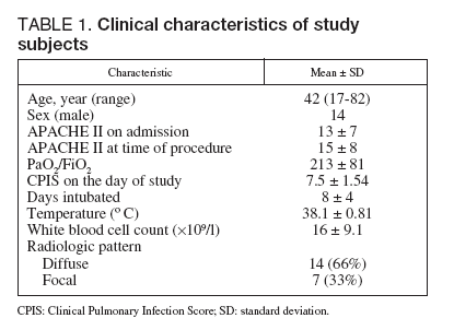
<body><![CDATA[<P>&nbsp;</P>     <P><font face="Verdana"><B>Results</B></font></P>     <P><font face="Verdana" size="2">Twenty-five eligible patients were included in the study, with 4 patients being excluded from analysis due to contamination of microbiology cultures. The 21 subjects (14 men and 7 women) had a mean age of 42 years-old (range 17-82), with mean APACHE II score of 15 (± 8) and a CPIS of 7.5 on the day of the procedures (<a href="#t1">table 1</a>). The reasons for hospital admission were pneumonia (5), trauma (4), sepsis (2), stroke (1), burns (1), severe pancreatitis (1), eclampsia (1), brain tumor (1), empyema (1), aortic aneurysm (1), pulmonary hemorrhage (1), hypovolemic shock (1), and respiratory failure (1). All patients were intubated, and 15 of 21 had been receiving antibiotics for the reason that prompted their admission to the ICU.</font></P>     <P align="center"><a name="t1"><IMG src="/img/revistas/medinte/v32n3/64v32n03-13117801tab01.gif" border=0></a></P>      <P><font face="Verdana" size="2">Every patient underwent both bronchoscopic and non-bronchoscopic procedures. The mean volume of fluid recovered from lavage was 42 ± 8 ml in the bronchoscopic technique and 40 ± 10 ml in the nonbronchoscopic technique. With the bronchoscopic technique, significant growth was found in 66.7% (n = 14) of the samples and non-significant growth in 33.3% (n = 7). With the non-bronchoscopic technique significant growth was found in 71.4% (n = 15) of the samples, non-significant growth in 28.6% (n = 6) (tables <a target="_blank" href="/img/revistas/medinte/v32n3/64v32n03-13117801tab02.gif">2</a> and <a target="_blank" href="/img/revistas/medinte/v32n3/64v32n03-13117801tab03.gif">3</a>).</font></P>     <P><font face="Verdana" size="2">The quantitative cultures obtained through either technique are shown in <a target="_blank" href="/img/revistas/medinte/v32n3/64v32n03-13117801tab03.gif">table 3</a>. Only two patients had discordant cultures, the non-bronchoscopic technique failed to provide quantitative evidence of infection in one case, and it identified a second organism inanother patient, that was not isolated with the bronchoscopic technique.</font></P>     <P><font face="Verdana" size="2">The most common isolated organisms were <I>Staphylococcus aureus</I>, <I>Acinetobacter baumannii</I>, and <I>Pseudomonas aeruginosa</I> with polymicrobial infection present in 16 (76.2%) cases, and single organism infection present in 14.3% of the cases. The Spearman’s coefficient of rank correlation (r) for number of colonies showed a positive correlation at 0.90 (confidence interval &#091;CI&#093; 0.77-0.96; p = 0.0001)between the two techniques. The sensitivity of the non-bronchoscopic technique was 93%, and the specificity was 85% when compared to the bronchoscopic-guided bronchoalveolar lavage (<a target="_blank" href="/img/revistas/medinte/v32n3/64v32n03-13117801tab04.gif">table 4</a>). We calculated the likelihood ratios<SUP>16</SUP> or how many times more likely patients with the disease are to have that particular result than patients without the disease. The positive and negative likelihood ratios showed strong evidence to rule in or out the presence of VAP in this group of patients. The procedures were tolerated well with no episodes of desaturation below 88% with either technique.</font></P>     <P>&nbsp;</P>     <P><font face="Verdana"><B>Discussion</B></font></P>     <P><font face="Verdana" size="2">In this single center, prospective study we demonstrated the excellent operating characteristics of a non-bronchoscopic BAL technique using a nasogastric tube compared with the frequently cited fiberoptic bronchoscope-guided BAL.</font></P>     ]]></body>
<body><![CDATA[<P><font face="Verdana" size="2">There is currently no gold standard for the diagnosis of VAP and clinicians rely on clinical and bacteriologic strategies to manage patients with VAP<SUP>4</SUP>. The bacteriologic strategy uses quantitative cultures of lower respiratory secretions and has been associated with less use of antibiotics<SUP>4</SUP>. In a seminal paper, Fagon et al showed that an invasive strategy using FOB with quantitative cultures improves survival (14 days), and decreases antibiotic use<SUP>7</SUP>. Eventhough the bronchoscopically-guided BAL has several advantages, the most important being the ability to direct sampling into the desired lobe, it is important to emphasize its limitations in resource constrained settings. Fiberoptic bronchoscopes and qualified operators are not always readily available, thus potentially delaying pathogen-directed treatment with its harmful consequences<SUP>17,18</SUP>. Previous reports of «blind» invasive procedures have yield conflicting evidence, mostly because of variable methodologies, different thresholds of the quantitative studies, and reference standards<SUP>11,19-26</SUP>. Minutoli et al reported in the late 1980s the use of a nasogastric tube to do bronchoalveolar lavages in patients with the acquired immunodeficiency syndrome<SUP>9</SUP>. We extended their experience using this technique to obtain distal airway sample for bacterial cultures of patients with high clinical suspicion of VAP.</font></P>     <P><font face="Verdana" size="2">In an attempt to standardize a technique that should be simple, widely available, inexpensive, and with low risk of complications, we analyzed the performance of the nasogastric tube with quantitative cultures side-by-side with the bronchoscopy-directed BAL in the same patient with excellent results. By using the exact same lavage volume, quantitative threshold, and discarding the first aspirate, we obtained an excellent correlation of results between the two techniques. Furthermore, by using the same patient, and randomizing which technique to use first, as well as blinding the laboratory technician reading the cultures, we minimized bias. Our study also suggested that VAP is a diffuse disease involving multiple lobes, and samples obtained blindly have a comparable performance to FOB-guided samples<SUP>11,20,21,27-30</SUP>. Furthermore, histology-based reports suggest VAP is predominantly a dependent lung segment disease<SUP>24</SUP> where is more likely that a nasogastric tube will go. Because the nasogastric tube has roughly the same size as a fiberoptic bronchoscope and unable to reach peripheral sections of the lung, we avoided complications such as pneumothorax.</font></P>     <P><font face="Verdana" size="2">Our study has several limitations, the two most important being that like any single center study, its results may not be generalizable to other settings. The second limitation of the study is the small sample size did not allow for subgroup analysis for specific admission diagnosis. However, the main objective of the study was to compare the microbiologic findings of the two techniques and we achieved that objective.</font></P>     <P><font face="Verdana" size="2">The results of our study have important implications in the care of patients with VAP in resourceconstrained settings, where the availability of bronchoscopes to confirm the diagnosis of VAP is limited.</font></P>     <P><font face="Verdana" size="2">We believe this innovative and simple technique should be validated in larger clinical trials, where antibiotic use, organ dysfunction improvement, and ultimately survival should be used as outcome measures.</font></P>     <P>    <br><font face="Verdana" size="2"><b>Declaration of conflict of interest</b></font></P>     <P><font face="Verdana" size="2">All the authors reported no conflict of interest.</font></P>     <P>&nbsp;</P>     <P><font face="Verdana"><B>References</B></font></P>     ]]></body>
<body><![CDATA[<!-- ref --><P><font face="Verdana" size="2">1. Warren DK, Shukla SJ, Olsen MA, Kollef MH, Hollenbeak CS, Cox MJ, et al. Outcome and attributable cost of ventilator-associated pneumonia among intensive care unit patients in a suburban medical center. Crit Care Med. 2003;31:1312-7.</font>&nbsp;&nbsp;&nbsp;&nbsp;&nbsp;&nbsp;&nbsp;&nbsp;[&#160;<a href="javascript:void(0);" onclick="javascript: window.open('/scielo.php?script=sci_nlinks&ref=2859793&pid=S0210-5691200800030000300001&lng=','','width=640,height=500,resizable=yes,scrollbars=1,menubar=yes,');">Links</a>&#160;]<!-- end-ref --><!-- ref --><p><font face="Verdana" size="2">2. Safdar N, Dezfulian C, Collard HR, Saint S. Clinical and economic consequences of ventilator-associated pneumonia: a systematic review. Crit Care Med. 2005; 33: 2184-93.</font>&nbsp;&nbsp;&nbsp;&nbsp;&nbsp;&nbsp;&nbsp;&nbsp;[&#160;<a href="javascript:void(0);" onclick="javascript: window.open('/scielo.php?script=sci_nlinks&ref=2859794&pid=S0210-5691200800030000300002&lng=','','width=640,height=500,resizable=yes,scrollbars=1,menubar=yes,');">Links</a>&#160;]<!-- end-ref --><!-- ref --><p><font face="Verdana" size="2">3. Kollef MH, Shorr A, Tabak YP, Gupta V, Liu LZ, Johannes RS. Epidemiology and outcomes of health-care-associated pneumonia: results from a large US database of culture-positive pneumonia. Chest. 2005;128:3854-62.</font>&nbsp;&nbsp;&nbsp;&nbsp;&nbsp;&nbsp;&nbsp;&nbsp;[&#160;<a href="javascript:void(0);" onclick="javascript: window.open('/scielo.php?script=sci_nlinks&ref=2859795&pid=S0210-5691200800030000300003&lng=','','width=640,height=500,resizable=yes,scrollbars=1,menubar=yes,');">Links</a>&#160;]<!-- end-ref --><!-- ref --><p><font face="Verdana" size="2">4. American Thoracic Society; Infectious Diseases Society of America. Guidelines for the management of adults with hospital-acquired, ventilator-associated and healthcare-associated pneumonia. Am J Respir Crit Care Med. 2005;171:388-416.</font>&nbsp;&nbsp;&nbsp;&nbsp;&nbsp;&nbsp;&nbsp;&nbsp;[&#160;<a href="javascript:void(0);" onclick="javascript: window.open('/scielo.php?script=sci_nlinks&ref=2859796&pid=S0210-5691200800030000300004&lng=','','width=640,height=500,resizable=yes,scrollbars=1,menubar=yes,');">Links</a>&#160;]<!-- end-ref --><!-- ref --><p><font face="Verdana" size="2">5. Ranes JL, Gordon SM, Chen P, Fatica C, Hammel J, Gonzales JP, et al. Predictors of long-term mortality in patients with ventilator-associated pneumonia. Am J Med. 2006;119:897. e13-9.</font>&nbsp;&nbsp;&nbsp;&nbsp;&nbsp;&nbsp;&nbsp;&nbsp;[&#160;<a href="javascript:void(0);" onclick="javascript: window.open('/scielo.php?script=sci_nlinks&ref=2859797&pid=S0210-5691200800030000300005&lng=','','width=640,height=500,resizable=yes,scrollbars=1,menubar=yes,');">Links</a>&#160;]<!-- end-ref --><!-- ref --><p><font face="Verdana" size="2">6. Heyland DK, Cook DJ, Marshall J, Heule M, Guslits B, Lang J, et al. The clinical utility of invasive diagnostic techniques in the setting of ventilator-associated pneumonia. Chest. 1999;115:1076-84.</font>&nbsp;&nbsp;&nbsp;&nbsp;&nbsp;&nbsp;&nbsp;&nbsp;[&#160;<a href="javascript:void(0);" onclick="javascript: window.open('/scielo.php?script=sci_nlinks&ref=2859798&pid=S0210-5691200800030000300006&lng=','','width=640,height=500,resizable=yes,scrollbars=1,menubar=yes,');">Links</a>&#160;]<!-- end-ref --><!-- ref --><p><font face="Verdana" size="2">7. Fagon JY, Chastre J, Wolff M, Gervais C, Parer-Aubas S, Stéphan F, et al. Invasive and noninvasive strategies for management for suspected ventilator-associated pneumonia. A randomized trial. Ann Intern Med. 2000;132:621-30.</font>&nbsp;&nbsp;&nbsp;&nbsp;&nbsp;&nbsp;&nbsp;&nbsp;[&#160;<a href="javascript:void(0);" onclick="javascript: window.open('/scielo.php?script=sci_nlinks&ref=2859799&pid=S0210-5691200800030000300007&lng=','','width=640,height=500,resizable=yes,scrollbars=1,menubar=yes,');">Links</a>&#160;]<!-- end-ref --><!-- ref --><p><font face="Verdana" size="2">8. Ost DE, Hall CS, Joseph G, Ginocchio C, Condon S, Kao E, et al. Decision analysis of antibiotic and diagnostic strategies in ventilator-associated pneumonia. Am J Respir Crit Care Med. 2003;168:1060-7.</font>&nbsp;&nbsp;&nbsp;&nbsp;&nbsp;&nbsp;&nbsp;&nbsp;[&#160;<a href="javascript:void(0);" onclick="javascript: window.open('/scielo.php?script=sci_nlinks&ref=2859800&pid=S0210-5691200800030000300008&lng=','','width=640,height=500,resizable=yes,scrollbars=1,menubar=yes,');">Links</a>&#160;]<!-- end-ref --><!-- ref --><p><font face="Verdana" size="2">9. Minutoli R, Eden E, Brachfeld C. Bronchoalveolar lavage via a modified stomach tube in intubated patients with the acquired immunodeficiency syndrome and diffuse pneumonia. Thorax. 1990;45:771-3.</font>&nbsp;&nbsp;&nbsp;&nbsp;&nbsp;&nbsp;&nbsp;&nbsp;[&#160;<a href="javascript:void(0);" onclick="javascript: window.open('/scielo.php?script=sci_nlinks&ref=2859801&pid=S0210-5691200800030000300009&lng=','','width=640,height=500,resizable=yes,scrollbars=1,menubar=yes,');">Links</a>&#160;]<!-- end-ref --><!-- ref --><p><font face="Verdana" size="2">10. Knaus WA, Draper EA, Wagner DP, Zimmerman JE. APACHE II: a severity of disease classification system. Crit Care Med. 1985;13:818-29.</font>&nbsp;&nbsp;&nbsp;&nbsp;&nbsp;&nbsp;&nbsp;&nbsp;[&#160;<a href="javascript:void(0);" onclick="javascript: window.open('/scielo.php?script=sci_nlinks&ref=2859802&pid=S0210-5691200800030000300010&lng=','','width=640,height=500,resizable=yes,scrollbars=1,menubar=yes,');">Links</a>&#160;]<!-- end-ref --><!-- ref --><p><font face="Verdana" size="2">11. Pugin J, Auckenthaler R, Mili N, Janssens JP, Lew PD, Suter PM. Diagnosis of ventilator-associated pneumonia by bacteriologic analysis of bronchoscopic and nonbronchoscopic “blind” bronchoalveolar lavage fluid. Am Rev Respir Dis. 1991;143:1121-9.</font>&nbsp;&nbsp;&nbsp;&nbsp;&nbsp;&nbsp;&nbsp;&nbsp;[&#160;<a href="javascript:void(0);" onclick="javascript: window.open('/scielo.php?script=sci_nlinks&ref=2859803&pid=S0210-5691200800030000300011&lng=','','width=640,height=500,resizable=yes,scrollbars=1,menubar=yes,');">Links</a>&#160;]<!-- end-ref --><!-- ref --><p><font face="Verdana" size="2">12. Baselski VS, el-Torky M, Coalson JJ, Griffin JP. The standardization of criteria for processing and interpreting laboratory specimens in patients with suspected ventilator-associated pneumonia. Chest. 1992;102 (5 Suppl 1):571S-579S.</font>&nbsp;&nbsp;&nbsp;&nbsp;&nbsp;&nbsp;&nbsp;&nbsp;[&#160;<a href="javascript:void(0);" onclick="javascript: window.open('/scielo.php?script=sci_nlinks&ref=2859804&pid=S0210-5691200800030000300012&lng=','','width=640,height=500,resizable=yes,scrollbars=1,menubar=yes,');">Links</a>&#160;]<!-- end-ref --><!-- ref --><p><font face="Verdana" size="2">13. Meduri GU, Chastre J. The standardization of bronchoscopic techniques for ventilator-associated pneumonia. Chest. 1992;102 (5 Suppl 1):557S-564S.</font>&nbsp;&nbsp;&nbsp;&nbsp;&nbsp;&nbsp;&nbsp;&nbsp;[&#160;<a href="javascript:void(0);" onclick="javascript: window.open('/scielo.php?script=sci_nlinks&ref=2859805&pid=S0210-5691200800030000300013&lng=','','width=640,height=500,resizable=yes,scrollbars=1,menubar=yes,');">Links</a>&#160;]<!-- end-ref --><!-- ref --><p><font face="Verdana" size="2">14. Baker AM, Bowton DL, Haponik EF. Decision making in nosocomial pneumonia. An analytic approach to the interpretation of quantitative bronchoscopic cultures. Chest. 1995;107:85-95.</font>&nbsp;&nbsp;&nbsp;&nbsp;&nbsp;&nbsp;&nbsp;&nbsp;[&#160;<a href="javascript:void(0);" onclick="javascript: window.open('/scielo.php?script=sci_nlinks&ref=2859806&pid=S0210-5691200800030000300014&lng=','','width=640,height=500,resizable=yes,scrollbars=1,menubar=yes,');">Links</a>&#160;]<!-- end-ref --><!-- ref --><p><font face="Verdana" size="2">15. Timsit JF, Misset B, Goldstein FW, Vaury P, Carlet J. Reappraisal of distal diagnostic testing in the diagnosis of ICU-acquired pneumonia. Chest. 1995;108:1632-9.</font>&nbsp;&nbsp;&nbsp;&nbsp;&nbsp;&nbsp;&nbsp;&nbsp;[&#160;<a href="javascript:void(0);" onclick="javascript: window.open('/scielo.php?script=sci_nlinks&ref=2859807&pid=S0210-5691200800030000300015&lng=','','width=640,height=500,resizable=yes,scrollbars=1,menubar=yes,');">Links</a>&#160;]<!-- end-ref --><!-- ref --><p><font face="Verdana" size="2">16. Deeks JJ, Altman DG. Diagnostic Tests 4: likelihood ratios. BMJ. 2004;329:168-9.</font>&nbsp;&nbsp;&nbsp;&nbsp;&nbsp;&nbsp;&nbsp;&nbsp;[&#160;<a href="javascript:void(0);" onclick="javascript: window.open('/scielo.php?script=sci_nlinks&ref=2859808&pid=S0210-5691200800030000300016&lng=','','width=640,height=500,resizable=yes,scrollbars=1,menubar=yes,');">Links</a>&#160;]<!-- end-ref --><!-- ref --><p><font face="Verdana" size="2">17. Luna CM, Vujacich P, Niederman MS, Vay C, Gherardi C, Matera J, et al. Impact of BAL data on the therapy and outcome of ventilator-associated pneumonia. Chest. 1997;111:676-85.</font>&nbsp;&nbsp;&nbsp;&nbsp;&nbsp;&nbsp;&nbsp;&nbsp;[&#160;<a href="javascript:void(0);" onclick="javascript: window.open('/scielo.php?script=sci_nlinks&ref=2859809&pid=S0210-5691200800030000300017&lng=','','width=640,height=500,resizable=yes,scrollbars=1,menubar=yes,');">Links</a>&#160;]<!-- end-ref --><!-- ref --><p><font face="Verdana" size="2">18. Kollef MH, Ward S. The influence of mini-BAL cultures on patient outcomes: implications for the antibiotic management of ventilator-associated pneumonia. Chest. 1998;113:412-20.</font>&nbsp;&nbsp;&nbsp;&nbsp;&nbsp;&nbsp;&nbsp;&nbsp;[&#160;<a href="javascript:void(0);" onclick="javascript: window.open('/scielo.php?script=sci_nlinks&ref=2859810&pid=S0210-5691200800030000300018&lng=','','width=640,height=500,resizable=yes,scrollbars=1,menubar=yes,');">Links</a>&#160;]<!-- end-ref --><!-- ref --><p><font face="Verdana" size="2">19. Pham LH, Brun-Buisson C, Legrand P, Rauss A, Verra F, Brochard L, et al. Diagnosis of nosocomial pneumonia in mechanically ventilated patients. Comparison of a plugged telescoping catheter with the protected specimen brush. Am Rev Respir Dis. 1991;143(5 Pt 1):1055-61.</font>&nbsp;&nbsp;&nbsp;&nbsp;&nbsp;&nbsp;&nbsp;&nbsp;[&#160;<a href="javascript:void(0);" onclick="javascript: window.open('/scielo.php?script=sci_nlinks&ref=2859811&pid=S0210-5691200800030000300019&lng=','','width=640,height=500,resizable=yes,scrollbars=1,menubar=yes,');">Links</a>&#160;]<!-- end-ref --><!-- ref --><p><font face="Verdana" size="2">20. Marik PE, Brown WJ. A comparison of bronchoscopic vs. blind protected specimen brush sampling in patients with suspected ventilator-associated pneumonia. Chest. 1995;108:203-7.</font>&nbsp;&nbsp;&nbsp;&nbsp;&nbsp;&nbsp;&nbsp;&nbsp;[&#160;<a href="javascript:void(0);" onclick="javascript: window.open('/scielo.php?script=sci_nlinks&ref=2859812&pid=S0210-5691200800030000300020&lng=','','width=640,height=500,resizable=yes,scrollbars=1,menubar=yes,');">Links</a>&#160;]<!-- end-ref --><!-- ref --><p><font face="Verdana" size="2">21. Papazian L, Thomas P, Garbe L, Guignon I, Thirion X, Charrel J, et al. Bronchoscopic or blind sampling techniques for the diagnosis of ventilator-associated pneumonia. Am J Respir Crit Care Med. 1995;152(6 Pt 1):1982-91.</font>&nbsp;&nbsp;&nbsp;&nbsp;&nbsp;&nbsp;&nbsp;&nbsp;[&#160;<a href="javascript:void(0);" onclick="javascript: window.open('/scielo.php?script=sci_nlinks&ref=2859813&pid=S0210-5691200800030000300021&lng=','','width=640,height=500,resizable=yes,scrollbars=1,menubar=yes,');">Links</a>&#160;]<!-- end-ref --><!-- ref --><p><font face="Verdana" size="2">22. Kollef MH, Bock KR, Richards RD, Hearns ML. The safety and diagnostic accuracy of minibronchoalveolar lavage in patients with suspected ventilator-associated pneumonia. Ann Intern Med. 1995;122:743-8.</font>&nbsp;&nbsp;&nbsp;&nbsp;&nbsp;&nbsp;&nbsp;&nbsp;[&#160;<a href="javascript:void(0);" onclick="javascript: window.open('/scielo.php?script=sci_nlinks&ref=2859814&pid=S0210-5691200800030000300022&lng=','','width=640,height=500,resizable=yes,scrollbars=1,menubar=yes,');">Links</a>&#160;]<!-- end-ref --><!-- ref --><p><font face="Verdana" size="2">23. Fujitani S, Yu VL. Diagnosis of ventilator-associated pneumonia: focus on nonbronchoscopic techniques (nonbronchoscopic bronchoalveolar lavage, including mini-BAL, blinded protected specimen brush, and blinded bronchial sampling) and endotracheal aspirates. J Intensive Care Med. 2006;21:17-21.</font>&nbsp;&nbsp;&nbsp;&nbsp;&nbsp;&nbsp;&nbsp;&nbsp;[&#160;<a href="javascript:void(0);" onclick="javascript: window.open('/scielo.php?script=sci_nlinks&ref=2859815&pid=S0210-5691200800030000300023&lng=','','width=640,height=500,resizable=yes,scrollbars=1,menubar=yes,');">Links</a>&#160;]<!-- end-ref --><!-- ref --><p><font face="Verdana" size="2">24. Rouby JJ, Martin de Lassale E, Poete P, Nicolas MH, Bodin L, Jarlier V, et al. Nosocomial bronchopneumonia in the critically ill. Histologic and bacteriologic aspects. Am Rev Respir Dis. 1992;146: 1059-66.</font>&nbsp;&nbsp;&nbsp;&nbsp;&nbsp;&nbsp;&nbsp;&nbsp;[&#160;<a href="javascript:void(0);" onclick="javascript: window.open('/scielo.php?script=sci_nlinks&ref=2859816&pid=S0210-5691200800030000300024&lng=','','width=640,height=500,resizable=yes,scrollbars=1,menubar=yes,');">Links</a>&#160;]<!-- end-ref --><!-- ref --><p><font face="Verdana" size="2">25. A’Court CH, Garrard CS, Crook D, Bowler I, Conlon C, Peto T, et al. Microbiological lung surveillance in mechanically ventilated patients, using non-directed bronchial lavage and quantitative culture. Q J Med. 1993;86:635-48.</font>&nbsp;&nbsp;&nbsp;&nbsp;&nbsp;&nbsp;&nbsp;&nbsp;[&#160;<a href="javascript:void(0);" onclick="javascript: window.open('/scielo.php?script=sci_nlinks&ref=2859817&pid=S0210-5691200800030000300025&lng=','','width=640,height=500,resizable=yes,scrollbars=1,menubar=yes,');">Links</a>&#160;]<!-- end-ref --><!-- ref --><p><font face="Verdana" size="2">26. Rouby JJ, Rossignon MD, Nicolas MH, Martin de Lassale E, Cristin S, Grosset J, et al. A prospective study of protected bronchoalveolar lavage in the diagnosis of nosocomial pneumonia. Anesthesiology. 1989;71:679-85.</font>&nbsp;&nbsp;&nbsp;&nbsp;&nbsp;&nbsp;&nbsp;&nbsp;[&#160;<a href="javascript:void(0);" onclick="javascript: window.open('/scielo.php?script=sci_nlinks&ref=2859818&pid=S0210-5691200800030000300026&lng=','','width=640,height=500,resizable=yes,scrollbars=1,menubar=yes,');">Links</a>&#160;]<!-- end-ref --><!-- ref --><p><font face="Verdana" size="2">27. Bello S, Tajada A, Chacón E, Villuendas MC, Senar A, Gascón M, et al. “Blind” protected specimen brushing versus bronchoscopic techniques in the aetiological diagnosis of ventilator-associated pneumonia. Eur Respir J. 1996;9:1494-9.</font>&nbsp;&nbsp;&nbsp;&nbsp;&nbsp;&nbsp;&nbsp;&nbsp;[&#160;<a href="javascript:void(0);" onclick="javascript: window.open('/scielo.php?script=sci_nlinks&ref=2859819&pid=S0210-5691200800030000300027&lng=','','width=640,height=500,resizable=yes,scrollbars=1,menubar=yes,');">Links</a>&#160;]<!-- end-ref --><!-- ref --><p><font face="Verdana" size="2">28. Papazian L, Autillo-Touati A, Thomas P, Bregeon F, Garbe K, Saux P, et al. Diagnosis of ventilator-associated pneumonia: an evaluation of direct examination and presence of intracellular organisms. Anesthesiology. 1997;87:268-76.</font>&nbsp;&nbsp;&nbsp;&nbsp;&nbsp;&nbsp;&nbsp;&nbsp;[&#160;<a href="javascript:void(0);" onclick="javascript: window.open('/scielo.php?script=sci_nlinks&ref=2859820&pid=S0210-5691200800030000300028&lng=','','width=640,height=500,resizable=yes,scrollbars=1,menubar=yes,');">Links</a>&#160;]<!-- end-ref --><!-- ref --><p><font face="Verdana" size="2">29. Flanagan PG, Findlay GP, Magee JT, Ionescu A, Barnes RA, Smithies M. The diagnosis of ventilator-associated pneumonia using non-bronchoscopic, non-directed lung lavages. Intensive Care Med. 2000;26:20-30.</font>&nbsp;&nbsp;&nbsp;&nbsp;&nbsp;&nbsp;&nbsp;&nbsp;[&#160;<a href="javascript:void(0);" onclick="javascript: window.open('/scielo.php?script=sci_nlinks&ref=2859821&pid=S0210-5691200800030000300029&lng=','','width=640,height=500,resizable=yes,scrollbars=1,menubar=yes,');">Links</a>&#160;]<!-- end-ref --><!-- ref --><p><font face="Verdana" size="2">30. Leal-Noval SR, Alfaro-Rodríguez E, Murillo-Cabeza F, Garnacho-Montero J, Rey-Pérez J, Múñoz-Sánchez MA. Diagnostic value of the blind brush in mechanically ventilated patients with nosocomial pneumonia. Intensive Care Med. 1992;18: 410-4.</font>&nbsp;&nbsp;&nbsp;&nbsp;&nbsp;&nbsp;&nbsp;&nbsp;[&#160;<a href="javascript:void(0);" onclick="javascript: window.open('/scielo.php?script=sci_nlinks&ref=2859822&pid=S0210-5691200800030000300030&lng=','','width=640,height=500,resizable=yes,scrollbars=1,menubar=yes,');">Links</a>&#160;]<!-- end-ref --><p>&nbsp;</p>     <p>&nbsp;</p>     <p><a href="#top"><img border="0" src="/img/revistas/medinte/v32n3/seta.gif" width="15" height="17"></a><font face="Verdana" size="2"><B><a name="back"></a>Correspondence:</B>    <BR> Dr. A.C. Arroliga.    <BR> Scott &amp; White Hospital.    <BR> 2401 South 31st Street.    <BR> Temple, TX 76508. USA    <BR> e-mail: <a href="mailto:aarroliga@swmail.sw.org">aarroliga@swmail.sw.org</a></font></p>     <p><font face="Verdana" size="2">Manuscript accepted on 8-VIII-2007.</font></p>       ]]></body><back>
<ref-list>
<ref id="B1">
<label>1</label><nlm-citation citation-type="journal">
<person-group person-group-type="author">
<name>
<surname><![CDATA[Warren]]></surname>
<given-names><![CDATA[DK]]></given-names>
</name>
<name>
<surname><![CDATA[Shukla]]></surname>
<given-names><![CDATA[SJ]]></given-names>
</name>
<name>
<surname><![CDATA[Olsen]]></surname>
<given-names><![CDATA[MA]]></given-names>
</name>
<name>
<surname><![CDATA[Kollef]]></surname>
<given-names><![CDATA[MH]]></given-names>
</name>
<name>
<surname><![CDATA[Hollenbeak]]></surname>
<given-names><![CDATA[CS]]></given-names>
</name>
<name>
<surname><![CDATA[Cox]]></surname>
<given-names><![CDATA[MJ]]></given-names>
</name>
</person-group>
<article-title xml:lang="en"><![CDATA[Outcome and attributable cost of ventilator-associated pneumonia among intensive care unit patients in a suburban medical center]]></article-title>
<source><![CDATA[Crit Care Med]]></source>
<year>2003</year>
<volume>31</volume>
<page-range>1312-7</page-range></nlm-citation>
</ref>
<ref id="B2">
<label>2</label><nlm-citation citation-type="journal">
<person-group person-group-type="author">
<name>
<surname><![CDATA[Safdar]]></surname>
<given-names><![CDATA[N]]></given-names>
</name>
<name>
<surname><![CDATA[Dezfulian]]></surname>
<given-names><![CDATA[C]]></given-names>
</name>
<name>
<surname><![CDATA[Collard]]></surname>
<given-names><![CDATA[HR]]></given-names>
</name>
<name>
<surname><![CDATA[Saint]]></surname>
<given-names><![CDATA[S]]></given-names>
</name>
</person-group>
<article-title xml:lang="en"><![CDATA[Clinical and economic consequences of ventilator-associated pneumonia: a systematic review]]></article-title>
<source><![CDATA[Crit Care Med]]></source>
<year>2005</year>
<volume>33</volume>
<page-range>2184-93</page-range></nlm-citation>
</ref>
<ref id="B3">
<label>3</label><nlm-citation citation-type="journal">
<person-group person-group-type="author">
<name>
<surname><![CDATA[Kollef]]></surname>
<given-names><![CDATA[MH]]></given-names>
</name>
<name>
<surname><![CDATA[Shorr]]></surname>
<given-names><![CDATA[A]]></given-names>
</name>
<name>
<surname><![CDATA[Tabak]]></surname>
<given-names><![CDATA[YP]]></given-names>
</name>
<name>
<surname><![CDATA[Gupta]]></surname>
<given-names><![CDATA[V]]></given-names>
</name>
<name>
<surname><![CDATA[Liu]]></surname>
<given-names><![CDATA[LZ]]></given-names>
</name>
<name>
<surname><![CDATA[Johannes]]></surname>
<given-names><![CDATA[RS]]></given-names>
</name>
</person-group>
<article-title xml:lang="en"><![CDATA[Epidemiology and outcomes of health-care-associated pneumonia: results from a large US database of culture-positive pneumonia]]></article-title>
<source><![CDATA[Chest]]></source>
<year>2005</year>
<volume>128</volume>
<page-range>3854-62</page-range></nlm-citation>
</ref>
<ref id="B4">
<label>4</label><nlm-citation citation-type="journal">
<collab>American Thoracic Society</collab>
<article-title xml:lang="en"><![CDATA[Infectious Diseases Society of America: Guidelines for the management of adults with hospital-acquired, ventilator-associated and healthcare-associated pneumonia]]></article-title>
<source><![CDATA[Am J Respir Crit Care Med]]></source>
<year>2005</year>
<volume>171</volume>
<page-range>388-416</page-range></nlm-citation>
</ref>
<ref id="B5">
<label>5</label><nlm-citation citation-type="journal">
<person-group person-group-type="author">
<name>
<surname><![CDATA[Ranes]]></surname>
<given-names><![CDATA[JL]]></given-names>
</name>
<name>
<surname><![CDATA[Gordon]]></surname>
<given-names><![CDATA[SM]]></given-names>
</name>
<name>
<surname><![CDATA[Chen]]></surname>
<given-names><![CDATA[P]]></given-names>
</name>
<name>
<surname><![CDATA[Fatica]]></surname>
<given-names><![CDATA[C]]></given-names>
</name>
<name>
<surname><![CDATA[Hammel]]></surname>
<given-names><![CDATA[J]]></given-names>
</name>
<name>
<surname><![CDATA[Gonzales]]></surname>
<given-names><![CDATA[JP]]></given-names>
</name>
</person-group>
<article-title xml:lang="en"><![CDATA[Predictors of long-term mortality in patients with ventilator-associated pneumonia]]></article-title>
<source><![CDATA[Am J Med]]></source>
<year>2006</year>
<volume>119</volume>
<page-range>897</page-range></nlm-citation>
</ref>
<ref id="B6">
<label>6</label><nlm-citation citation-type="journal">
<person-group person-group-type="author">
<name>
<surname><![CDATA[Heyland]]></surname>
<given-names><![CDATA[DK]]></given-names>
</name>
<name>
<surname><![CDATA[Cook]]></surname>
<given-names><![CDATA[DJ]]></given-names>
</name>
<name>
<surname><![CDATA[Marshall]]></surname>
<given-names><![CDATA[J]]></given-names>
</name>
<name>
<surname><![CDATA[Heule]]></surname>
<given-names><![CDATA[M]]></given-names>
</name>
<name>
<surname><![CDATA[Guslits]]></surname>
<given-names><![CDATA[B]]></given-names>
</name>
<name>
<surname><![CDATA[Lang]]></surname>
<given-names><![CDATA[J]]></given-names>
</name>
</person-group>
<article-title xml:lang="en"><![CDATA[The clinical utility of invasive diagnostic techniques in the setting of ventilator-associated pneumonia]]></article-title>
<source><![CDATA[Chest]]></source>
<year>1999</year>
<volume>115</volume>
<page-range>1076-84</page-range></nlm-citation>
</ref>
<ref id="B7">
<label>7</label><nlm-citation citation-type="journal">
<person-group person-group-type="author">
<name>
<surname><![CDATA[Fagon]]></surname>
<given-names><![CDATA[JY]]></given-names>
</name>
<name>
<surname><![CDATA[Chastre]]></surname>
<given-names><![CDATA[J]]></given-names>
</name>
<name>
<surname><![CDATA[Wolff]]></surname>
<given-names><![CDATA[M]]></given-names>
</name>
<name>
<surname><![CDATA[Gervais]]></surname>
<given-names><![CDATA[C]]></given-names>
</name>
<name>
<surname><![CDATA[Parer-Aubas]]></surname>
<given-names><![CDATA[S]]></given-names>
</name>
<name>
<surname><![CDATA[Stéphan]]></surname>
<given-names><![CDATA[F]]></given-names>
</name>
</person-group>
<article-title xml:lang="en"><![CDATA[Invasive and noninvasive strategies for management for suspected ventilator-associated pneumonia: A randomized trial]]></article-title>
<source><![CDATA[Ann Intern Med]]></source>
<year>2000</year>
<volume>132</volume>
<page-range>621-30</page-range></nlm-citation>
</ref>
<ref id="B8">
<label>8</label><nlm-citation citation-type="journal">
<person-group person-group-type="author">
<name>
<surname><![CDATA[Ost]]></surname>
<given-names><![CDATA[DE]]></given-names>
</name>
<name>
<surname><![CDATA[Hall]]></surname>
<given-names><![CDATA[CS]]></given-names>
</name>
<name>
<surname><![CDATA[Joseph]]></surname>
<given-names><![CDATA[G]]></given-names>
</name>
<name>
<surname><![CDATA[Ginocchio]]></surname>
<given-names><![CDATA[C]]></given-names>
</name>
<name>
<surname><![CDATA[Condon]]></surname>
<given-names><![CDATA[S]]></given-names>
</name>
<name>
<surname><![CDATA[Kao]]></surname>
<given-names><![CDATA[E]]></given-names>
</name>
</person-group>
<article-title xml:lang="en"><![CDATA[Decision analysis of antibiotic and diagnostic strategies in ventilator-associated pneumonia]]></article-title>
<source><![CDATA[Am J Respir Crit Care Med]]></source>
<year>2003</year>
<volume>168</volume>
<page-range>1060-7</page-range></nlm-citation>
</ref>
<ref id="B9">
<label>9</label><nlm-citation citation-type="journal">
<person-group person-group-type="author">
<name>
<surname><![CDATA[Minutoli]]></surname>
<given-names><![CDATA[R]]></given-names>
</name>
<name>
<surname><![CDATA[Eden]]></surname>
<given-names><![CDATA[E]]></given-names>
</name>
<name>
<surname><![CDATA[Brachfeld]]></surname>
<given-names><![CDATA[C]]></given-names>
</name>
</person-group>
<article-title xml:lang="en"><![CDATA[Bronchoalveolar lavage via a modified stomach tube in intubated patients with the acquired immunodeficiency syndrome and diffuse pneumonia]]></article-title>
<source><![CDATA[Thorax]]></source>
<year>1990</year>
<volume>45</volume>
<page-range>771-3</page-range></nlm-citation>
</ref>
<ref id="B10">
<label>10</label><nlm-citation citation-type="journal">
<person-group person-group-type="author">
<name>
<surname><![CDATA[Knaus]]></surname>
<given-names><![CDATA[WA]]></given-names>
</name>
<name>
<surname><![CDATA[Draper]]></surname>
<given-names><![CDATA[EA]]></given-names>
</name>
<name>
<surname><![CDATA[Wagner]]></surname>
<given-names><![CDATA[DP]]></given-names>
</name>
<name>
<surname><![CDATA[Zimmerman]]></surname>
<given-names><![CDATA[JE]]></given-names>
</name>
</person-group>
<article-title xml:lang="en"><![CDATA[APACHE II: a severity of disease classification system]]></article-title>
<source><![CDATA[Crit Care Med]]></source>
<year>1985</year>
<volume>13</volume>
<page-range>818-29</page-range></nlm-citation>
</ref>
<ref id="B11">
<label>11</label><nlm-citation citation-type="journal">
<person-group person-group-type="author">
<name>
<surname><![CDATA[Pugin]]></surname>
<given-names><![CDATA[J]]></given-names>
</name>
<name>
<surname><![CDATA[Auckenthaler]]></surname>
<given-names><![CDATA[R]]></given-names>
</name>
<name>
<surname><![CDATA[Mili]]></surname>
<given-names><![CDATA[N]]></given-names>
</name>
<name>
<surname><![CDATA[Janssens]]></surname>
<given-names><![CDATA[JP]]></given-names>
</name>
<name>
<surname><![CDATA[Lew]]></surname>
<given-names><![CDATA[PD]]></given-names>
</name>
<name>
<surname><![CDATA[Suter]]></surname>
<given-names><![CDATA[PM]]></given-names>
</name>
</person-group>
<article-title xml:lang="en"><![CDATA[Diagnosis of ventilator-associated pneumonia by bacteriologic analysis of bronchoscopic and nonbronchoscopic "blind" bronchoalveolar lavage fluid]]></article-title>
<source><![CDATA[Am Rev Respir Dis]]></source>
<year>1991</year>
<volume>143</volume>
<page-range>1121-9</page-range></nlm-citation>
</ref>
<ref id="B12">
<label>12</label><nlm-citation citation-type="journal">
<person-group person-group-type="author">
<name>
<surname><![CDATA[Baselski]]></surname>
<given-names><![CDATA[VS]]></given-names>
</name>
<name>
<surname><![CDATA[el-Torky]]></surname>
<given-names><![CDATA[M]]></given-names>
</name>
<name>
<surname><![CDATA[Coalson]]></surname>
<given-names><![CDATA[JJ]]></given-names>
</name>
<name>
<surname><![CDATA[Griffin]]></surname>
<given-names><![CDATA[JP]]></given-names>
</name>
</person-group>
<article-title xml:lang="en"><![CDATA[The standardization of criteria for processing and interpreting laboratory specimens in patients with suspected ventilator-associated pneumonia]]></article-title>
<source><![CDATA[Chest]]></source>
<year>1992</year>
<volume>102</volume>
<numero>5^s1</numero>
<issue>5^s1</issue>
<supplement>1</supplement>
<page-range>571S-579S</page-range></nlm-citation>
</ref>
<ref id="B13">
<label>13</label><nlm-citation citation-type="journal">
<person-group person-group-type="author">
<name>
<surname><![CDATA[Meduri]]></surname>
<given-names><![CDATA[GU]]></given-names>
</name>
<name>
<surname><![CDATA[Chastre]]></surname>
<given-names><![CDATA[J]]></given-names>
</name>
</person-group>
<article-title xml:lang="en"><![CDATA[The standardization of bronchoscopic techniques for ventilator-associated pneumonia]]></article-title>
<source><![CDATA[Chest]]></source>
<year>1992</year>
<volume>102</volume>
<numero>5^s1</numero>
<issue>5^s1</issue>
<supplement>1</supplement>
<page-range>557S-564S</page-range></nlm-citation>
</ref>
<ref id="B14">
<label>14</label><nlm-citation citation-type="journal">
<person-group person-group-type="author">
<name>
<surname><![CDATA[Baker]]></surname>
<given-names><![CDATA[AM]]></given-names>
</name>
<name>
<surname><![CDATA[Bowton]]></surname>
<given-names><![CDATA[DL]]></given-names>
</name>
<name>
<surname><![CDATA[Haponik]]></surname>
<given-names><![CDATA[EF]]></given-names>
</name>
</person-group>
<article-title xml:lang="en"><![CDATA[Decision making in nosocomial pneumonia: An analytic approach to the interpretation of quantitative bronchoscopic cultures]]></article-title>
<source><![CDATA[Chest]]></source>
<year>1995</year>
<volume>107</volume>
<page-range>85-95</page-range></nlm-citation>
</ref>
<ref id="B15">
<label>15</label><nlm-citation citation-type="journal">
<person-group person-group-type="author">
<name>
<surname><![CDATA[Timsit]]></surname>
<given-names><![CDATA[JF]]></given-names>
</name>
<name>
<surname><![CDATA[Misset]]></surname>
<given-names><![CDATA[B]]></given-names>
</name>
<name>
<surname><![CDATA[Goldstein]]></surname>
<given-names><![CDATA[FW]]></given-names>
</name>
<name>
<surname><![CDATA[Vaury]]></surname>
<given-names><![CDATA[P]]></given-names>
</name>
<name>
<surname><![CDATA[Carlet]]></surname>
<given-names><![CDATA[J]]></given-names>
</name>
</person-group>
<article-title xml:lang="en"><![CDATA[Reappraisal of distal diagnostic testing in the diagnosis of ICU-acquired pneumonia]]></article-title>
<source><![CDATA[Chest]]></source>
<year>1995</year>
<volume>108</volume>
<page-range>1632-9</page-range></nlm-citation>
</ref>
<ref id="B16">
<label>16</label><nlm-citation citation-type="journal">
<person-group person-group-type="author">
<name>
<surname><![CDATA[Deeks]]></surname>
<given-names><![CDATA[JJ]]></given-names>
</name>
<name>
<surname><![CDATA[Altman]]></surname>
<given-names><![CDATA[DG]]></given-names>
</name>
</person-group>
<article-title xml:lang="en"><![CDATA[Diagnostic Tests 4: likelihood ratios]]></article-title>
<source><![CDATA[BMJ]]></source>
<year>2004</year>
<volume>329</volume>
<page-range>168-9</page-range></nlm-citation>
</ref>
<ref id="B17">
<label>17</label><nlm-citation citation-type="journal">
<person-group person-group-type="author">
<name>
<surname><![CDATA[Luna]]></surname>
<given-names><![CDATA[CM]]></given-names>
</name>
<name>
<surname><![CDATA[Vujacich]]></surname>
<given-names><![CDATA[P]]></given-names>
</name>
<name>
<surname><![CDATA[Niederman]]></surname>
<given-names><![CDATA[MS]]></given-names>
</name>
<name>
<surname><![CDATA[Vay]]></surname>
<given-names><![CDATA[C]]></given-names>
</name>
<name>
<surname><![CDATA[Gherardi]]></surname>
<given-names><![CDATA[C]]></given-names>
</name>
<name>
<surname><![CDATA[Matera]]></surname>
<given-names><![CDATA[J]]></given-names>
</name>
</person-group>
<article-title xml:lang="en"><![CDATA[Impact of BAL data on the therapy and outcome of ventilator-associated pneumonia]]></article-title>
<source><![CDATA[Chest]]></source>
<year>1997</year>
<volume>111</volume>
<page-range>676-85</page-range></nlm-citation>
</ref>
<ref id="B18">
<label>18</label><nlm-citation citation-type="journal">
<person-group person-group-type="author">
<name>
<surname><![CDATA[Kollef]]></surname>
<given-names><![CDATA[MH]]></given-names>
</name>
<name>
<surname><![CDATA[Ward]]></surname>
<given-names><![CDATA[S]]></given-names>
</name>
</person-group>
<article-title xml:lang="en"><![CDATA[The influence of mini-BAL cultures on patient outcomes: implications for the antibiotic management of ventilator-associated pneumonia]]></article-title>
<source><![CDATA[Chest]]></source>
<year>1998</year>
<volume>113</volume>
<page-range>412-20</page-range></nlm-citation>
</ref>
<ref id="B19">
<label>19</label><nlm-citation citation-type="journal">
<person-group person-group-type="author">
<name>
<surname><![CDATA[Pham]]></surname>
<given-names><![CDATA[LH]]></given-names>
</name>
<name>
<surname><![CDATA[Brun-Buisson]]></surname>
<given-names><![CDATA[C]]></given-names>
</name>
<name>
<surname><![CDATA[Legrand]]></surname>
<given-names><![CDATA[P]]></given-names>
</name>
<name>
<surname><![CDATA[Rauss]]></surname>
<given-names><![CDATA[A]]></given-names>
</name>
<name>
<surname><![CDATA[Verra]]></surname>
<given-names><![CDATA[F]]></given-names>
</name>
<name>
<surname><![CDATA[Brochard]]></surname>
<given-names><![CDATA[L]]></given-names>
</name>
</person-group>
<article-title xml:lang="en"><![CDATA[Diagnosis of nosocomial pneumonia in mechanically ventilated patients: Comparison of a plugged telescoping catheter with the protected specimen brush]]></article-title>
<source><![CDATA[Am Rev Respir Dis]]></source>
<year>1991</year>
<volume>143</volume>
<numero>5 Pt 1</numero>
<issue>5 Pt 1</issue>
<page-range>1055-61</page-range></nlm-citation>
</ref>
<ref id="B20">
<label>20</label><nlm-citation citation-type="journal">
<person-group person-group-type="author">
<name>
<surname><![CDATA[Marik]]></surname>
<given-names><![CDATA[PE]]></given-names>
</name>
<name>
<surname><![CDATA[Brown]]></surname>
<given-names><![CDATA[WJ]]></given-names>
</name>
</person-group>
<article-title xml:lang="en"><![CDATA[A comparison of bronchoscopic vs. blind protected specimen brush sampling in patients with suspected ventilator-associated pneumonia]]></article-title>
<source><![CDATA[Chest]]></source>
<year>1995</year>
<volume>108</volume>
<page-range>203-7</page-range></nlm-citation>
</ref>
<ref id="B21">
<label>21</label><nlm-citation citation-type="journal">
<person-group person-group-type="author">
<name>
<surname><![CDATA[Papazian]]></surname>
<given-names><![CDATA[L]]></given-names>
</name>
<name>
<surname><![CDATA[Thomas]]></surname>
<given-names><![CDATA[P]]></given-names>
</name>
<name>
<surname><![CDATA[Garbe]]></surname>
<given-names><![CDATA[L]]></given-names>
</name>
<name>
<surname><![CDATA[Guignon]]></surname>
<given-names><![CDATA[I]]></given-names>
</name>
<name>
<surname><![CDATA[Thirion]]></surname>
<given-names><![CDATA[X]]></given-names>
</name>
<name>
<surname><![CDATA[Charrel]]></surname>
<given-names><![CDATA[J]]></given-names>
</name>
</person-group>
<article-title xml:lang="en"><![CDATA[Bronchoscopic or blind sampling techniques for the diagnosis of ventilator-associated pneumonia]]></article-title>
<source><![CDATA[Am J Respir Crit Care Med]]></source>
<year>1995</year>
<volume>152</volume>
<numero>6 Pt 1</numero>
<issue>6 Pt 1</issue>
<page-range>1982-91</page-range></nlm-citation>
</ref>
<ref id="B22">
<label>22</label><nlm-citation citation-type="journal">
<person-group person-group-type="author">
<name>
<surname><![CDATA[Kollef]]></surname>
<given-names><![CDATA[MH]]></given-names>
</name>
<name>
<surname><![CDATA[Bock]]></surname>
<given-names><![CDATA[KR]]></given-names>
</name>
<name>
<surname><![CDATA[Richards]]></surname>
<given-names><![CDATA[RD]]></given-names>
</name>
<name>
<surname><![CDATA[Hearns]]></surname>
<given-names><![CDATA[ML]]></given-names>
</name>
</person-group>
<article-title xml:lang="en"><![CDATA[The safety and diagnostic accuracy of minibronchoalveolar lavage in patients with suspected ventilator-associated pneumonia]]></article-title>
<source><![CDATA[Ann Intern Med]]></source>
<year>1995</year>
<volume>122</volume>
<page-range>743-8</page-range></nlm-citation>
</ref>
<ref id="B23">
<label>23</label><nlm-citation citation-type="journal">
<person-group person-group-type="author">
<name>
<surname><![CDATA[Fujitani]]></surname>
<given-names><![CDATA[S]]></given-names>
</name>
<name>
<surname><![CDATA[Yu]]></surname>
<given-names><![CDATA[VL]]></given-names>
</name>
</person-group>
<article-title xml:lang="en"><![CDATA[Diagnosis of ventilator-associated pneumonia: focus on nonbronchoscopic techniques (nonbronchoscopic bronchoalveolar lavage, including mini-BAL, blinded protected specimen brush, and blinded bronchial sampling) and endotracheal aspirates]]></article-title>
<source><![CDATA[J Intensive Care Med]]></source>
<year>2006</year>
<volume>21</volume>
<page-range>17-21</page-range></nlm-citation>
</ref>
<ref id="B24">
<label>24</label><nlm-citation citation-type="journal">
<person-group person-group-type="author">
<name>
<surname><![CDATA[Rouby]]></surname>
<given-names><![CDATA[JJ]]></given-names>
</name>
<name>
<surname><![CDATA[Martin de Lassale]]></surname>
<given-names><![CDATA[E]]></given-names>
</name>
<name>
<surname><![CDATA[Poete]]></surname>
<given-names><![CDATA[P]]></given-names>
</name>
<name>
<surname><![CDATA[Nicolas]]></surname>
<given-names><![CDATA[MH]]></given-names>
</name>
<name>
<surname><![CDATA[Bodin]]></surname>
<given-names><![CDATA[L]]></given-names>
</name>
<name>
<surname><![CDATA[Jarlier]]></surname>
<given-names><![CDATA[V]]></given-names>
</name>
</person-group>
<article-title xml:lang="en"><![CDATA[Nosocomial bronchopneumonia in the critically ill: Histologic and bacteriologic aspects]]></article-title>
<source><![CDATA[Am Rev Respir Dis]]></source>
<year>1992</year>
<volume>146</volume>
<page-range>1059-66</page-range></nlm-citation>
</ref>
<ref id="B25">
<label>25</label><nlm-citation citation-type="journal">
<person-group person-group-type="author">
<name>
<surname><![CDATA[A’Court]]></surname>
<given-names><![CDATA[CH]]></given-names>
</name>
<name>
<surname><![CDATA[Garrard]]></surname>
<given-names><![CDATA[CS]]></given-names>
</name>
<name>
<surname><![CDATA[Crook]]></surname>
<given-names><![CDATA[D]]></given-names>
</name>
<name>
<surname><![CDATA[Bowler]]></surname>
<given-names><![CDATA[I]]></given-names>
</name>
<name>
<surname><![CDATA[Conlon]]></surname>
<given-names><![CDATA[C]]></given-names>
</name>
<name>
<surname><![CDATA[Peto]]></surname>
<given-names><![CDATA[T]]></given-names>
</name>
</person-group>
<article-title xml:lang="en"><![CDATA[Microbiological lung surveillance in mechanically ventilated patients, using non-directed bronchial lavage and quantitative culture]]></article-title>
<source><![CDATA[Q J Med]]></source>
<year>1993</year>
<volume>86</volume>
<page-range>635-48</page-range></nlm-citation>
</ref>
<ref id="B26">
<label>26</label><nlm-citation citation-type="journal">
<person-group person-group-type="author">
<name>
<surname><![CDATA[Rouby]]></surname>
<given-names><![CDATA[JJ]]></given-names>
</name>
<name>
<surname><![CDATA[Rossignon]]></surname>
<given-names><![CDATA[MD]]></given-names>
</name>
<name>
<surname><![CDATA[Nicolas]]></surname>
<given-names><![CDATA[MH]]></given-names>
</name>
<name>
<surname><![CDATA[Martin de Lassale]]></surname>
<given-names><![CDATA[E]]></given-names>
</name>
<name>
<surname><![CDATA[Cristin]]></surname>
<given-names><![CDATA[S]]></given-names>
</name>
<name>
<surname><![CDATA[Grosset]]></surname>
<given-names><![CDATA[J]]></given-names>
</name>
</person-group>
<article-title xml:lang="en"><![CDATA[A prospective study of protected bronchoalveolar lavage in the diagnosis of nosocomial pneumonia]]></article-title>
<source><![CDATA[Anesthesiology]]></source>
<year>1989</year>
<volume>71</volume>
<page-range>679-85</page-range></nlm-citation>
</ref>
<ref id="B27">
<label>27</label><nlm-citation citation-type="journal">
<person-group person-group-type="author">
<name>
<surname><![CDATA[Bello]]></surname>
<given-names><![CDATA[S]]></given-names>
</name>
<name>
<surname><![CDATA[Tajada]]></surname>
<given-names><![CDATA[A]]></given-names>
</name>
<name>
<surname><![CDATA[Chacón]]></surname>
<given-names><![CDATA[E]]></given-names>
</name>
<name>
<surname><![CDATA[Villuendas]]></surname>
<given-names><![CDATA[MC]]></given-names>
</name>
<name>
<surname><![CDATA[Senar]]></surname>
<given-names><![CDATA[A]]></given-names>
</name>
<name>
<surname><![CDATA[Gascón]]></surname>
<given-names><![CDATA[M]]></given-names>
</name>
</person-group>
<article-title xml:lang="en"><![CDATA["Blind" protected specimen brushing versus bronchoscopic techniques in the aetiological diagnosis of ventilator-associated pneumonia]]></article-title>
<source><![CDATA[Eur Respir J]]></source>
<year>1996</year>
<volume>9</volume>
<page-range>1494-9</page-range></nlm-citation>
</ref>
<ref id="B28">
<label>28</label><nlm-citation citation-type="journal">
<person-group person-group-type="author">
<name>
<surname><![CDATA[Papazian]]></surname>
<given-names><![CDATA[L]]></given-names>
</name>
<name>
<surname><![CDATA[AutilloTouati]]></surname>
<given-names><![CDATA[A]]></given-names>
</name>
<name>
<surname><![CDATA[Thomas]]></surname>
<given-names><![CDATA[P]]></given-names>
</name>
<name>
<surname><![CDATA[Bregeon]]></surname>
<given-names><![CDATA[F]]></given-names>
</name>
<name>
<surname><![CDATA[Garbe]]></surname>
<given-names><![CDATA[K]]></given-names>
</name>
<name>
<surname><![CDATA[Saux]]></surname>
<given-names><![CDATA[P]]></given-names>
</name>
</person-group>
<article-title xml:lang="en"><![CDATA[Diagnosis of ventilator-associated pneumonia: an evaluation of direct examination and presence of intracellular organisms]]></article-title>
<source><![CDATA[Anesthesiology]]></source>
<year>1997</year>
<volume>87</volume>
<page-range>268-76</page-range></nlm-citation>
</ref>
<ref id="B29">
<label>29</label><nlm-citation citation-type="journal">
<person-group person-group-type="author">
<name>
<surname><![CDATA[Flanagan]]></surname>
<given-names><![CDATA[PG]]></given-names>
</name>
<name>
<surname><![CDATA[Findlay]]></surname>
<given-names><![CDATA[GP]]></given-names>
</name>
<name>
<surname><![CDATA[Magee]]></surname>
<given-names><![CDATA[JT]]></given-names>
</name>
<name>
<surname><![CDATA[Ionescu]]></surname>
<given-names><![CDATA[A]]></given-names>
</name>
<name>
<surname><![CDATA[Barnes]]></surname>
<given-names><![CDATA[RA]]></given-names>
</name>
<name>
<surname><![CDATA[Smithies]]></surname>
<given-names><![CDATA[M]]></given-names>
</name>
</person-group>
<article-title xml:lang="en"><![CDATA[The diagnosis of ventilator-associated pneumonia using non-bronchoscopic, non-directed lung lavages]]></article-title>
<source><![CDATA[Intensive Care Med]]></source>
<year>2000</year>
<volume>26</volume>
<page-range>20-30</page-range></nlm-citation>
</ref>
<ref id="B30">
<label>30</label><nlm-citation citation-type="journal">
<person-group person-group-type="author">
<name>
<surname><![CDATA[Leal-Noval]]></surname>
<given-names><![CDATA[SR]]></given-names>
</name>
<name>
<surname><![CDATA[Alfaro-Rodríguez]]></surname>
<given-names><![CDATA[E]]></given-names>
</name>
<name>
<surname><![CDATA[Murillo-Cabeza]]></surname>
<given-names><![CDATA[F]]></given-names>
</name>
<name>
<surname><![CDATA[Garnacho-Montero]]></surname>
<given-names><![CDATA[J]]></given-names>
</name>
<name>
<surname><![CDATA[Rey-Pérez]]></surname>
<given-names><![CDATA[J]]></given-names>
</name>
<name>
<surname><![CDATA[Múñoz-Sánchez]]></surname>
<given-names><![CDATA[MA]]></given-names>
</name>
</person-group>
<article-title xml:lang="en"><![CDATA[Diagnostic value of the blind brush in mechanically ventilated patients with nosocomial pneumonia]]></article-title>
<source><![CDATA[Intensive Care Med]]></source>
<year>1992</year>
<volume>18</volume>
<page-range>410-4</page-range></nlm-citation>
</ref>
</ref-list>
</back>
</article>
