<?xml version="1.0" encoding="ISO-8859-1"?><article xmlns:mml="http://www.w3.org/1998/Math/MathML" xmlns:xlink="http://www.w3.org/1999/xlink" xmlns:xsi="http://www.w3.org/2001/XMLSchema-instance">
<front>
<journal-meta>
<journal-id>1130-0108</journal-id>
<journal-title><![CDATA[Revista Española de Enfermedades Digestivas]]></journal-title>
<abbrev-journal-title><![CDATA[Rev. esp. enferm. dig.]]></abbrev-journal-title>
<issn>1130-0108</issn>
<publisher>
<publisher-name><![CDATA[Sociedad Española de Patología Digestiva]]></publisher-name>
</publisher>
</journal-meta>
<article-meta>
<article-id>S1130-01082005000500008</article-id>
<title-group>
<article-title xml:lang="en"><![CDATA[Anal incontinence: evaluation of the anal sphincter by endoscopic ultrasound]]></article-title>
<article-title xml:lang="es"><![CDATA[Incontinencia anal: evaluación del canal anal por ultrasonografía endoscópica]]></article-title>
</title-group>
<contrib-group>
<contrib contrib-type="author">
<name>
<surname><![CDATA[Tejada Cabrera]]></surname>
<given-names><![CDATA[M.]]></given-names>
</name>
<xref ref-type="aff" rid="A01"/>
</contrib>
<contrib contrib-type="author">
<name>
<surname><![CDATA[López Cano]]></surname>
<given-names><![CDATA[A.]]></given-names>
</name>
<xref ref-type="aff" rid="A01"/>
</contrib>
<contrib contrib-type="author">
<name>
<surname><![CDATA[Muñoz Benvenuty]]></surname>
<given-names><![CDATA[A.]]></given-names>
</name>
<xref ref-type="aff" rid="A01"/>
</contrib>
</contrib-group>
<aff id="A01">
<institution><![CDATA[,Clínica Nuestra Señora de la Salud Unit of Diagnosis and Ecographic and Endoscopic Treatment ]]></institution>
<addr-line><![CDATA[Cádiz ]]></addr-line>
<country>Spain</country>
</aff>
<pub-date pub-type="pub">
<day>00</day>
<month>05</month>
<year>2005</year>
</pub-date>
<pub-date pub-type="epub">
<day>00</day>
<month>05</month>
<year>2005</year>
</pub-date>
<volume>97</volume>
<numero>5</numero>
<fpage>375</fpage>
<lpage>376</lpage>
<copyright-statement/>
<copyright-year/>
<self-uri xlink:href="http://scielo.isciii.es/scielo.php?script=sci_arttext&amp;pid=S1130-01082005000500008&amp;lng=en&amp;nrm=iso"></self-uri><self-uri xlink:href="http://scielo.isciii.es/scielo.php?script=sci_abstract&amp;pid=S1130-01082005000500008&amp;lng=en&amp;nrm=iso"></self-uri><self-uri xlink:href="http://scielo.isciii.es/scielo.php?script=sci_pdf&amp;pid=S1130-01082005000500008&amp;lng=en&amp;nrm=iso"></self-uri></article-meta>
</front><body><![CDATA[ <div align="center">       <center>   <table border="1" width="40%">     <tr>       <td width="100%" align="center"><font face="Arial"><b>PICTURES IN         DIGESTIVE PATHOLOGY</b></font></td>     </tr>   </table>   </center> </div>     <p><b>    <br> <font size=5>Anal incontinence: evaluation of the anal sphincter by endoscopic ultrasound</font></b></p>     <p><b>M. Tejada Cabrera, A. L&oacute;pez Cano and A. Mu&ntilde;oz Benvenuty</b></p>     <p><i>Unit of Diagnosis and Ecographic and Endoscopic Treatment. Cl&iacute;nica Nuestra Se&ntilde;ora de la Salud. C&aacute;diz, Spain</i></p>     <p>&nbsp;</p>     <p align="center"><a name="f1"><img src="/img/diges/v97n5/imagenes/imagenes_fig1.gif" width="324" height="355"></a>&nbsp;&nbsp;&nbsp;&nbsp;&nbsp;&nbsp;&nbsp;&nbsp;&nbsp;&nbsp;&nbsp;&nbsp; <a name="f2"><img src="/img/diges/v97n5/imagenes/imagenes_fig2.gif" width="318" height="355"></a></p>     <p>    <br> A 39-year-old woman was referred by her surgeon for assessment of her anal canal. The patient began to have fecal incontinence (gas and liquid stools) shortly after proctologic surgery (fistulectomy). She did not report any obstetric trauma.</p>     ]]></body>
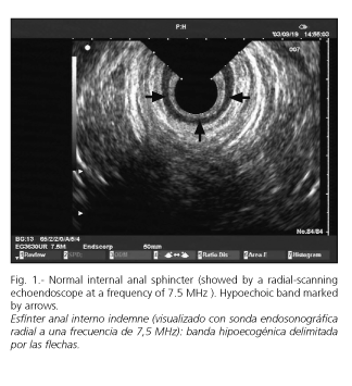
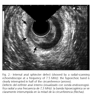
<body><![CDATA[<p>We performed an anal inspection and a digital rectal examination. There were no anatomical defects, but a severe hypotonic anal sphincter was encountered. Since there is a radial-scanning ultrasound endoscope with frontal vision (Pentax EG3630UR) available in our Unit, an endoscopic review of the rectal and sigmoid mucosa was carried out, with no abnormalities being detected. A radial endoscopic ultrasonography was performed using a frequency of 7.5 MHz during the same exploration. At the middle of the anal canal, the internal anal sphincter is seen as a hypoechoic ring under normal conditions (<a href="#f1">Fig. 1</a>). In this patient there was a 50% defect at this hypoechoic band representing the internal anal sphincter (<a href="#f2">Fig. 2</a>). No defects in the external anal sphincter were seen, other than it was slightly thin. The patient had fecal incontinence due to proctologic surgery with internal and sphincter damage. Therefore, the patient had fecal incontinence due to her proctologic surgery having damaged the internal anal sphincter. An anorectal manometry was performed, and it confirmed the internal anal sphincter hypotonia and also showed a lower pressure at the external anal sphincter.</p>     <p>Endoscopic ultrasonography of the anal canal is an accurate technique for the evaluation of the external and internal anal sphincters (1-3). The sensitivity for the detection of abnormalities at both sphincters is about 100%, since the specificity for defects in the external sphincter is 100%, and 95.5% regarding the internal sphincter (4). Endoanal ultrasound is essential in order to plan fecal incontinence surgery, and mandatory along with a functional study of the anal canal in Units where pelvic floor surgery is performed.</p>     <p><b>REFERENCES</b></p>     <p>1. Segura Cabral JM, Olveira Mart&iacute;n A, del Valle Hern&aacute;ndez E. Ecograf&iacute;a endoanal y endorrectal. Gastroenterol Hepatol 2001; 24 (3): 135-42.</p>     <p>2. Ram&iacute;rez Rodr&iacute;guez JM, Palacios Fanlo MJ, Artigas Marco V, Aguilella Diago V, Mart&iacute;nez D&iacute;ez M. Ecograf&iacute;a endoanal en el estudio de la anatom&iacute;a del canal anal: normalidad y variantes ecogr&aacute;ficas. Rev Esp Enferm Dig 2001; 93 (6): 364-7.</p>     <p>3. Schwartz DA, Harewood GC, Wiersema MJ. EUS for rectal disease. Gastrointest Endosc 2002; 56 (1): 100-9.</p>     <p>4. Deek K, Kumar D, Williams J, Olliff J, Keighley M. Anal sphincter defects. Correlation between endoanal ultrasound and surgery. Ann Surg 1993; 218 (2): 201-5.</p>      ]]></body>
</article>
