<?xml version="1.0" encoding="ISO-8859-1"?><article xmlns:mml="http://www.w3.org/1998/Math/MathML" xmlns:xlink="http://www.w3.org/1999/xlink" xmlns:xsi="http://www.w3.org/2001/XMLSchema-instance">
<front>
<journal-meta>
<journal-id>1130-0108</journal-id>
<journal-title><![CDATA[Revista Española de Enfermedades Digestivas]]></journal-title>
<abbrev-journal-title><![CDATA[Rev. esp. enferm. dig.]]></abbrev-journal-title>
<issn>1130-0108</issn>
<publisher>
<publisher-name><![CDATA[Sociedad Española de Patología Digestiva]]></publisher-name>
</publisher>
</journal-meta>
<article-meta>
<article-id>S1130-01082009001000007</article-id>
<title-group>
<article-title xml:lang="en"><![CDATA[Endoscopic ultrasound (EUS) elastography of the liver]]></article-title>
<article-title xml:lang="es"><![CDATA[Elastografía hepática guiada por ultrasonografía endoscópica]]></article-title>
</title-group>
<contrib-group>
<contrib contrib-type="author">
<name>
<surname><![CDATA[Iglesias García J]]></surname>
<given-names><![CDATA[J.]]></given-names>
</name>
<xref ref-type="aff" rid="A01"/>
<xref ref-type="aff" rid="A02"/>
</contrib>
<contrib contrib-type="author">
<name>
<surname><![CDATA[Lariño Noia]]></surname>
<given-names><![CDATA[J.]]></given-names>
</name>
<xref ref-type="aff" rid="A01"/>
<xref ref-type="aff" rid="A02"/>
</contrib>
<contrib contrib-type="author">
<name>
<surname><![CDATA[Souto]]></surname>
<given-names><![CDATA[R.]]></given-names>
</name>
<xref ref-type="aff" rid="A01"/>
<xref ref-type="aff" rid="A02"/>
</contrib>
<contrib contrib-type="author">
<name>
<surname><![CDATA[Álvarez Castro]]></surname>
<given-names><![CDATA[A.]]></given-names>
</name>
<xref ref-type="aff" rid="A01"/>
<xref ref-type="aff" rid="A02"/>
</contrib>
<contrib contrib-type="author">
<name>
<surname><![CDATA[Cigarrán]]></surname>
<given-names><![CDATA[B.]]></given-names>
</name>
<xref ref-type="aff" rid="A02"/>
<xref ref-type="aff" rid="A03"/>
</contrib>
<contrib contrib-type="author">
<name>
<surname><![CDATA[Domínguez Muñoz]]></surname>
<given-names><![CDATA[J. E.]]></given-names>
</name>
<xref ref-type="aff" rid="A01"/>
<xref ref-type="aff" rid="A02"/>
</contrib>
</contrib-group>
<aff id="A01">
<institution><![CDATA[,University Hospital of Santiago de Compostela Gastroenterology Department ]]></institution>
<addr-line><![CDATA[ ]]></addr-line>
</aff>
<aff id="A02">
<institution><![CDATA[,University Hospital of Santiago de Compostela Foundation for Research in Digestive Diseases ]]></institution>
<addr-line><![CDATA[Santiago de Compostela ]]></addr-line>
<country>Spain</country>
</aff>
<aff id="A03">
<institution><![CDATA[,Virxe da Xunqueira Hospital Department of Internal Medicine ]]></institution>
<addr-line><![CDATA[Cee ]]></addr-line>
<country>Spain</country>
</aff>
<pub-date pub-type="pub">
<day>00</day>
<month>10</month>
<year>2009</year>
</pub-date>
<pub-date pub-type="epub">
<day>00</day>
<month>10</month>
<year>2009</year>
</pub-date>
<volume>101</volume>
<numero>10</numero>
<fpage>717</fpage>
<lpage>718</lpage>
<copyright-statement/>
<copyright-year/>
<self-uri xlink:href="http://scielo.isciii.es/scielo.php?script=sci_arttext&amp;pid=S1130-01082009001000007&amp;lng=en&amp;nrm=iso"></self-uri><self-uri xlink:href="http://scielo.isciii.es/scielo.php?script=sci_abstract&amp;pid=S1130-01082009001000007&amp;lng=en&amp;nrm=iso"></self-uri><self-uri xlink:href="http://scielo.isciii.es/scielo.php?script=sci_pdf&amp;pid=S1130-01082009001000007&amp;lng=en&amp;nrm=iso"></self-uri></article-meta>
</front><body><![CDATA[ <p><font face="Verdana" size="2"><b>PICTURES IN DIGESTIVE PATHOLOGY</b></font></p>     <p>&nbsp;</p>     <p><font face="Verdana" size="4"><b>Endoscopic ultrasound (EUS) elastography of the liver</b></font></p>     <p><font face="Verdana" size="4"><b>Elastograf&iacute;a hep&aacute;tica guiada por ultrasonograf&iacute;a endosc&oacute;pica</b></font></p>     <p>&nbsp;</p>     <p>&nbsp;</p>     <p><font face="Verdana" size="2"><b>J. Iglesias Garc&iacute;a J<sup>1,2</sup>, J. Lari&ntilde;o Noia<sup>1,2</sup>, R. Souto<sup>1,2</sup>, A. &Aacute;lvarez Castro<sup>1,2</sup>, B. Cigarr&aacute;n<sup>2,3</sup> and J. E. Dom&iacute;nguez Mu&ntilde;oz<sup>1,2</sup></b></font></p>     <p><font face="Verdana" size="2"><sup>1</sup>Gastroenterology Department, and <sup>2</sup>Foundation for Research in Digestive Diseases. University Hospital of Santiago de Compostela. A Coru&ntilde;a, Spain. <sup>3</sup>Department of Internal Medicine. Virxe da Xunqueira Hospital. Cee. A Coru&ntilde;a, Spain</font></p>     <p>&nbsp;</p>     <p>&nbsp;</p>     ]]></body>
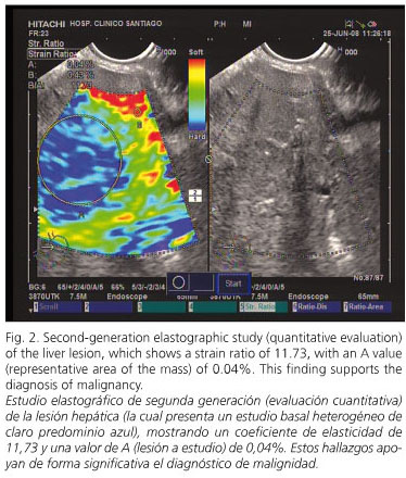
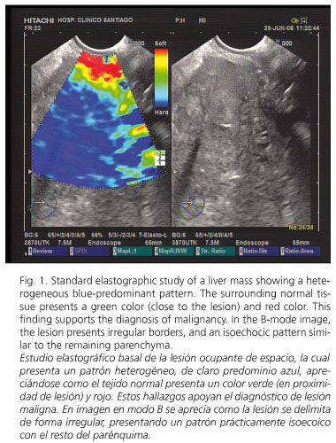
<body><![CDATA[<p><font face="Verdana" size="2"><b>Case report</b></font></p>     <p><font face="Verdana" size="2">A 68-year-old woman with no previous medical history, except a lumbar vertebra broken at the age of 35, was first admitted because of abdominal pain in the right hypochondria for the last few weeks, together with anorexia and weight loss of 6 kg. A lab workup showed a slight increase of bilirubin (2.34 mg/dL) as well as liver and cholestasis enzymes &#091;AST (245), ALT (375), GGT (636) and alkaline phosphatase (694)&#093;. The remaining parameters, including tumor markers, were completely normal. A helical CT scan identified an enlarged choledocus with an associated dilatation of the intrahepatic biliary tree. A liver solid lesion in segment IVB, 6 x 5.1 x 3.5 cm in size, was also detected, which had poorly defined borders and exofitic growing, suggesting metastasis. We obtained an endoscopic ultrasonogram (EUS) with the linear EUS probe (EG 3870 UTK; Pentax Europe GmbH, Hamburg, Germany) attached to a platform 900 (Hitachi Medical Systems GmbH, Wiesbaden, Germany), which includes the elastography module. During EUS we performed an elastographic evaluation of the solid liver lesion, and obtained a heterogeneous blue-predominant pattern with slight green areas and red lines resulting in a geographic appearance (characteristic of malignant tumors); the mass was clearly differentiated from normal tissue (<a href="#fig1">Fig. 1</a>). In the second-generation elastography study, the strain ratio obtained was 11.76, with a value for area A (corresponding to the lesion) of 0.04% (<a href="#fig2">Fig. 2</a>). A EUS-guided fine-needle aspiration of the liver lesion with a 22-gauge needle (Echo-Tip from Wilson-cook Medical, Winston-Salem, North Carolina, USA) was performed. This biopsy confirmed the diagnosis of cholangiocarcinoma. Finally, the patient underwent an endoscopic retrograde cholangio-pancreatography that confirmed the presence of a 2-cm filiform stenosis in the middle portion of the common bile duct (<a href="#fig3">Fig. 3</a>); a Wallstent<sup>&reg; </sup>(10 mm diameter, 60 mm length) was inserted for palliative treatment.</font></p>     <p align="center"><font face="Verdana" size="2"><a name="fig1"><img border="0" src="/img/revistas/diges/v101n10/imagenes1_fig1.jpg" width="380" height="500"></a></font></p>     <p align="center"><font face="Verdana" size="2"><a name="fig2"><img border="0" src="/img/revistas/diges/v101n10/imagenes1_fig2.jpg" width="374" height="440"></a></font></p>     <p align="center"><font face="Verdana" size="2"><a name="fig3"><img border="0" src="/img/revistas/diges/v101n10/imagenes1_fig3.jpg" width="378" height="380"></a></font></p>     <p align="center">&nbsp;</p>     <p><font face="Verdana" size="2"><b>Discussion</b></font></p>     <p><font face="Verdana" size="2">Transient liver elastography is mainly used for the quantification of liver fibrosis. In this setting, Fibroscan<sup>&reg;</sup> has been the method most commonly used to perform this quantification, allowing an easy, quick and a non-invasive measurement of tissue stiffness. Many publications analyze the usefulness of this technique, which demonstrates a very high diagnostic accuracy, close to 90%, for the determination of cirrhosis, but more limited for the evaluation of fibrosis in early stages (1). However, there are few studies evaluating its usefulness in the differential diagnosis of solid liver lesions.</font></p>     <p><font face="Verdana" size="2">A new model of elastographic equipment has been developed by Hitachi<sup>&reg;</sup>, which allows a real-time evaluation of tissue stiffness; it has been used for the analysis of superficial organ lesions, associated with percutaneous ultrasounds (2), and of pancreatic lesions and lymph nodes, associated with EUS (3,4). The technology is based on the detection of small structure deformations within the B-mode image as caused by compression, so that strain is smaller in hard tissue versus soft tissue. The degree of deformation is used as an indicator of tissue stiffness. Different elasticity values are marked with different colors resulting in different tissue elasticity patterns.</font></p>     <p><font face="Verdana" size="2">Tissue elasticity is usually uniform in benign lesions, with stiffness ranging from soft to intermediate (colors yellow and green); on the other hand, malignant tumors grow in a disorganized manner, and show heterogeneous patterns with hard tissue (blue color) (4,5). In order to obviate the bias related to the subjective interpretation of the color map, a quantitative analysis is possible with second-generation elastogragphy. With this method, two different areas (A and B) from the region of interest are selected: area A is representative of the mass, and area B refers to a soft reference area; finally, a B/A ratio (strain ratio) is considered the result of the elastographic evaluation. Data published on this new technique are scarce, but malignant lesions show higher strain ratios when compared to benign lesions (5).</font></p>     ]]></body>
<body><![CDATA[<p><font face="Verdana" size="2">We present two images demonstrating the feasibility of a EUS-guided elastographic study of the liver, both standard and second-generation. This is possible due to the proximity of the liver (mainly left liver lobe) to the gastric and duodenal wall. The probe is pressed against the wall with only the pressure needed for an optimal and stable B-mode image at 7.5 MHz. In conclusion, liver EUS-guided elastography could be a useful tool in the evaluation of some patients with solid liver tumors.</font></p>     <p>&nbsp;</p>     <p><font face="Verdana" size="2"><b>References</b></font></p>     <!-- ref --><p><font face="Verdana" size="2">1. Talwalkar JA. Elastography for detecting hepatic fibrosis: options and considerations. Gastroenterology 2008; 135: 299-302.</font>&nbsp;&nbsp;&nbsp;&nbsp;&nbsp;&nbsp;&nbsp;&nbsp;[&#160;<a href="javascript:void(0);" onclick="javascript: window.open('/scielo.php?script=sci_nlinks&ref=5325016&pid=S1130-0108200900100000700001&lng=','','width=640,height=500,resizable=yes,scrollbars=1,menubar=yes,');">Links</a>&#160;]<!-- end-ref --><!-- ref --><p><font face="Verdana" size="2">2. Gao L, Parker KJ, Lerner RM, Levinson SF. Imaging of the elasticity properties of tissue-a review. Ultrasound Med Biol 1996; 22: 959-77.</font>&nbsp;&nbsp;&nbsp;&nbsp;&nbsp;&nbsp;&nbsp;&nbsp;[&#160;<a href="javascript:void(0);" onclick="javascript: window.open('/scielo.php?script=sci_nlinks&ref=5325017&pid=S1130-0108200900100000700002&lng=','','width=640,height=500,resizable=yes,scrollbars=1,menubar=yes,');">Links</a>&#160;]<!-- end-ref --><!-- ref --><p><font face="Verdana" size="2">3. Saftoiu A, Vilman P. Endoscopic ultrasound elastography - a new imaging technique for the visualization of tissue elasticity distribution. J Gastrointestin Liver Dis 2006; 15: 161-5.</font>&nbsp;&nbsp;&nbsp;&nbsp;&nbsp;&nbsp;&nbsp;&nbsp;[&#160;<a href="javascript:void(0);" onclick="javascript: window.open('/scielo.php?script=sci_nlinks&ref=5325018&pid=S1130-0108200900100000700003&lng=','','width=640,height=500,resizable=yes,scrollbars=1,menubar=yes,');">Links</a>&#160;]<!-- end-ref --><!-- ref --><p><font face="Verdana" size="2">4. Giovannini M, Hookey LC, Bories E, Pesenti C, Monges G, Delpero JR. Endoscopic ultrasound elastography: the first step towards virtual biopsy? Preliminary results in 49 patients. Endoscopy 2006; 38(4): 344-8.</font>&nbsp;&nbsp;&nbsp;&nbsp;&nbsp;&nbsp;&nbsp;&nbsp;[&#160;<a href="javascript:void(0);" onclick="javascript: window.open('/scielo.php?script=sci_nlinks&ref=5325019&pid=S1130-0108200900100000700004&lng=','','width=640,height=500,resizable=yes,scrollbars=1,menubar=yes,');">Links</a>&#160;]<!-- end-ref --><!-- ref --><p><font face="Verdana" size="2">5. Iglesias-Garc&iacute;a J, Lari&ntilde;o-Noia J, Dominguez-Mu&ntilde;oz JE. Elastograf&iacute;a pancre&aacute;tica. Gastroenterolog&iacute;a y Hepatolog&iacute;a Continuada 2009 (en prensa).</font>&nbsp;&nbsp;&nbsp;&nbsp;&nbsp;&nbsp;&nbsp;&nbsp;[&#160;<a href="javascript:void(0);" onclick="javascript: window.open('/scielo.php?script=sci_nlinks&ref=5325020&pid=S1130-0108200900100000700005&lng=','','width=640,height=500,resizable=yes,scrollbars=1,menubar=yes,');">Links</a>&#160;]<!-- end-ref --> ]]></body><back>
<ref-list>
<ref id="B1">
<label>1</label><nlm-citation citation-type="journal">
<person-group person-group-type="author">
<name>
<surname><![CDATA[Talwalkar]]></surname>
<given-names><![CDATA[JA]]></given-names>
</name>
</person-group>
<article-title xml:lang="en"><![CDATA[Elastography for detecting hepatic fibrosis: options and considerations]]></article-title>
<source><![CDATA[Gastroenterology]]></source>
<year>2008</year>
<volume>135</volume>
<page-range>299-302</page-range></nlm-citation>
</ref>
<ref id="B2">
<label>2</label><nlm-citation citation-type="journal">
<person-group person-group-type="author">
<name>
<surname><![CDATA[Gao]]></surname>
<given-names><![CDATA[L]]></given-names>
</name>
<name>
<surname><![CDATA[Parker]]></surname>
<given-names><![CDATA[KJ]]></given-names>
</name>
<name>
<surname><![CDATA[Lerner]]></surname>
<given-names><![CDATA[RM]]></given-names>
</name>
<name>
<surname><![CDATA[Levinson]]></surname>
<given-names><![CDATA[SF]]></given-names>
</name>
</person-group>
<article-title xml:lang="en"><![CDATA[Imaging of the elasticity properties of tissue: a review]]></article-title>
<source><![CDATA[Ultrasound Med Biol]]></source>
<year>1996</year>
<volume>22</volume>
<page-range>959-77</page-range></nlm-citation>
</ref>
<ref id="B3">
<label>3</label><nlm-citation citation-type="journal">
<person-group person-group-type="author">
<name>
<surname><![CDATA[Saftoiu]]></surname>
<given-names><![CDATA[A]]></given-names>
</name>
<name>
<surname><![CDATA[Vilman]]></surname>
<given-names><![CDATA[P]]></given-names>
</name>
</person-group>
<article-title xml:lang="en"><![CDATA[Endoscopic ultrasound elastography: a new imaging technique for the visualization of tissue elasticity distribution]]></article-title>
<source><![CDATA[J Gastrointestin Liver Dis]]></source>
<year>2006</year>
<volume>15</volume>
<page-range>161-5</page-range></nlm-citation>
</ref>
<ref id="B4">
<label>4</label><nlm-citation citation-type="journal">
<person-group person-group-type="author">
<name>
<surname><![CDATA[Giovannini]]></surname>
<given-names><![CDATA[M]]></given-names>
</name>
<name>
<surname><![CDATA[Hookey]]></surname>
<given-names><![CDATA[LC]]></given-names>
</name>
<name>
<surname><![CDATA[Bories]]></surname>
<given-names><![CDATA[E]]></given-names>
</name>
<name>
<surname><![CDATA[Pesenti]]></surname>
<given-names><![CDATA[C]]></given-names>
</name>
<name>
<surname><![CDATA[Monges]]></surname>
<given-names><![CDATA[G]]></given-names>
</name>
<name>
<surname><![CDATA[Delpero]]></surname>
<given-names><![CDATA[JR]]></given-names>
</name>
</person-group>
<article-title xml:lang="en"><![CDATA[Endoscopic ultrasound elastography: the first step towards virtual biopsy? Preliminary results in 49 patients]]></article-title>
<source><![CDATA[Endoscopy]]></source>
<year>2006</year>
<volume>38</volume>
<numero>4</numero>
<issue>4</issue>
<page-range>344-8</page-range></nlm-citation>
</ref>
<ref id="B5">
<label>5</label><nlm-citation citation-type="journal">
<person-group person-group-type="author">
<name>
<surname><![CDATA[Iglesias-García]]></surname>
<given-names><![CDATA[J]]></given-names>
</name>
<name>
<surname><![CDATA[Lariño-Noia]]></surname>
<given-names><![CDATA[J]]></given-names>
</name>
<name>
<surname><![CDATA[Dominguez-Muñoz]]></surname>
<given-names><![CDATA[JE]]></given-names>
</name>
</person-group>
<article-title xml:lang="es"><![CDATA[Elastografía pancreática]]></article-title>
<source><![CDATA[Gastroenterología y Hepatología Continuada]]></source>
<year>2009</year>
</nlm-citation>
</ref>
</ref-list>
</back>
</article>