<?xml version="1.0" encoding="ISO-8859-1"?><article xmlns:mml="http://www.w3.org/1998/Math/MathML" xmlns:xlink="http://www.w3.org/1999/xlink" xmlns:xsi="http://www.w3.org/2001/XMLSchema-instance">
<front>
<journal-meta>
<journal-id>1130-0108</journal-id>
<journal-title><![CDATA[Revista Española de Enfermedades Digestivas]]></journal-title>
<abbrev-journal-title><![CDATA[Rev. esp. enferm. dig.]]></abbrev-journal-title>
<issn>1130-0108</issn>
<publisher>
<publisher-name><![CDATA[Sociedad Española de Patología Digestiva]]></publisher-name>
</publisher>
</journal-meta>
<article-meta>
<article-id>S1130-01082010001000013</article-id>
<title-group>
<article-title xml:lang="en"><![CDATA[Incidental finding of cystadenocarcinoma of the appendix]]></article-title>
<article-title xml:lang="es"><![CDATA[Hallazgo incidental de cistoadenocarcinoma apendicular]]></article-title>
</title-group>
<contrib-group>
<contrib contrib-type="author">
<name>
<surname><![CDATA[Cases-Baldó]]></surname>
<given-names><![CDATA[M. J.]]></given-names>
</name>
<xref ref-type="aff" rid="A01"/>
</contrib>
<contrib contrib-type="author">
<name>
<surname><![CDATA[Alcalá Martínez-Gómez]]></surname>
<given-names><![CDATA[D. de]]></given-names>
</name>
<xref ref-type="aff" rid="A01"/>
</contrib>
<contrib contrib-type="author">
<name>
<surname><![CDATA[García-García]]></surname>
<given-names><![CDATA[M. L.]]></given-names>
</name>
<xref ref-type="aff" rid="A01"/>
</contrib>
<contrib contrib-type="author">
<name>
<surname><![CDATA[Pérez Ramos]]></surname>
<given-names><![CDATA[M.]]></given-names>
</name>
<xref ref-type="aff" rid="A01"/>
</contrib>
<contrib contrib-type="author">
<name>
<surname><![CDATA[Aguayo-Albasini]]></surname>
<given-names><![CDATA[J. L.]]></given-names>
</name>
<xref ref-type="aff" rid="A02"/>
</contrib>
</contrib-group>
<aff id="A01">
<institution><![CDATA[,Hospital General Universitario Morales Meseguer Service of Pathological Anatomy ]]></institution>
<addr-line><![CDATA[ ]]></addr-line>
</aff>
<aff id="A02">
<institution><![CDATA[,Hospital General Universitario Morales Meseguer Service of General Surgery y Digestive System ]]></institution>
<addr-line><![CDATA[Murcia ]]></addr-line>
<country>Spain</country>
</aff>
<pub-date pub-type="pub">
<day>00</day>
<month>10</month>
<year>2010</year>
</pub-date>
<pub-date pub-type="epub">
<day>00</day>
<month>10</month>
<year>2010</year>
</pub-date>
<volume>102</volume>
<numero>10</numero>
<fpage>613</fpage>
<lpage>614</lpage>
<copyright-statement/>
<copyright-year/>
<self-uri xlink:href="http://scielo.isciii.es/scielo.php?script=sci_arttext&amp;pid=S1130-01082010001000013&amp;lng=en&amp;nrm=iso"></self-uri><self-uri xlink:href="http://scielo.isciii.es/scielo.php?script=sci_abstract&amp;pid=S1130-01082010001000013&amp;lng=en&amp;nrm=iso"></self-uri><self-uri xlink:href="http://scielo.isciii.es/scielo.php?script=sci_pdf&amp;pid=S1130-01082010001000013&amp;lng=en&amp;nrm=iso"></self-uri></article-meta>
</front><body><![CDATA[ <p><font face="Verdana" size="2"><b>LETTERS TO THE EDITOR</b></font></p>     <p>&nbsp;</p>     <p><font face="Verdana" size="4"><b>Incidental finding of cystadenocarcinoma of the appendix</b></font></p>     <p><font face="Verdana" size="4"><b>Hallazgo incidental de cistoadenocarcinoma apendicular</b></font></p>     <p>&nbsp;</p>     <p>&nbsp;</p> <hr size="1">     <p><font face="Verdana" size="2"><i>Key words: Incidental cystadenocarcinoma. Carcinoma of the appendix. Cystadenocarcinoma of the appendix.</i></font></p>     <p><font face="Verdana" size="2"><i>Palabras clave: Cistoadenocarcinoma incidental. Tumores apendiculares. Ciastoadenocarcinoma apendicular.</i></font></p> <hr size="1">     <p>&nbsp;</p>     <p>&nbsp;</p>     ]]></body>
<body><![CDATA[<p><font face="Verdana" size="2"><i>Dear Editor,</i></font></p>     <p><font face="Verdana" size="2">The incidence of appendicular tumours is relatively low (0.5%). Preoperative diagnosis is difficult and in as many as one-half of cases, the macroscopic appearance of the appendix during surgery suggests a diagnosis of appendicular neoplasia. Below, we describe the case of a patient who underwent surgery for infiltrating neoplasia of the bladder with an incidental finding of mucinous adenocarcinoma of the appendix.</font></p>     <p>&nbsp;</p>     <p><font face="Verdana" size="2"><b>Case report</b></font></p>     <p><font face="Verdana" size="2">
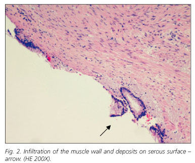
A 73 year-old hypertensive male being followed by urology for hematuria and vesicular neoformation. The findings on abdominal CT with intravenous contrast identify a diffuse parietal enlargement of the bladder wall that extends to the trigone consistent with bladder neoplasia infiltrating the left meatus and secondary obstructive uropathy of the left kidney with no related adenopathies or other tumoral dissemination. With a diagnosis of infiltrating bladder neoplasia, surgery is scheduled. During the surgery, prostatectomy and radical cystectomy with Bricker shunt are performed. The macroscopic appearance of the appendix is normal, performed as part of the appendicectomy. The pathological anatomy of the lesion confirms the diagnosis of transitional cell carcinoma of the bladder and the incidental finding in the appendicular vertex of a cystadenocarinoma of the appendix that infiltrates the serosal membranes with free resection margins and without related adenopathies (Figs. <a href="#f1">1</a> and <a href="#f2">2</a>).</font></p>     <p align="center"><font face="Verdana" size="2"><a name="f1"><img border="0" src="/img/revistas/diges/v102n10/carta3_f1.jpg" width="394" height="332"></a></font></p>     <p align="center"><font face="Verdana" size="2"><a name="f2"><img border="0" src="/img/revistas/diges/v102n10/carta3_f2.jpg" width="386" height="322"></a></font></p>     <p>&nbsp;</p>     <p><font face="Verdana" size="2">Post-operatory recovery without complications and the patient is released eight days after the surgery. After consulting with the hospital's General Surgery Service for histological finding on the appendix, it is decided to perform a colonoscopy and monitor the patient clinically.</font></p>     <p><font face="Verdana" size="2">Appendicular tumours represent 0.5% of all intestinal tumours. Fifteen to twenty per cent of these tumours are associated with other malignant tumours of the gastrointestinal tract. The most frequent of these are carcinoid tumours (80-90% of appendicular tumours) followed by mucinous cystoadenocarcinoma and adenocarcinoma of the colon.</font></p>     ]]></body>
<body><![CDATA[<p><font face="Verdana" size="2">Preoperative diagnosis is difficult and its clinical presentation mimics acute appendicitis in most patients. Other symptoms are abdominal pain, palpable mass on the lower abdominal quadrant or rectorrhagia. The tomographic finding of a solid-cystic mass dependent upon the appendix with peripheral enhancement or irregularities on the wall and locoregional adenopathies can support the preoperative diagnosis of appendicular tumours (1,2).</font></p>     <p><font face="Verdana" size="2">Carcinoid tumours less than 1 cm rarely metastasize and simple appendectomy is sufficient. In tumours larger than 2 cm found macroscopically during appendectomy, it is necessary to look for adenopathies and remove them. If metastasis is histologically confirmed, hemicolectomy is recommended (3). In adenocarcinomas of a colonic origin, right hemicolectomy should be performed systematically.</font></p>     <p><font face="Verdana" size="2">Mucinous cystoadenocarcinoma rarely disseminates lymphatically and grows slower than adenocarcinomas of a colonic origin. However, up to 50% of patients present intraabdominal dissemination, metastasis or pseudomyxoma peritonei. Incidental diagnosis, as occurred in this case, is an infrequent finding and unlike carcinoid tumours, there is no consensus regarding treatment (4,5).</font></p>     <p><font face="Verdana" size="2">Gonz&aacute;lez-Moreno and Sugarbaker (5) retrospectively analysed the long-term survival of 501 patients with epithelial tumours of the appendix. In 198 patients, the surgical technique used was appendicectomy while in 280 patients (55.9%) the technique was right hemicolectomy. According to this study, there were no significant differences between survival and the surgical technique (appendicectomy vs. hemicolectomy) and survival was higher in those cases where intraoperative chemotherapy was applied following the appendicectomy or right hemicolectomy. The authors concluded that right hemicolectomy should be reserved for cases where the mesoappendix or ileocolic ganglia are affected or cases of affected resection margins.</font></p>     <p><font face="Verdana" size="2">In the case described, the tumour was localised in the appendix; no related adenopathies were identified and the resection margin was free. We consider that oncological surgery is ideal and should be used to rule out other synchronous gastrointestinal tumours.</font></p>     <p>&nbsp;</p>     <p align="right"><font face="Verdana" size="2"><b>M. J. Cases-Bald&oacute;, D. de Alcal&aacute; Mart&iacute;nez-G&oacute;mez, M. L. Garc&iacute;a-Garc&iacute;a, M. P&eacute;rez Ramos<sup>1</sup> and J. L. Aguayo-Albasini<sup>2</sup></b>    <br>Services of <sup>1</sup>Pathological Anatomy and <sup>2</sup>General Surgery y Digestive System. Hospital General Universitario Morales Meseguer. Murcia, Spain</font></p>     <p>&nbsp;</p>     <p><font face="Verdana" size="2"><b>References</b></font></p>     ]]></body>
<body><![CDATA[<!-- ref --><p><font face="Verdana" size="2">1. Pickhardt PJ, Levy AP, Rohrmann CA Jr, Dende AI. Primary neoplasm of the appendix: radiologic spectrum of disease with pathologic correlation. Radiographics 2003; 23: 645-62.</font>&nbsp;&nbsp;&nbsp;&nbsp;&nbsp;&nbsp;&nbsp;&nbsp;[&#160;<a href="javascript:void(0);" onclick="javascript: window.open('/scielo.php?script=sci_nlinks&ref=5302846&pid=S1130-0108201000100001300001&lng=','','width=640,height=500,resizable=yes,scrollbars=1,menubar=yes,');">Links</a>&#160;]<!-- end-ref --><!-- ref --><p><font face="Verdana" size="2">2. Pitiakoudis M, Argyropoulou PI, Tsaroucha AK, Prassopoulos P, Simopoulos C. Cystadenocarcinoma of the appendix: an incidental imaging finding in a patient with adenocarcinomas of the ascending and the sigmoid colon. BMC Gastroenterology 2003; 3: 30.</font>&nbsp;&nbsp;&nbsp;&nbsp;&nbsp;&nbsp;&nbsp;&nbsp;[&#160;<a href="javascript:void(0);" onclick="javascript: window.open('/scielo.php?script=sci_nlinks&ref=5302847&pid=S1130-0108201000100001300002&lng=','','width=640,height=500,resizable=yes,scrollbars=1,menubar=yes,');">Links</a>&#160;]<!-- end-ref --><!-- ref --><p><font face="Verdana" size="2">3. Marrie A. Chirurgie de l&aacute;ppendice il&eacute;o-caecal. France: Encycl. M&eacute;d. Chir. Elsevier ; 1991.</font>&nbsp;&nbsp;&nbsp;&nbsp;&nbsp;&nbsp;&nbsp;&nbsp;[&#160;<a href="javascript:void(0);" onclick="javascript: window.open('/scielo.php?script=sci_nlinks&ref=5302848&pid=S1130-0108201000100001300003&lng=','','width=640,height=500,resizable=yes,scrollbars=1,menubar=yes,');">Links</a>&#160;]<!-- end-ref --><!-- ref --><p><font face="Verdana" size="2">4. Lee J, Gardiner KR, Wilson BG. Incidental mucinous cystadenocarcinoma of the appendix. Postgrad Med J 1996; 72: 55-6.</font>&nbsp;&nbsp;&nbsp;&nbsp;&nbsp;&nbsp;&nbsp;&nbsp;[&#160;<a href="javascript:void(0);" onclick="javascript: window.open('/scielo.php?script=sci_nlinks&ref=5302849&pid=S1130-0108201000100001300004&lng=','','width=640,height=500,resizable=yes,scrollbars=1,menubar=yes,');">Links</a>&#160;]<!-- end-ref --><!-- ref --><p><font face="Verdana" size="2">5. Gonz&aacute;lez-Moreno S, Sugarbaker PH. Right hemicolectomy does not confer a survival advantage in patients with mucinous carcinoma of the appendix and peritoneal seeding. BJ Surg 2004; 91: 304-11.</font>&nbsp;&nbsp;&nbsp;&nbsp;&nbsp;&nbsp;&nbsp;&nbsp;[&#160;<a href="javascript:void(0);" onclick="javascript: window.open('/scielo.php?script=sci_nlinks&ref=5302850&pid=S1130-0108201000100001300005&lng=','','width=640,height=500,resizable=yes,scrollbars=1,menubar=yes,');">Links</a>&#160;]<!-- end-ref --> ]]></body><back>
<ref-list>
<ref id="B1">
<label>1</label><nlm-citation citation-type="journal">
<person-group person-group-type="author">
<name>
<surname><![CDATA[Pickhardt]]></surname>
<given-names><![CDATA[PJ]]></given-names>
</name>
<name>
<surname><![CDATA[Levy]]></surname>
<given-names><![CDATA[AP]]></given-names>
</name>
<name>
<surname><![CDATA[Rohrmann]]></surname>
<given-names><![CDATA[CA Jr]]></given-names>
</name>
<name>
<surname><![CDATA[Dende]]></surname>
<given-names><![CDATA[AI]]></given-names>
</name>
</person-group>
<article-title xml:lang="en"><![CDATA[Primary neoplasm of the appendix: radiologic spectrum of disease with pathologic correlation]]></article-title>
<source><![CDATA[Radiographics]]></source>
<year>2003</year>
<volume>23</volume>
<page-range>645-62</page-range></nlm-citation>
</ref>
<ref id="B2">
<label>2</label><nlm-citation citation-type="journal">
<person-group person-group-type="author">
<name>
<surname><![CDATA[Pitiakoudis]]></surname>
<given-names><![CDATA[M]]></given-names>
</name>
<name>
<surname><![CDATA[Argyropoulou]]></surname>
<given-names><![CDATA[PI]]></given-names>
</name>
<name>
<surname><![CDATA[Tsaroucha]]></surname>
<given-names><![CDATA[AK]]></given-names>
</name>
<name>
<surname><![CDATA[Prassopoulos]]></surname>
<given-names><![CDATA[P]]></given-names>
</name>
<name>
<surname><![CDATA[Simopoulos]]></surname>
<given-names><![CDATA[C]]></given-names>
</name>
</person-group>
<article-title xml:lang="en"><![CDATA[Cystadenocarcinoma of the appendix: an incidental imaging finding in a patient with adenocarcinomas of the ascending and the sigmoid colon]]></article-title>
<source><![CDATA[BMC Gastroenterology]]></source>
<year>2003</year>
<volume>3</volume>
<page-range>30</page-range></nlm-citation>
</ref>
<ref id="B3">
<label>3</label><nlm-citation citation-type="book">
<person-group person-group-type="author">
<name>
<surname><![CDATA[Marrie]]></surname>
<given-names><![CDATA[A]]></given-names>
</name>
</person-group>
<article-title xml:lang="fr"><![CDATA[Chirurgie de láppendice iléo-caecal]]></article-title>
<source><![CDATA[Encycl. Méd. Chir]]></source>
<year>1991</year>
<publisher-name><![CDATA[Elsevier]]></publisher-name>
</nlm-citation>
</ref>
<ref id="B4">
<label>4</label><nlm-citation citation-type="journal">
<person-group person-group-type="author">
<name>
<surname><![CDATA[Lee]]></surname>
<given-names><![CDATA[J]]></given-names>
</name>
<name>
<surname><![CDATA[Gardiner]]></surname>
<given-names><![CDATA[KR]]></given-names>
</name>
<name>
<surname><![CDATA[Wilson]]></surname>
<given-names><![CDATA[BG]]></given-names>
</name>
</person-group>
<article-title xml:lang="en"><![CDATA[Incidental mucinous cystadenocarcinoma of the appendix]]></article-title>
<source><![CDATA[Postgrad Med J]]></source>
<year>1996</year>
<volume>72</volume>
<page-range>55-6</page-range></nlm-citation>
</ref>
<ref id="B5">
<label>5</label><nlm-citation citation-type="journal">
<person-group person-group-type="author">
<name>
<surname><![CDATA[González-Moreno]]></surname>
<given-names><![CDATA[S]]></given-names>
</name>
<name>
<surname><![CDATA[Sugarbaker]]></surname>
<given-names><![CDATA[PH]]></given-names>
</name>
</person-group>
<article-title xml:lang="en"><![CDATA[Right hemicolectomy does not confer a survival advantage in patients with mucinous carcinoma of the appendix and peritoneal seeding]]></article-title>
<source><![CDATA[BJ Surg]]></source>
<year>2004</year>
<volume>91</volume>
<page-range>304-11</page-range></nlm-citation>
</ref>
</ref-list>
</back>
</article>
