<?xml version="1.0" encoding="ISO-8859-1"?><article xmlns:mml="http://www.w3.org/1998/Math/MathML" xmlns:xlink="http://www.w3.org/1999/xlink" xmlns:xsi="http://www.w3.org/2001/XMLSchema-instance">
<front>
<journal-meta>
<journal-id>1130-0108</journal-id>
<journal-title><![CDATA[Revista Española de Enfermedades Digestivas]]></journal-title>
<abbrev-journal-title><![CDATA[Rev. esp. enferm. dig.]]></abbrev-journal-title>
<issn>1130-0108</issn>
<publisher>
<publisher-name><![CDATA[Sociedad Española de Patología Digestiva]]></publisher-name>
</publisher>
</journal-meta>
<article-meta>
<article-id>S1130-01082014000700007</article-id>
<title-group>
<article-title xml:lang="en"><![CDATA[Pancreatic dysontogenetic cysts]]></article-title>
<article-title xml:lang="es"><![CDATA[Quistes disontogénicos pancreáticos]]></article-title>
</title-group>
<contrib-group>
<contrib contrib-type="author">
<name>
<surname><![CDATA[Rodrigues-Pinto]]></surname>
<given-names><![CDATA[Eduardo]]></given-names>
</name>
<xref ref-type="aff" rid="A01"/>
</contrib>
<contrib contrib-type="author">
<name>
<surname><![CDATA[Pereira]]></surname>
<given-names><![CDATA[Pedro]]></given-names>
</name>
<xref ref-type="aff" rid="A01"/>
</contrib>
<contrib contrib-type="author">
<name>
<surname><![CDATA[Cunha]]></surname>
<given-names><![CDATA[Rui]]></given-names>
</name>
<xref ref-type="aff" rid="A02"/>
</contrib>
<contrib contrib-type="author">
<name>
<surname><![CDATA[Macedo]]></surname>
<given-names><![CDATA[Guilherme]]></given-names>
</name>
<xref ref-type="aff" rid="A01"/>
</contrib>
</contrib-group>
<aff id="A01">
<institution><![CDATA[,Centro Hospitalar São João Gastroenterology Department ]]></institution>
<addr-line><![CDATA[ ]]></addr-line>
</aff>
<aff id="A02">
<institution><![CDATA[,Centro Hospitalar São João Radiology Department ]]></institution>
<addr-line><![CDATA[São João ]]></addr-line>
<country>Portugal</country>
</aff>
<pub-date pub-type="pub">
<day>00</day>
<month>08</month>
<year>2014</year>
</pub-date>
<pub-date pub-type="epub">
<day>00</day>
<month>08</month>
<year>2014</year>
</pub-date>
<volume>106</volume>
<numero>7</numero>
<fpage>477</fpage>
<lpage>478</lpage>
<copyright-statement/>
<copyright-year/>
<self-uri xlink:href="http://scielo.isciii.es/scielo.php?script=sci_arttext&amp;pid=S1130-01082014000700007&amp;lng=en&amp;nrm=iso"></self-uri><self-uri xlink:href="http://scielo.isciii.es/scielo.php?script=sci_abstract&amp;pid=S1130-01082014000700007&amp;lng=en&amp;nrm=iso"></self-uri><self-uri xlink:href="http://scielo.isciii.es/scielo.php?script=sci_pdf&amp;pid=S1130-01082014000700007&amp;lng=en&amp;nrm=iso"></self-uri></article-meta>
</front><body><![CDATA[ 
    <p><font face="Verdana" size="2"><b>PICTURES IN DIGESTIVE PATHOLOGY</b></font></p>
    <p>&nbsp;</p>
    <p><font face="Verdana" size="4"><b>Pancreatic dysontogenetic cysts</b></font></p>
    <p><font face="Verdana" size="4"><b>Quistes disontogénicos pancreáticos</b></font></p>
    <p>&nbsp;</p>
    <p>&nbsp;</p>
    <p><font face="Verdana" size="2"><b>Eduardo Rodrigues-Pinto<sup>1</sup>, Pedro Pereira<sup>1</sup>, Rui Cunha<sup>2</sup> and Guilherme Macedo<sup>1</sup></b></font></p>
    <p><font face="Verdana" size="2"><sup>1</sup>Gastroenterology Department and <sup>2</sup>Radiology Department. Centro Hospitalar S&atilde;o Jo&atilde;o. Porto, Portugal</font></p>
    <p>&nbsp;</p>
    ]]></body>
<body><![CDATA[<p>&nbsp;</p>
    <p><font face="Verdana" size="2"><b>Case report</b></font></p>
    <p><font face="Verdana" size="2">
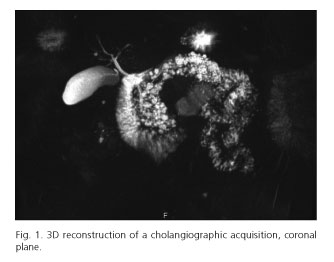
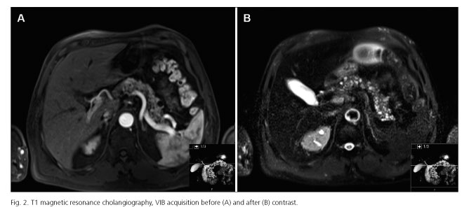
A male patient, 57-years-old, was submitted to abdominal imaging for study of urinary complaints. He had no complaints suggestive of pancreatic disease, no history of smoking, alcohol consumption or family history of pancreatic diseases. Abdominal imaging revealed multiple hypodense lesions mainly on the body and tail of the pancreas, consistent with cystic pancreatic formations (Figs. <a href="#f1">1</a> and <a href="#f2">2</a>) with associated parenchymatous calcifications without Wirsung dilatation or solid nodular lesions suggestive of malignancy. He also had multiple renal cysts. Remaining exams revealed no other changes. Pancreatic cysts are present in 2.4-13.5 % of patients without known pancreatic disease (1). Accurate categorization is important since some of them have malignant potential and should be resected. Pancreatic cysts are broadly classified as congenital-developmental cysts, retention cysts, duplication cysts, pseudocysts, neoplastic cysts and parasitic cysts (2). An important differential diagnosis is branch duct intraductal papillary mucinous neoplasm (IPMN). It is accepted that true cysts occur as a result of developmental anomalies related to the sequestration of primitive pancreatic ducts. In the presenting case, there was no previous history of pancreatitis or other pancreatic diseases, the patient was asymptomatic and also had renal involvement by cysts. His pancreatic cysts were multiple, small and diffusely involve the body and tail of pancreas. IPMNs are an important differential diagnosis, due to prognosis implications; however, Wirsung had a normal calibre, with no dilatation. Dysontogenetic pancreatic cysts are a very rare entity that may occur singly or in association with renal cysts, liver, central nervous system or retinal abnormalities (3). Currently there are no well-defined diagnostic criteria for dysontogenetic pancreatic cysts. Therefore it is important to always consider differential diagnosis, namely IPMNs, due to different needs of follow-up.</font></p>
    <p>&nbsp;</p>
    <p align="center"><font face="Verdana" size="2"><a name="f1"><img src="/img/revistas/diges/v106n7/imagenes1_fig1.jpg"></a></font></p>
    <p>&nbsp;</p>
    <p align="center"><font face="Verdana" size="2"><a name="f2"><img src="/img/revistas/diges/v106n7/imagenes1_fig2.jpg"></a></font></p>
    <p>&nbsp;</p>
    <p><font face="Verdana" size="2"><b>References</b></font></p>
    <!-- ref --><p><font face="Verdana" size="2">1. de Jong K, Nio CY, Mearadji B, Phoa SS, Engelbrecht MR, Dijkgraaf MG, et al. Disappointing interobserver agreement among radiologists for a classifying diagnosis of pancreatic cysts using magnetic resonance imaging. Pancreas 2012;41:278-82.    &nbsp;&nbsp;&nbsp;&nbsp;&nbsp;&nbsp;&nbsp;&nbsp;[&#160;<a href="javascript:void(0);" onclick="javascript: window.open('/scielo.php?script=sci_nlinks&ref=5380295&pid=S1130-0108201400070000700001&lng=','','width=640,height=500,resizable=yes,scrollbars=1,menubar=yes,');">Links</a>&#160;]<!-- end-ref --></font></p>    <!-- ref --><p><font face="Verdana" size="2">2. Rowe MI, O'Neill JA Jr, Grosfeld JL, Fonkalsrud EW, Coran AG, editors. Essentials of pediatric surgery. Disorders of the pancreas. Louis MO: MosbyYear Book; 1995. p. 663-74.    &nbsp;&nbsp;&nbsp;&nbsp;&nbsp;&nbsp;&nbsp;&nbsp;[&#160;<a href="javascript:void(0);" onclick="javascript: window.open('/scielo.php?script=sci_nlinks&ref=5380297&pid=S1130-0108201400070000700002&lng=','','width=640,height=500,resizable=yes,scrollbars=1,menubar=yes,');">Links</a>&#160;]<!-- end-ref --></font></p>    <!-- ref --><p><font face="Verdana" size="2">3. Nygaard KK, Walters W. Polycystic disease of the pancreas (Dysontogenetic cysts): Report of a case with partial pancreatectomy. Ann Surg 1937;106:49-53.    &nbsp;&nbsp;&nbsp;&nbsp;&nbsp;&nbsp;&nbsp;&nbsp;[&#160;<a href="javascript:void(0);" onclick="javascript: window.open('/scielo.php?script=sci_nlinks&ref=5380299&pid=S1130-0108201400070000700003&lng=','','width=640,height=500,resizable=yes,scrollbars=1,menubar=yes,');">Links</a>&#160;]<!-- end-ref --></font></p>
     ]]></body><back>
<ref-list>
<ref id="B1">
<label>1</label><nlm-citation citation-type="journal">
<person-group person-group-type="author">
<name>
<surname><![CDATA[de Jong]]></surname>
<given-names><![CDATA[K]]></given-names>
</name>
<name>
<surname><![CDATA[Nio]]></surname>
<given-names><![CDATA[CY]]></given-names>
</name>
<name>
<surname><![CDATA[Mearadji]]></surname>
<given-names><![CDATA[B]]></given-names>
</name>
<name>
<surname><![CDATA[Phoa]]></surname>
<given-names><![CDATA[SS]]></given-names>
</name>
<name>
<surname><![CDATA[Engelbrecht]]></surname>
<given-names><![CDATA[MR]]></given-names>
</name>
<name>
<surname><![CDATA[Dijkgraaf]]></surname>
<given-names><![CDATA[MG]]></given-names>
</name>
</person-group>
<article-title xml:lang="en"><![CDATA[Disappointing interobserver agreement among radiologists for a classifying diagnosis of pancreatic cysts using magnetic resonance imaging]]></article-title>
<source><![CDATA[Pancreas]]></source>
<year>2012</year>
<volume>41</volume>
<page-range>278-82</page-range></nlm-citation>
</ref>
<ref id="B2">
<label>2</label><nlm-citation citation-type="book">
<person-group person-group-type="author">
<name>
<surname><![CDATA[Rowe]]></surname>
<given-names><![CDATA[MI]]></given-names>
</name>
<name>
<surname><![CDATA[O'Neill]]></surname>
<given-names><![CDATA[JA Jr]]></given-names>
</name>
<name>
<surname><![CDATA[Grosfeld]]></surname>
<given-names><![CDATA[JL]]></given-names>
</name>
<name>
<surname><![CDATA[Fonkalsrud]]></surname>
<given-names><![CDATA[EW]]></given-names>
</name>
<name>
<surname><![CDATA[Coran]]></surname>
<given-names><![CDATA[AG]]></given-names>
</name>
</person-group>
<source><![CDATA[Essentials of pediatric surgery: Disorders of the pancreas]]></source>
<year>1995</year>
<page-range>663-74</page-range><publisher-loc><![CDATA[Louis^eMO MO]]></publisher-loc>
<publisher-name><![CDATA[MosbyYear Book]]></publisher-name>
</nlm-citation>
</ref>
<ref id="B3">
<label>3</label><nlm-citation citation-type="journal">
<person-group person-group-type="author">
<name>
<surname><![CDATA[Nygaard]]></surname>
<given-names><![CDATA[KK]]></given-names>
</name>
<name>
<surname><![CDATA[Walters]]></surname>
<given-names><![CDATA[W]]></given-names>
</name>
</person-group>
<article-title xml:lang="en"><![CDATA[Polycystic disease of the pancreas (Dysontogenetic cysts): Report of a case with partial pancreatectomy]]></article-title>
<source><![CDATA[Ann Surg]]></source>
<year>1937</year>
<volume>106</volume>
<page-range>49-53</page-range></nlm-citation>
</ref>
</ref-list>
</back>
</article>
