<?xml version="1.0" encoding="ISO-8859-1"?><article xmlns:mml="http://www.w3.org/1998/Math/MathML" xmlns:xlink="http://www.w3.org/1999/xlink" xmlns:xsi="http://www.w3.org/2001/XMLSchema-instance">
<front>
<journal-meta>
<journal-id>1130-0108</journal-id>
<journal-title><![CDATA[Revista Española de Enfermedades Digestivas]]></journal-title>
<abbrev-journal-title><![CDATA[Rev. esp. enferm. dig.]]></abbrev-journal-title>
<issn>1130-0108</issn>
<publisher>
<publisher-name><![CDATA[Sociedad Española de Patología Digestiva]]></publisher-name>
</publisher>
</journal-meta>
<article-meta>
<article-id>S1130-01082016001200003</article-id>
<article-id pub-id-type="doi">10.17235/reed.2016.4194/2016</article-id>
<title-group>
<article-title xml:lang="en"><![CDATA[Regression of gastric intestinal metaplasia after the eradication of Helicobacter pylori infection in a hospital in Mexico]]></article-title>
<article-title xml:lang="es"><![CDATA[Regresión de la metaplasia intestinal gástrica tras la erradicación de la infección por Helicobacter pylori en un hospital de México]]></article-title>
</title-group>
<contrib-group>
<contrib contrib-type="author">
<name>
<surname><![CDATA[Sánchez-Cuén]]></surname>
<given-names><![CDATA[Jaime Alberto]]></given-names>
</name>
<xref ref-type="aff" rid="A01"/>
<xref ref-type="aff" rid="A02"/>
</contrib>
<contrib contrib-type="author">
<name>
<surname><![CDATA[Irineo-Cabrales]]></surname>
<given-names><![CDATA[Ana Bertha]]></given-names>
</name>
<xref ref-type="aff" rid="A01"/>
<xref ref-type="aff" rid="A02"/>
</contrib>
<contrib contrib-type="author">
<name>
<surname><![CDATA[Bernal-Magaña]]></surname>
<given-names><![CDATA[Gregorio]]></given-names>
</name>
<xref ref-type="aff" rid="A01"/>
</contrib>
<contrib contrib-type="author">
<name>
<surname><![CDATA[Peraza-Garay]]></surname>
<given-names><![CDATA[Felipe de Jesús]]></given-names>
</name>
<xref ref-type="aff" rid="A03"/>
</contrib>
</contrib-group>
<aff id="A01">
<institution><![CDATA[,Instituto de Seguridad y Servicios Sociales de los Trabajadores del Estado Hospital Regional Department of Digestive Diseases]]></institution>
<addr-line><![CDATA[Culiacán Sinaloa]]></addr-line>
<country>Mexico</country>
</aff>
<aff id="A02">
<institution><![CDATA[,Universidad Autónoma de Sinaloa School of Medicine Department of Postgraduate Studies]]></institution>
<addr-line><![CDATA[Culiacán Sinaloa]]></addr-line>
<country>Mexico</country>
</aff>
<aff id="A03">
<institution><![CDATA[,Universidad Autónoma de Sinaloa Centro de Investigación y Docencia en Ciencias de la Salud ]]></institution>
<addr-line><![CDATA[Culiacán Sinaloa]]></addr-line>
<country>Mexico</country>
</aff>
<pub-date pub-type="pub">
<day>00</day>
<month>12</month>
<year>2016</year>
</pub-date>
<pub-date pub-type="epub">
<day>00</day>
<month>12</month>
<year>2016</year>
</pub-date>
<volume>108</volume>
<numero>12</numero>
<fpage>770</fpage>
<lpage>775</lpage>
<copyright-statement/>
<copyright-year/>
<self-uri xlink:href="http://scielo.isciii.es/scielo.php?script=sci_arttext&amp;pid=S1130-01082016001200003&amp;lng=en&amp;nrm=iso"></self-uri><self-uri xlink:href="http://scielo.isciii.es/scielo.php?script=sci_abstract&amp;pid=S1130-01082016001200003&amp;lng=en&amp;nrm=iso"></self-uri><self-uri xlink:href="http://scielo.isciii.es/scielo.php?script=sci_pdf&amp;pid=S1130-01082016001200003&amp;lng=en&amp;nrm=iso"></self-uri><abstract abstract-type="short" xml:lang="en"><p><![CDATA[Background: Intestinal metaplasia is a precursor lesion of gastric cancer. Infection by Helicobacter pylori is the principal cause of metaplasia. While evidence of the regression of metaplasia after treatment to eradicate this infection has been demonstrated, controversy remains with regard to this subject. Objective: The objective of this study was to determine the frequency of the regression of gastric intestinal metaplasia one year after the eradication of Helicobacter pylori. Methods: A prospective longitudinal designed study was carried out. The population studied in this research consisted of patients attending the Endoscopy Unit to undergo an upper endoscopy, in whom various symptoms indicated intestinal metaplasia associated with Helicobacter pylori, and who received standard empiric triple therapy to eradicate the bacteria. An upper endoscopy was performed in which four gastric biopsy samples were taken (two from the antrum and two from the body) before and after the eradicating treatment, with the endoscopic and histological findings studied after one year of monitoring. The statistical analysis was conducted using the Fisher's exact test and the McNemar's test. Results: Forty-six patients were studied, of whom 20 (43.5%) were men and 26 (56.5%) were women, with an average age of 58.9 (DE 11.2). Intestinal metaplasia was found in 46 (100%) patients before treatment and in 21 (45.7%) patients post-eradication. Complete intestinal metaplasia (type I) was found in 35 patients (76.1%) before treatment and in 11 (23.9%) patients post-eradication (p = 0.000), while incomplete intestinal metaplasia (type II) was found in 10 (21.7%) patients before treatment and in 10 (21.7%) patients post-eradication. Non-atrophic chronic gastritis was found in 35 (76.1%) patients before treatment and in 32 (69.6%) patients post-eradication. Conclusions: In this study, gastric intestinal metaplasia associated with Helicobacter pylori infection showed a regression of 54.3% one year after the eradication of this microorganism. This treatment could modify the natural history of the development of gastric cancer.]]></p></abstract>
<abstract abstract-type="short" xml:lang="es"><p><![CDATA[Introducción: la metaplasia intestinal es una lesión precursora del cáncer gástrico. La infección por Helicobacter pylori es la principal causa de metaplasia. Aunque se ha demostrado evidencia de regresión de metaplasia intestinal tras el tratamiento erradicador de esta infección, aún existen controversias al respecto. Objetivo: el objetivo de este estudio fue determinar la frecuencia de la regresión de la metaplasia intestinal gástrica al año de haber erradicado Helicobacter pylori. Métodos: se realizó un diseño prospectivo, longitudinal. La población estudiada fueron pacientes que acudieron a la Unidad de Endoscopia para la realización de una endoscopia digestiva alta por distintas indicaciones, comprobándose metaplasia intestinal asociada a Helicobacter pylori, y que recibieron terapia triple estándar empírica con erradicación de la bacteria. Se realizó una endoscopia digestiva alta con toma de cuatro muestras de biopsias gástricas (dos en antro y dos en cuerpo) antes y después del tratamiento erradicador, estudiándose los hallazgos endoscópicos e histológicos al año de seguimiento. Análisis estadístico con prueba exacta de Fisher y McNemar. Resultados: fueron estudiados 46 pacientes, 20 (43,5%) hombres y 26 (56,5%) mujeres, con edad media de 58,9 (DE 11,2) años. Se encontró metaplasia intestinal en 46 (100%) pacientes antes del tratamiento y en 21 (45,7%) tras la erradicación; metaplasia intestinal completa (tipo I) en 35 pacientes (76,1%) antes del tratamiento y en 11 (23,9%) posterior a la erradicación (p = 0,000); metaplasia intestinal incompleta (tipo II) en 10 (21,7%) pacientes antes del tratamiento y en 10 (21,7%) posterior a la erradicación; y gastritis crónica no atrófica en 35 (76,1%) pacientes antes del tratamiento y en 32 (69,6%) pacientes posterior a la erradicación. Conclusiones: en este estudio la metaplasia intestinal gástrica asociada a la infección por Helicobacter pylori presentó una regresión al año del 54,3% posterior a la erradicación de este microorganismo. Este tratamiento podría modificar la historia natural del desarrollo del cáncer gástrico.]]></p></abstract>
<kwd-group>
<kwd lng="en"><![CDATA[Metaplasia]]></kwd>
<kwd lng="en"><![CDATA[Helicobacter pylori]]></kwd>
<kwd lng="en"><![CDATA[Precancerous lesions]]></kwd>
<kwd lng="es"><![CDATA[Metaplasia]]></kwd>
<kwd lng="es"><![CDATA[Helicobacter pylori]]></kwd>
<kwd lng="es"><![CDATA[Lesiones precancerosas]]></kwd>
</kwd-group>
</article-meta>
</front><body><![CDATA[ <p><a name="top"></a><font face="Verdana" size="2"><b>ORIGINAL PAPERS</b></font></p>     <p>&nbsp;</p>     <p><font face="Verdana" size="4"><b>Regression of gastric intestinal metaplasia after the eradication of <i>Helicobacter pylori</i> infection in a hospital in Mexico</b></font></p>     <p><font face="Verdana" size="4"><b>Regresi&oacute;n de la metaplasia intestinal g&aacute;strica tras la erradicaci&oacute;n de la infecci&oacute;n por <i>Helicobacter pylori</i> en un hospital de M&eacute;xico</b></font></p>     <p>&nbsp;</p>     <p>&nbsp;</p>     <p><font face="Verdana" size="2"><b>Jaime Alberto S&aacute;nchez-Cu&eacute;n<sup>1,2</sup>, Ana Bertha Irineo-Cabrales<sup>1,2</sup>, Gregorio Bernal-Maga&ntilde;a<sup>1</sup> and Felipe de Jes&uacute;s Peraza-Garay<sup>3</sup></b></font></p>     <p><font face="Verdana" size="2"><sup>1</sup>Department of Digestive Diseases. Hospital Regional del Instituto de Seguridad y Servicios Sociales de los Trabajadores del Estado. Culiac&aacute;n, Sinaloa. Mexico.    <br><sup>2</sup>Department of Postgraduate Studies. School of Medicine. Universidad Aut&oacute;noma de Sinaloa. Culiac&aacute;n, Sinaloa. Mexico.    <br><sup>3</sup>Centro de Investigaci&oacute;n y Docencia en Ciencias de la Salud. Universidad Aut&oacute;noma de Sinaloa. Culiac&aacute;n, Sinaloa. Mexico</font></p>     ]]></body>
<body><![CDATA[<p><font face="Verdana" size="2"><a href="#bajo">Correspondence</a></font></p>     <p>&nbsp;</p>     <p>&nbsp;</p> <hr size="1">     <p><font face="Verdana" size="2"><b>ABSTRACT</b></font></p>     <p><font face="Verdana" size="2"><b>Background:</b> Intestinal metaplasia is a precursor lesion of gastric cancer. Infection by <i>Helicobacter pylori</i> is the principal cause of metaplasia. While evidence of the regression of metaplasia after treatment to eradicate this infection has been demonstrated, controversy remains with regard to this subject.    <br><b>Objective:</b> The objective of this study was to determine the frequency of the regression of gastric intestinal metaplasia one year after the eradication of <i>Helicobacter pylori</i>.    <br><b>Methods:</b> A prospective longitudinal designed study was carried out. The population studied in this research consisted of patients attending the Endoscopy Unit to undergo an upper endoscopy, in whom various symptoms indicated intestinal metaplasia associated with <i>Helicobacter pylori</i>, and who received standard empiric triple therapy to eradicate the bacteria. An upper endoscopy was performed in which four gastric biopsy samples were taken (two from the antrum and two from the body) before and after the eradicating treatment, with the endoscopic and histological findings studied after one year of monitoring. The statistical analysis was conducted using the Fisher's exact test and the McNemar's test.    <br><b>Results:</b> Forty-six patients were studied, of whom 20 (43.5%) were men and 26 (56.5%) were women, with an average age of 58.9 (DE 11.2). Intestinal metaplasia was found in 46 (100%) patients before treatment and in 21 (45.7%) patients post-eradication. Complete intestinal metaplasia (type I) was found in 35 patients (76.1%) before treatment and in 11 (23.9%) patients post-eradication (p = 0.000), while incomplete intestinal metaplasia (type II) was found in 10 (21.7%) patients before treatment and in 10 (21.7%) patients post-eradication. Non-atrophic chronic gastritis was found in 35 (76.1%) patients before treatment and in 32 (69.6%) patients post-eradication.    <br><b>Conclusions:</b> In this study, gastric intestinal metaplasia associated with <i>Helicobacter pylori</i> infection showed a regression of 54.3% one year after the eradication of this microorganism. This treatment could modify the natural history of the development of gastric cancer.</font></p>     <p><font face="Verdana" size="2"><b>Key words:</b> Metaplasia. <i>Helicobacter pylori</i>. Precancerous lesions.</font></p> <hr size="1">     ]]></body>
<body><![CDATA[<p><font face="Verdana" size="2"><b>RESUMEN</b></font></p>     <p><font face="Verdana" size="2"><b>Introducci&oacute;n:</b> la metaplasia intestinal es una lesi&oacute;n precursora del c&aacute;ncer g&aacute;strico. La infecci&oacute;n por <i>Helicobacter pylori</i> es la principal causa de metaplasia. Aunque se ha demostrado evidencia de regresi&oacute;n de metaplasia intestinal tras el tratamiento erradicador de esta infecci&oacute;n, a&uacute;n existen controversias al respecto.    <br><b>Objetivo:</b> el objetivo de este estudio fue determinar la frecuencia de la regresi&oacute;n de la metaplasia intestinal g&aacute;strica al a&ntilde;o de haber erradicado <i>Helicobacter pylori</i>.    <br><b>M&eacute;todos:</b> se realiz&oacute; un dise&ntilde;o prospectivo, longitudinal. La poblaci&oacute;n estudiada fueron pacientes que acudieron a la Unidad de Endoscopia para la realizaci&oacute;n de una endoscopia digestiva alta por distintas indicaciones, comprob&aacute;ndose metaplasia intestinal asociada a <i>Helicobacter pylori</i>, y que recibieron terapia triple est&aacute;ndar emp&iacute;rica con erradicaci&oacute;n de la bacteria. Se realiz&oacute; una endoscopia digestiva alta con toma de cuatro muestras de biopsias g&aacute;stricas (dos en antro y dos en cuerpo) antes y despu&eacute;s del tratamiento erradicador, estudi&aacute;ndose los hallazgos endosc&oacute;picos e histol&oacute;gicos al a&ntilde;o de seguimiento. An&aacute;lisis estad&iacute;stico con prueba exacta de Fisher y McNemar.    <br><b>Resultados:</b> fueron estudiados 46 pacientes, 20 (43,5%) hombres y 26 (56,5%) mujeres, con edad media de 58,9 (DE 11,2) a&ntilde;os. Se encontr&oacute; metaplasia intestinal en 46 (100%) pacientes antes del tratamiento y en 21 (45,7%) tras la erradicaci&oacute;n; metaplasia intestinal completa (tipo I) en 35 pacientes (76,1%) antes del tratamiento y en 11 (23,9%) posterior a la erradicaci&oacute;n (p = 0,000); metaplasia intestinal incompleta (tipo II) en 10 (21,7%) pacientes antes del tratamiento y en 10 (21,7%) posterior a la erradicaci&oacute;n; y gastritis cr&oacute;nica no atr&oacute;fica en 35 (76,1%) pacientes antes del tratamiento y en 32 (69,6%) pacientes posterior a la erradicaci&oacute;n.    <br><b>Conclusiones:</b> en este estudio la metaplasia intestinal g&aacute;strica asociada a la infecci&oacute;n por <i>Helicobacter pylori</i> present&oacute; una regresi&oacute;n al a&ntilde;o del 54,3% posterior a la erradicaci&oacute;n de este microorganismo. Este tratamiento podr&iacute;a modificar la historia natural del desarrollo del c&aacute;ncer g&aacute;strico.</font></p>     <p><font face="Verdana" size="2"><b>Palabras clave:</b> Metaplasia. <i>Helicobacter pylori</i>. Lesiones precancerosas.</font></p> <hr size="1">     <p>&nbsp;</p>     <p><font face="Verdana" size="2"><b>Introducci&oacute;n</b></font></p>     <p><font face="Verdana" size="2"><i>Helicobacter pylori (H. pylori)</i> infection is common, with a global prevalence of more than 50% and distinct geographical variations (1). When <i>H. pylori</i> colonize the stomach, the initial immune response of the gastric mucosa varies considerably according to the virulence of the strain (2). Reports describe an evolving succession of pre-neoplasic lesions (atrophic gastritis, intestinal metaplasia, displasia) developing from <i>H. pylori</i> infection and the development of gastric cancer (3). In the initial pre-cancerous lesion, atrophic gastritis is characterized by the death of the parietal cells and the loss of the glandular epithelium that covers the mucosa of the stomach (4). Intestinal metaplasia involves the replacement of glandular and/or foveolar epithelium by intestinal epithelium and is characterized by its morphological similarity to enterocytes. There are many classifications for intestinal metaplasia, with no universally accepted classification. Histopathological and histochemical studies have identified two types of metaplasia: type I, or complete, which is characterized by the presence of non-secretory absorption cells with well delineated brush-borders, Paneth cells, and cells that secrete sialomucins, and which corresponds to a phenotype from the small intestine; and type II, or incomplete or colonic, including types IIA and IIB, which are characterized by the presence of columnar cells and cells which secrete sialomucins (type IIA) and sulphomucins (type IIB) (5). Intestinal metaplasia is associated with <i>H. pylori</i> infection and bile reflux, although <i>H. pylori</i> is cited as the principal cause of metaplasia (6,7).</font></p>     ]]></body>
<body><![CDATA[<p><font face="Verdana" size="2">Some studies have demonstrated the following as risk factors for developing intestinal metaplasia: an age of &ge; 61, <i>H. pylori</i> infection, the ingestion of spicy food, a history of smoking (8), male gender, a family history of gastric cancer, a below-university educational level, the consumption of dairy products (9), and dietary factors, such as excessive salt consumption, deficient ascorbic acid, and insufficient ingestion of carotene (10). However, among the risk factors, <i>H. pylori</i> infection is considered to be the most significant risk factor for intestinal metaplasia (11). As few studies have been conducted that estimate the prevalence of intestinal metaplasia in the general asymptomatic population, it is necessary to carry out an upper endoscopy and a histological examination. The prevalence of intestinal metaplasia varies from 7.1% to 42.5% depending on country and diagnostic method (12).</font></p>     <p><font face="Verdana" size="2">Controversy has arisen with regard to the impact of the eradication of <i>H. pylori</i> and the risk of gastric carcinoma. There is evidence of the regression of precancerous lesions such as intestinal metaplasia after the eradication treatment (13). However, although some authors have observed a regression of gastric atrophy and low grade dysplasia (14), the reversibility of the intestinal metaplasia continues to be controversial (15,16). The objective of this study was to determine the frequency with which gastric intestinal metaplasia regresses one year after the eradication of <i>H. pylori</i> infection.</font></p>     <p>&nbsp;</p>     <p><font face="Verdana" size="2"><b>Material and methods</b></font></p>     <p><font face="Verdana" size="2">This research consisted of a longitudinal and prospective quasi-experimental study, with pre and post-test readings taken from solely one group. Approved by the Commission for Research and Ethics at the Hospital Regional of the Instituto de Seguridad y Servicios Sociales de los Trabajadores del Estado, or ISSSTE, in Culiac&aacute;n, Sinaloa, this study did not violate concepts of authorship, plagiarism, conflict of interests, or informed consent.</font></p>     <p><font face="Verdana" size="2">This research studied a population of adult patients attending the Endoscopy Unit to undergo an upper endoscopy for various symptoms (a dyspepsia test, gastrointestinal bleeding, anemia, and dysphagia, among others), and in whom the presence of gastric metaplasia associated with <i>H. pylori</i> infection has been identified. All the patients included in the study were treated with standard empiric triple therapy (40 mg omeprazole, 500 g clarithromycin, and 1 g amoxicillin, twice daily for two weeks). The study was conducted at the Department of Digestive Diseases at the ISSSTE Hospital Regional in the city of Culiacan, Sinaloa, in Northeastern Mexico, from June 2013 to June 2015. The patients were monitored for one year, with an upper endoscopy conducted one year after the eradication of <i>H. pylori</i>, and the findings of the stomach endoscopy were evaluated, as were the findings of the histological analysis performed on the gastric mucosa. The inclusion criteria were: both male and female subjects, aged between 30 and 80, in whom the eradication of <i>H. pylori</i> had been confirmed using the carbon-14 urea breath test and who had provided their informed consent. The exclusion criteria included patients with a history of two or more treatments for the eradication of <i>H. pylori</i> infection, pregnant women, and patients with a history of gastrointestinal surgery or a diagnosis of gastric cancer. The elimination criteria included patients who had not been monitored for a year, patients for whom the presence of <i>H. pylori</i> bacillus had been confirmed via histology during monitoring, and patients with errors in data collection.</font></p>     <p><font face="Verdana" size="2"><b>Definition of the variables</b></font></p>     <p><font face="Verdana" size="2">The histological and endoscopic findings of the gastric mucosa and <i>H. pylori</i> infection were defined according to the Sidney system (18-21). Gastric intestinal metaplasia was defined according to the Filipe and Jass classification (5). Gastrointestinal bleeding was defined according to the Forrest classification (22).</font></p>     <p><font face="Verdana" size="2"><b>Sample</b></font></p>     <p><font face="Verdana" size="2">The sample size was set at 49 patients, a power of 80% to detect a difference among the hypothetical proportions and the alternative of 0.20 (Delta) was calculated. The proportion for the null hypothesis was 0.30, assumed by the review of a Spanish study (17), and under the alternative hypothesis of 0.50. The bilateral (two tails) Z test was applied with a level of significance of 0.05, with a 10% expected patient loss. Due to convenience and the number of consecutive patients, the type of sampling was not probabilistic.</font></p>     ]]></body>
<body><![CDATA[<p><font face="Verdana" size="2"><b>Collection of data</b></font></p>     <p><font face="Verdana" size="2">The data were collected from a primary source via the direct observation of the stomach (fundus, body and antrum) via two gastric antrum samples, and two body samples taken via an upper video endoscopy, as well as findings from the histological study of the gastric mucosa samples.</font></p>     <p><font face="Verdana" size="2"><b>Equipment management, biological samples</b></font></p>     <p><font face="Verdana" size="2"><i><b>Upper endoscopy techniques and gastric biopsies</b></i></font></p>     <p><font face="Verdana" size="2">Oropharyngeal lidocaine spray was applied in a left lateral decubitus position and a mouthpiece was placed. The endoscopy was performed under intravenous sedation using an Olympus EG 29-90I video endoscope. The macroscopic characteristics of the stomach were registered. Four biopsies were taken (two in the antrum and two in the body) using FB 25K-1 series biopsy forceps. The endoscopies were evaluated by two endoscopy specialists.</font></p>     <p><font face="Verdana" size="2"><b>Identification of gastric intestinal metaplasia</b></font></p>     <p><font face="Verdana" size="2"><b><i>Histological study</i></b></font></p>     <p><font face="Verdana" size="2">The gastric mucosa biopsies (two from the antrum and two from the body) were processed using standard paraffin techniques with histological sections colored with hematoxylin and eosin and Giemsa stain. Complementary stains with toluidine blue were used, with a sensitivity and specificity of 96 and 99, respectively (23). The sections were then evaluated by two pathologists. Type I metaplasia was identified by the presence of non-secretory absorption cells with well delineated brush-borders and Paneth cells (enterocytes), while the presence of columnar cells were used to identify type II (24).</font></p>     <p><font face="Verdana" size="2"><i><b>Identification of H. pylori in gastric biopsy samples</b></i></font></p>     <p><font face="Verdana" size="2">Histological studies were performed with sections colored with hematoxylin and eosin, according to the criteria from the modified Sydney system and Giemsa stain (23).</font></p>     ]]></body>
<body><![CDATA[<p><font face="Verdana" size="2"><b>Statistical analysis</b></font></p>     <p><font face="Verdana" size="2">The data were analyzed with the SPSS v 16 software package (SPSS, Inc., Chicago IL, USA). Average and standard deviation were used for the quantitative variables, while the categorical variables were presented as frequencies and percentages. The Fisher's exact test and the McNemar's test with binomial distribution were used. The data are presented in tables and figures. A p value of 0.05 was considered as significant.</font></p>     <p>&nbsp;</p>     <p><font face="Verdana" size="2"><b>Results</b></font></p>     <p><font face="Verdana" size="2">
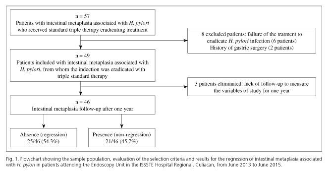
Of the 359 patients that underwent an upper endoscopy, 79 (22%) patients presented with intestinal metaplasia, 57 patients had metaplasia associated with <i>H. pylori</i>, 8 patients were excluded, 49 patients were included in the study, and 3 patients were finally eliminated (<a href="#f1">Fig. 1</a>).</font></p>     <p>&nbsp;</p>     <p align="center"><font face="Verdana" size="2"><a name="f1"></a><img src="/img/revistas/diges/v108n12/original2_figure_1.jpg"></font></p>     <p>&nbsp;</p>     <p><font face="Verdana" size="2">The average age of the patients was 58.9 (DE 11.2), of whom 20 (43.5%) were men and 26 (56.5%) were women (p = 0.207). Fifteen (32.6%) patients were referred from Primary Care, and 31 (67.4%) patients from the area of medical specialties (p = 0.002). With regard to educational level, 20 (43.5%) patients had a higher educational level and 26 (56.5%) patients had a basic or secondary level of education (p = 0.404).</font></p>     <p><font face="Verdana" size="2">The indications for upper endoscopy to be performed were dyspepsia tests in 33 (71.7%) patients, the consumption of non-steroid anti-inflammatory medication in 5 (10.8%) patients, a history of intestinal bleeding in 4 (8.7%) patients, iron-deficiency anemia in 2 (4.3%) patients and dysfagia in tests conducted on 2 (4.3%) patients.
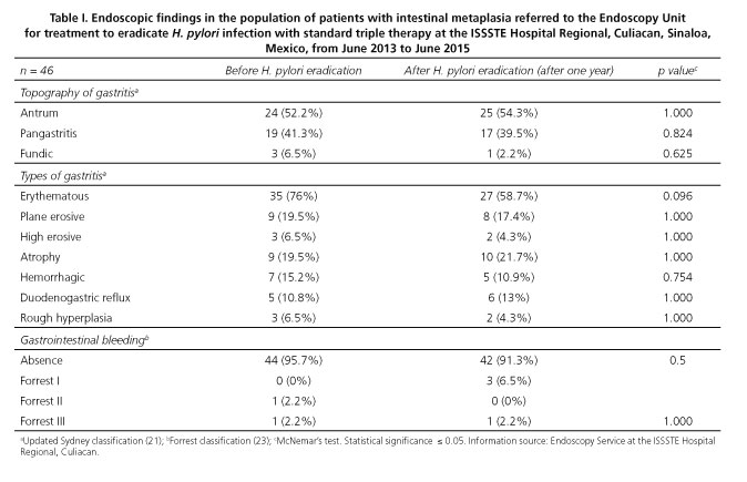
</font></p>     ]]></body>
<body><![CDATA[<p><font face="Verdana" size="2">Of the 57 patients who presented metaplasia associated with <i>H. pylori</i> and who were treated with standard triple therapy, the bacteria was not eradicated in 6 patients, resulting in an eradication rate of 89.4% (<a href="#f1">Fig. 1</a>).</font></p>     <p><font face="Verdana" size="2">There was no significant difference in the endoscopic findings from the gastric mucosa in terms of their topography, type of gastritis and intestinal bleeding both before treatment and after the eradication of <i>H. pylori</i> (p &gt; 0.05) (<a href="#t1">Table I</a>).</font></p>     <p>&nbsp;</p>     <p align="center"><font face="Verdana" size="2"><a name="t1"></a><img src="/img/revistas/diges/v108n12/original2_table_1.jpg"></font></p>     <p>&nbsp;</p>     <p><font face="Verdana" size="2">
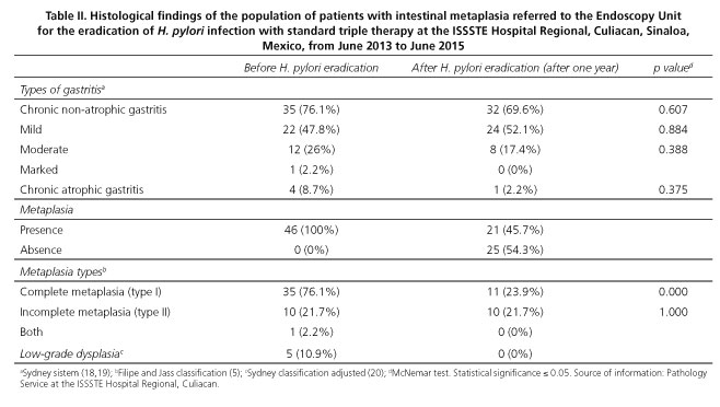
The most frequent histological finding was chronic non-atrophic gastritis, which occurred in 35 (76.1%) patients before treatment and in 32 (69.6%) patients after treatment to eradicate <i>H. pylori</i>. No significant difference was found between the histological findings for atrophic and non-atrophic chronic gastritis both before and after treatment to erradiacte <i>H. pylori</i> (p &gt; 0.05). Intestinal metaplasia was present in 46 (100%) patients before and in 21 (4.7%) patients after treatment to eradicate <i>H. pylori</i>. In 25 (54.3%) patients intestinal metaplasia was absent after the eradication of <i>H. pylori</i>. Complete intestinal metaplasia (type I) was present in 35 (76.1%) patients before and in 11 (23.9%) patients after the eradication of <i>H. pylori</i> (p = 0.000). Incomplete intestinal metaplasia (type II) was present in 10 (21.7%) patients before and after treatment to eradicate <i>H. pylori</i> (p = 1.000). Low grade dysplasia was present in 5 (10.9%) patients before treatment to eradicate <i>H. pylori</i> and was not present after treatment (<a href="#t2">Table II</a>).</font></p>     <p>&nbsp;</p>     <p align="center"><font face="Verdana" size="2"><a name="t2"></a><img src="/img/revistas/diges/v108n12/original2_table_2.jpg"></font></p>     <p>&nbsp;</p>     <p><font face="Verdana" size="2">The regression of intestinal metaplasia was observed one year after the eradication of <i>H. pylori</i> in 11 (55%) men and in 14 (53.8%) women (p = 0.588). Similarly, a regression of metaplasia was observed, after the eradication of <i>H. pylori</i>, in 15 (57.7%) of the patients with a basic or secondary educational level, compared to the 10 (50%) patients (p = 0.766) who had received higher education. The average age of the patients in whom the regression of metaplasia was observed was 58.5 (DE 12) years, compared to an average age of 59.4 (DE 10.4) (p = 0.768) in patients with no regression of the metaplasia after the eradication of <i>H. pylori</i>.</font></p>     ]]></body>
<body><![CDATA[<p>&nbsp;</p>     <p><font face="Verdana" size="2"><b>Discussion</b></font></p>     <p><font face="Verdana" size="2">Gastric intestinal metaplasia is a precursor lesion of gastric cancer, histological analysis shows the reduction or loss of the glandular component as it is replaced by enterocytes, and caliciform and Paneth cells. The Pelayo Correa "cascade" describes a sequence of histological lesions leading to the possible development of gastric cancer, beginning with chronic gastritis and evolving into chronic atrophic gastritis, intestinal metaplasia, dysplasia, and finally, cancer (2). The most important environmental factor related to the development of gastric intestinal metaplasia is <i>H. pylori</i> infection. In this study, the regression of intestinal metaplasia was observed in 25 (54.3%) patients one year after the eradication of <i>H. pylori</i>, a regression which was present preferentially in patients with type I metaplasia (complete). All 5 (10.9%) patients with low grade dysplasia from the population studied presented regression of the lesion before the eradication of <i>H. pylori</i>, although the results must be analyzed with caution given the reduced sample size included in the study.</font></p>     <p><font face="Verdana" size="2">While gastric intestinal metaplasia is prevalent, ranging from 7.1% to 42.5% in various countries around the world, it was found at a frequency of 22% in this study. The regression of gastric intestinal metaplasia subsequent to the eradication of <i>H. pylori</i> is clinically significant and represents an opportunity for the prevention of gastric cancer, which continues to be a public health problem. Moreover, prognosis is poor when this type of cancer is diagnosed in advanced stages despite treatment attempts, so the early detection of pre-neoplasic gastric lesions is important in at-risk patients, as well as monitoring and eradicating <i>H. pylori</i>. There is some controversy with regard to the type of pre-cancerous lesions that might lead to the evolution of gastric adenocarcinoma. Thus, Gonz&aacute;lez et al. (25) undertook a prospective study in 478 patients, monitored over the course of several years, who had been diagnosed with a type of pre-cancerous gastric lesion. They observed that a family history of gastric cancer and type II intestinal metaplasia (incomplete) were risk factors for progression to gastric adenocarcinoma. In this study, the regression of intestinal metaplasia type II (incomplete) was not observed in the patients when <i>H. pylori</i> had been eradicated.</font></p>     <p><font face="Verdana" size="2">In previous studies, various authors monitored patients with gastric intestinal metaplasia who were undergoing treatment to eradicate <i>H. pylori</i>, in order to evaluate the regression of the lesion. Mon&eacute;s et al. (26) presented a study of 37 patients with gastric intestinal metaplasia, and observed 100% regression of the lesion. V&aacute;zquez Romero et al. (17) studied 71 patients; in 50 (29.6%) of them the lesion regressed. Massarrat et al. (27) studied 30 patients, observing regression in 10 (21.7%) patients.</font></p>     <p><font face="Verdana" size="2">The discrepancies among the results described above could be related to the distinct designs of the various studies, the different methods for evaluating the grade of atrophy, the different number of gastric samples obtained, the number of patients included, the different monitoring times, the distinct strains of infectious <i>H. pylori</i> identified, and the types and number of treatment strategies used to eradicate <i>H. pylori</i>.</font></p>     <p><font face="Verdana" size="2">With regard to the endoscopic findings of this study, no significant difference was observed in the behavior of gastritis, in terms of type and topography, during the disease monitoring conducted a year after eradication treatment in this study (p &gt; 0.05). No association was found between the regression of intestinal metaplasia and age, gender and educational level of the patients studied (p &gt; 0.05).</font></p>     <p><font face="Verdana" size="2">The main limitations of this study include the fact that it did not determine the density of <i>H. pylori</i>, gastric biopsies were not taken from the <i>angular incisure</i>, and types IIA and IIB gastric intestinal metaplasia were not identified by histochemistry. Another limitation was not performing chromoendoscopy tests, which are more sensitive and thus precise for the identification of areas with metaplasia or dysplasia in the gastric mucosa. The methodology used in this study did not use a control group, which limited the ability of the research to establish evidence that the changes observed were due to the treatment being evaluated. However, this was a prospective study, and the data collection method used facilitated the control of the variables, while monitoring of the patients in the study enabled us to ensure losses of no more than 5% of the total sample population studied.</font></p>     <p><font face="Verdana" size="2">It can be concluded from this research that gastric intestinal metaplasia associated with <i>H. pylori</i> infection presented a 54.3% regression rate in those patients attending an endoscopy unit one year after their infection had been eradicated via standard triple therapy. The eradication of this micro-organism could modify the natural history of the progression of lesions in the gastric mucosa and their evolution into gastric carcinoma.</font></p>     <p>&nbsp;</p>     ]]></body>
<body><![CDATA[<p><font face="Verdana" size="2"><b>Acknowledgments</b></font></p>     <p><font face="Verdana" size="2">We would like to acknowledge the technical support received from the endoscopy service and the Pathological Anatomy service at the ISSSTE Regional Hospital, Culiacan.</font></p>     <p>&nbsp;</p>     <p><font face="Verdana" size="2"><b>References</b></font></p>     <!-- ref --><p><font face="Verdana" size="2">1. Suerbaum S, Michetti P. <i>Helicobacter pylori</i> infection. N Engl J Med 2002;347:175-86. DOI: 10.1056/NEJMra020542.    &nbsp;&nbsp;&nbsp;&nbsp;&nbsp;&nbsp;&nbsp;&nbsp;[&#160;<a href="javascript:void(0);" onclick="javascript: window.open('/scielo.php?script=sci_nlinks&ref=5435136&pid=S1130-0108201600120000300001&lng=','','width=640,height=500,resizable=yes,scrollbars=1,menubar=yes,');">Links</a>&#160;]<!-- end-ref --></font></p>    <!-- ref --><p><font face="Verdana" size="2">2. Correa P, Piazuelo MB, Camargo MC. Etiopathogenesis of gastric cancer. Scand J Surg 2006;95:218-24.    &nbsp;&nbsp;&nbsp;&nbsp;&nbsp;&nbsp;&nbsp;&nbsp;[&#160;<a href="javascript:void(0);" onclick="javascript: window.open('/scielo.php?script=sci_nlinks&ref=5435138&pid=S1130-0108201600120000300002&lng=','','width=640,height=500,resizable=yes,scrollbars=1,menubar=yes,');">Links</a>&#160;]<!-- end-ref --></font></p>    <!-- ref --><p><font face="Verdana" size="2">3. Conteduca V, Sansonno D, Lauletta G, et al. H. pylori infection and gastric cancer: State of the art (review). Int J Oncol 2013;42:5-18.    &nbsp;&nbsp;&nbsp;&nbsp;&nbsp;&nbsp;&nbsp;&nbsp;[&#160;<a href="javascript:void(0);" onclick="javascript: window.open('/scielo.php?script=sci_nlinks&ref=5435140&pid=S1130-0108201600120000300003&lng=','','width=640,height=500,resizable=yes,scrollbars=1,menubar=yes,');">Links</a>&#160;]<!-- end-ref --></font></p>    ]]></body>
<body><![CDATA[<!-- ref --><p><font face="Verdana" size="2">4. Fuentes-Panan&aacute; E, Camorlinga-Ponce M, Maldonado-Bernal C. Infection, inflammation and gastric cancer. Salud Publica Mex 2009;51:427-33. DOI: 10.1590/S0036-36342009000500010.    &nbsp;&nbsp;&nbsp;&nbsp;&nbsp;&nbsp;&nbsp;&nbsp;[&#160;<a href="javascript:void(0);" onclick="javascript: window.open('/scielo.php?script=sci_nlinks&ref=5435142&pid=S1130-0108201600120000300004&lng=','','width=640,height=500,resizable=yes,scrollbars=1,menubar=yes,');">Links</a>&#160;]<!-- end-ref --></font></p>    <!-- ref --><p><font face="Verdana" size="2">5. Jass JR, Filipe MI. The mucin profiles of normal gastric mucosa, intestinal metaplasia and its variants and gastric carcinoma. Histochem J 1981;13:931-9. DOI: 10.1007/BF01002633.    &nbsp;&nbsp;&nbsp;&nbsp;&nbsp;&nbsp;&nbsp;&nbsp;[&#160;<a href="javascript:void(0);" onclick="javascript: window.open('/scielo.php?script=sci_nlinks&ref=5435144&pid=S1130-0108201600120000300005&lng=','','width=640,height=500,resizable=yes,scrollbars=1,menubar=yes,');">Links</a>&#160;]<!-- end-ref --></font></p>    <!-- ref --><p><font face="Verdana" size="2">6. Quingalahua Flores Aldo, Mendoza Jaime Francisco. <i>Helicobacter pylori</i>, metaplasia intestinal de est&oacute;mago y c&aacute;ncer g&aacute;strico. Oncolog&iacute;a 2004;14:64-7.    &nbsp;&nbsp;&nbsp;&nbsp;&nbsp;&nbsp;&nbsp;&nbsp;[&#160;<a href="javascript:void(0);" onclick="javascript: window.open('/scielo.php?script=sci_nlinks&ref=5435146&pid=S1130-0108201600120000300006&lng=','','width=640,height=500,resizable=yes,scrollbars=1,menubar=yes,');">Links</a>&#160;]<!-- end-ref --></font></p>    <!-- ref --><p><font face="Verdana" size="2">7. Pi&ntilde;ol Jim&eacute;nez F, Paniagua Est&eacute;vez M, P&eacute;rez S&aacute;nchez G, et al. Metaplasia intestinal en pacientes con reflujo duodenog&aacute;strico y &aacute;cidos biliares totales elevados. Rev Cubana Med 2010;49:17-32.    &nbsp;&nbsp;&nbsp;&nbsp;&nbsp;&nbsp;&nbsp;&nbsp;[&#160;<a href="javascript:void(0);" onclick="javascript: window.open('/scielo.php?script=sci_nlinks&ref=5435148&pid=S1130-0108201600120000300007&lng=','','width=640,height=500,resizable=yes,scrollbars=1,menubar=yes,');">Links</a>&#160;]<!-- end-ref --></font></p>    <!-- ref --><p><font face="Verdana" size="2">8. Kim N, Park YS, Cho SI, et al. Prevalence and risk factors of atrophic gastritis and intestinal metaplasia in a Korean population without significant gastroduodenal disease. Helicobacter 2008;13:245-55. DOI: 10.1111/j.1523-5378.2008.00604.x.    &nbsp;&nbsp;&nbsp;&nbsp;&nbsp;&nbsp;&nbsp;&nbsp;[&#160;<a href="javascript:void(0);" onclick="javascript: window.open('/scielo.php?script=sci_nlinks&ref=5435150&pid=S1130-0108201600120000300008&lng=','','width=640,height=500,resizable=yes,scrollbars=1,menubar=yes,');">Links</a>&#160;]<!-- end-ref --></font></p>    ]]></body>
<body><![CDATA[<!-- ref --><p><font face="Verdana" size="2">9. Joo YE, Park HK, Myung DS, et al. Prevalence and risk factors of atrophic gastritis and intestinal metaplasia: A nationwide multicenter prospective study in Korea. Gut Liver 2013;7:303-10. DOI: 10.5009/gnl.2013.7.3.303.    &nbsp;&nbsp;&nbsp;&nbsp;&nbsp;&nbsp;&nbsp;&nbsp;[&#160;<a href="javascript:void(0);" onclick="javascript: window.open('/scielo.php?script=sci_nlinks&ref=5435152&pid=S1130-0108201600120000300009&lng=','','width=640,height=500,resizable=yes,scrollbars=1,menubar=yes,');">Links</a>&#160;]<!-- end-ref --></font></p>    <!-- ref --><p><font face="Verdana" size="2">10. Correa P. Human gastric carcinogenesis: A multistep and multifactorial process - First American Cancer Society Award Lecture on Cancer Epidemiology and Prevention. Cancer Res 1992;52:6735-40.    &nbsp;&nbsp;&nbsp;&nbsp;&nbsp;&nbsp;&nbsp;&nbsp;[&#160;<a href="javascript:void(0);" onclick="javascript: window.open('/scielo.php?script=sci_nlinks&ref=5435154&pid=S1130-0108201600120000300010&lng=','','width=640,height=500,resizable=yes,scrollbars=1,menubar=yes,');">Links</a>&#160;]<!-- end-ref --></font></p>    <!-- ref --><p><font face="Verdana" size="2">11. Kim N, Park RY, Cho SI, et al. <i>Helicobacter pylori</i> infection and development of gastric cancer in Korea: Long-term follow-up. J Clin Gastroenterol 2008;42:448-54. DOI: 10.1097/MCG.0b013e3180 46eac3.    &nbsp;&nbsp;&nbsp;&nbsp;&nbsp;&nbsp;&nbsp;&nbsp;[&#160;<a href="javascript:void(0);" onclick="javascript: window.open('/scielo.php?script=sci_nlinks&ref=5435156&pid=S1130-0108201600120000300011&lng=','','width=640,height=500,resizable=yes,scrollbars=1,menubar=yes,');">Links</a>&#160;]<!-- end-ref --></font></p>    <!-- ref --><p><font face="Verdana" size="2">12. Park YH, Kim N. Review of atrophic gastritis and intestinal metaplasia as a premalignant lesion of gastric cancer. J Cancer Prev 2015;20:25-40. DOI: 10.15430/JCP.2015.20.1.25.    &nbsp;&nbsp;&nbsp;&nbsp;&nbsp;&nbsp;&nbsp;&nbsp;[&#160;<a href="javascript:void(0);" onclick="javascript: window.open('/scielo.php?script=sci_nlinks&ref=5435158&pid=S1130-0108201600120000300012&lng=','','width=640,height=500,resizable=yes,scrollbars=1,menubar=yes,');">Links</a>&#160;]<!-- end-ref --></font></p>    <!-- ref --><p><font face="Verdana" size="2">13. A review of human carcinogens. B. Biological agents. IARC Working Group on the Evaluation of Carcinogenic Risks to Humans. Lyon, 24 February-3 March 2009. IARC Monogr Eval Carcinog Risks Hum 2012;100:385-423.    &nbsp;&nbsp;&nbsp;&nbsp;&nbsp;&nbsp;&nbsp;&nbsp;[&#160;<a href="javascript:void(0);" onclick="javascript: window.open('/scielo.php?script=sci_nlinks&ref=5435160&pid=S1130-0108201600120000300013&lng=','','width=640,height=500,resizable=yes,scrollbars=1,menubar=yes,');">Links</a>&#160;]<!-- end-ref --></font></p>    ]]></body>
<body><![CDATA[<!-- ref --><p><font face="Verdana" size="2">14. Micu G, St&#259;niceanu F, Zurac S, et al. Regression of precancerous epithelial alteration in patients with <i>Helicobacter pylori</i> chronic gastritis. Rom J Intern Med 2010;48:89-99.    &nbsp;&nbsp;&nbsp;&nbsp;&nbsp;&nbsp;&nbsp;&nbsp;[&#160;<a href="javascript:void(0);" onclick="javascript: window.open('/scielo.php?script=sci_nlinks&ref=5435162&pid=S1130-0108201600120000300014&lng=','','width=640,height=500,resizable=yes,scrollbars=1,menubar=yes,');">Links</a>&#160;]<!-- end-ref --></font></p>    <!-- ref --><p><font face="Verdana" size="2">15. Lahner E, Bordi C, Cattaruzza MS, et al. Long term follow-up in atrophic body gastritis patients: Atrophy and intestinal metaplasia are persistent lesions irrespective of <i>Helicobacter pylori</i> infection. Aliment Pharmacol Ther 2005;22:471-81. DOI: 10.1111/j.1365-2036.2005.02582.x.    &nbsp;&nbsp;&nbsp;&nbsp;&nbsp;&nbsp;&nbsp;&nbsp;[&#160;<a href="javascript:void(0);" onclick="javascript: window.open('/scielo.php?script=sci_nlinks&ref=5435164&pid=S1130-0108201600120000300015&lng=','','width=640,height=500,resizable=yes,scrollbars=1,menubar=yes,');">Links</a>&#160;]<!-- end-ref --></font></p>    <!-- ref --><p><font face="Verdana" size="2">16. Wang J, Xu L, Shi R, et al. Gastric atrophy and intestinal metaplasia before and after <i>Helicobacter pylori</i> eradication: A meta-analysis. Digestion 2011;83:253-60. DOI: 10.1159/000280318.    &nbsp;&nbsp;&nbsp;&nbsp;&nbsp;&nbsp;&nbsp;&nbsp;[&#160;<a href="javascript:void(0);" onclick="javascript: window.open('/scielo.php?script=sci_nlinks&ref=5435166&pid=S1130-0108201600120000300016&lng=','','width=640,height=500,resizable=yes,scrollbars=1,menubar=yes,');">Links</a>&#160;]<!-- end-ref --></font></p>    <!-- ref --><p><font face="Verdana" size="2">17. V&aacute;zquez Romero M, Boixeda de Miquel D, Valer L&oacute;pez-Fando MP, et al. Intestinal metaplasia: Evolution after <i>Helicobacter pylori</i> eradication and influence in the success of eradicating therapy. Rev Esp Enferm Dig 2003;95:781-4.    &nbsp;&nbsp;&nbsp;&nbsp;&nbsp;&nbsp;&nbsp;&nbsp;[&#160;<a href="javascript:void(0);" onclick="javascript: window.open('/scielo.php?script=sci_nlinks&ref=5435168&pid=S1130-0108201600120000300017&lng=','','width=640,height=500,resizable=yes,scrollbars=1,menubar=yes,');">Links</a>&#160;]<!-- end-ref --></font></p>    <!-- ref --><p><font face="Verdana" size="2">18. Correa P, Bravo L. Investigaciones sobre c&aacute;ncer g&aacute;strico en Nari&ntilde;o. Rev Med Hospital Universitario Departamental de Nari&ntilde;o 2007;3:6-12.    &nbsp;&nbsp;&nbsp;&nbsp;&nbsp;&nbsp;&nbsp;&nbsp;[&#160;<a href="javascript:void(0);" onclick="javascript: window.open('/scielo.php?script=sci_nlinks&ref=5435170&pid=S1130-0108201600120000300018&lng=','','width=640,height=500,resizable=yes,scrollbars=1,menubar=yes,');">Links</a>&#160;]<!-- end-ref --></font></p>    ]]></body>
<body><![CDATA[<!-- ref --><p><font face="Verdana" size="2">19. Correa P, Piazuelo MB. The gastric precancerous cascade. J Dig Dis 2012;13:2-9. DOI: 10.1111/j.1751-2980.2011.00550.x.    &nbsp;&nbsp;&nbsp;&nbsp;&nbsp;&nbsp;&nbsp;&nbsp;[&#160;<a href="javascript:void(0);" onclick="javascript: window.open('/scielo.php?script=sci_nlinks&ref=5435172&pid=S1130-0108201600120000300019&lng=','','width=640,height=500,resizable=yes,scrollbars=1,menubar=yes,');">Links</a>&#160;]<!-- end-ref --></font></p>    <!-- ref --><p><font face="Verdana" size="2">20. Salas Caudevilla A. Evaluation of dysplasia in gastrointestinal diseases. Gastroenterol Hepatol 2007;30:602-11. DOI: 10.1157/13112598.    &nbsp;&nbsp;&nbsp;&nbsp;&nbsp;&nbsp;&nbsp;&nbsp;[&#160;<a href="javascript:void(0);" onclick="javascript: window.open('/scielo.php?script=sci_nlinks&ref=5435174&pid=S1130-0108201600120000300020&lng=','','width=640,height=500,resizable=yes,scrollbars=1,menubar=yes,');">Links</a>&#160;]<!-- end-ref --></font></p>    <!-- ref --><p><font face="Verdana" size="2">21. Valdivia Rold&aacute;n M. Gastritis and gastropathies. Rev Gastroenterol Per&uacute; 2011;31:38-48.    &nbsp;&nbsp;&nbsp;&nbsp;&nbsp;&nbsp;&nbsp;&nbsp;[&#160;<a href="javascript:void(0);" onclick="javascript: window.open('/scielo.php?script=sci_nlinks&ref=5435176&pid=S1130-0108201600120000300021&lng=','','width=640,height=500,resizable=yes,scrollbars=1,menubar=yes,');">Links</a>&#160;]<!-- end-ref --></font></p>    <!-- ref --><p><font face="Verdana" size="2">22. Peetsalu A, Kirsim&auml;gi U, Peetsalu M. Giant peptic ulcer hemorrhages: Epidemiology, treatment, and outcome in the teaching hospital of Tartu. Medicina (Kaunas) 2011;47(1):19-24.    &nbsp;&nbsp;&nbsp;&nbsp;&nbsp;&nbsp;&nbsp;&nbsp;[&#160;<a href="javascript:void(0);" onclick="javascript: window.open('/scielo.php?script=sci_nlinks&ref=5435178&pid=S1130-0108201600120000300022&lng=','','width=640,height=500,resizable=yes,scrollbars=1,menubar=yes,');">Links</a>&#160;]<!-- end-ref --></font></p>    <!-- ref --><p><font face="Verdana" size="2">23. Abdo-Francis JM, Uscanga-Dom&iacute;nguez LF, Sobrino-Cossio S, et al. Tercer consenso mexicano de <i>Helicobacter pylori</i>. Rev Gastroenterol Mex 2007;72(3):321-38.    &nbsp;&nbsp;&nbsp;&nbsp;&nbsp;&nbsp;&nbsp;&nbsp;[&#160;<a href="javascript:void(0);" onclick="javascript: window.open('/scielo.php?script=sci_nlinks&ref=5435180&pid=S1130-0108201600120000300023&lng=','','width=640,height=500,resizable=yes,scrollbars=1,menubar=yes,');">Links</a>&#160;]<!-- end-ref --></font></p>    ]]></body>
<body><![CDATA[<!-- ref --><p><font face="Verdana" size="2">24. Calle Astudillo G, Jerves T, Pes&aacute;ntez L, et al. Utility of routine gastric biopsies and staining with methylene blue in the diagnosis of intestinal metaplasia in patients over 40 years. Acta Gastroenterol Latinoam 2013;43(3):189-97.    &nbsp;&nbsp;&nbsp;&nbsp;&nbsp;&nbsp;&nbsp;&nbsp;[&#160;<a href="javascript:void(0);" onclick="javascript: window.open('/scielo.php?script=sci_nlinks&ref=5435182&pid=S1130-0108201600120000300024&lng=','','width=640,height=500,resizable=yes,scrollbars=1,menubar=yes,');">Links</a>&#160;]<!-- end-ref --></font></p>    <!-- ref --><p><font face="Verdana" size="2">25. Gonz&aacute;lez CA, Pardo ML, Liso JM, et al. Gastric cancer occurrence in preneoplastic lesions: A long-term follow-up in a high-risk area in Spain. Int J Cancer 2010;127:2654-60. DOI: 10.1002/ijc.25273.    &nbsp;&nbsp;&nbsp;&nbsp;&nbsp;&nbsp;&nbsp;&nbsp;[&#160;<a href="javascript:void(0);" onclick="javascript: window.open('/scielo.php?script=sci_nlinks&ref=5435184&pid=S1130-0108201600120000300025&lng=','','width=640,height=500,resizable=yes,scrollbars=1,menubar=yes,');">Links</a>&#160;]<!-- end-ref --></font></p>    <!-- ref --><p><font face="Verdana" size="2">26. Mon&eacute;s J, Rodrigo L, Sancho F, et al. <i>Helicobacter pylori</i> eradication versus one-year maintenance therapy: Effect on relapse and gastritis outcome. Rev Esp Enferm Dig 2001;93(6):372-89.    &nbsp;&nbsp;&nbsp;&nbsp;&nbsp;&nbsp;&nbsp;&nbsp;[&#160;<a href="javascript:void(0);" onclick="javascript: window.open('/scielo.php?script=sci_nlinks&ref=5435186&pid=S1130-0108201600120000300026&lng=','','width=640,height=500,resizable=yes,scrollbars=1,menubar=yes,');">Links</a>&#160;]<!-- end-ref --></font></p>    <!-- ref --><p><font face="Verdana" size="2">27. Massarrat S, Haj-Sheykholeslami A, Mohamadkhani A, et al. Precancerous conditions after H. pylori eradication: A randomized double blind study in first degree relatives of gastric cancer patients. Arch Iran Med 2012;15(11):664-9.    &nbsp;&nbsp;&nbsp;&nbsp;&nbsp;&nbsp;&nbsp;&nbsp;[&#160;<a href="javascript:void(0);" onclick="javascript: window.open('/scielo.php?script=sci_nlinks&ref=5435188&pid=S1130-0108201600120000300027&lng=','','width=640,height=500,resizable=yes,scrollbars=1,menubar=yes,');">Links</a>&#160;]<!-- end-ref --></font></p>     <p>&nbsp;</p>     <p>&nbsp;</p>     ]]></body>
<body><![CDATA[<p><font face="Verdana" size="2"><a href="#top"><img border="0" src="/img/revistas/diges/v108n12/seta.gif" width="15" height="17"></a><a name="bajo"></a><b>Correspondence:</b>    <br>Jaime Alberto S&aacute;nchez Cu&eacute;n.    <br>Department of Digestive Diseases.    <br>Hospital Regional ISSSTE de Culiac&aacute;n.    <br>Calzada Heroico Colegio Militar 875 Sur, colonia 5 de Mayo.    <br>80000 Culiac&aacute;n, Sinaloa. Mexico    <br>e-mail: <a href="mailto:sanchezcuen_jaime@hotmail.com">sanchezcuen_jaime@hotmail.com</a></font></p>     <p><font face="Verdana" size="2">Received: 13-01-2016    <br>Accepted: 03-06-2016</font></p>      ]]></body><back>
<ref-list>
<ref id="B1">
<label>1</label><nlm-citation citation-type="journal">
<person-group person-group-type="author">
<name>
<surname><![CDATA[Suerbaum]]></surname>
<given-names><![CDATA[S]]></given-names>
</name>
<name>
<surname><![CDATA[Michetti]]></surname>
<given-names><![CDATA[P]]></given-names>
</name>
</person-group>
<article-title xml:lang="en"><![CDATA[Helicobacter pylori infection]]></article-title>
<source><![CDATA[N Engl J Med]]></source>
<year>2002</year>
<volume>347</volume>
<page-range>175-86</page-range></nlm-citation>
</ref>
<ref id="B2">
<label>2</label><nlm-citation citation-type="journal">
<person-group person-group-type="author">
<name>
<surname><![CDATA[Correa]]></surname>
<given-names><![CDATA[P]]></given-names>
</name>
<name>
<surname><![CDATA[Piazuelo]]></surname>
<given-names><![CDATA[MB]]></given-names>
</name>
<name>
<surname><![CDATA[Camargo]]></surname>
<given-names><![CDATA[MC]]></given-names>
</name>
</person-group>
<article-title xml:lang="en"><![CDATA[Etiopathogenesis of gastric cancer]]></article-title>
<source><![CDATA[Scand J Surg]]></source>
<year>2006</year>
<volume>95</volume>
<page-range>218-24</page-range></nlm-citation>
</ref>
<ref id="B3">
<label>3</label><nlm-citation citation-type="journal">
<person-group person-group-type="author">
<name>
<surname><![CDATA[Conteduca]]></surname>
<given-names><![CDATA[V]]></given-names>
</name>
<name>
<surname><![CDATA[Sansonno]]></surname>
<given-names><![CDATA[D]]></given-names>
</name>
<name>
<surname><![CDATA[Lauletta]]></surname>
<given-names><![CDATA[G]]></given-names>
</name>
</person-group>
<article-title xml:lang="en"><![CDATA[H. pylori infection and gastric cancer: State of the art (review)]]></article-title>
<source><![CDATA[Int J Oncol]]></source>
<year>2013</year>
<volume>42</volume>
<page-range>5-18</page-range></nlm-citation>
</ref>
<ref id="B4">
<label>4</label><nlm-citation citation-type="journal">
<person-group person-group-type="author">
<name>
<surname><![CDATA[Fuentes-Pananá]]></surname>
<given-names><![CDATA[E]]></given-names>
</name>
<name>
<surname><![CDATA[Camorlinga-Ponce]]></surname>
<given-names><![CDATA[M]]></given-names>
</name>
<name>
<surname><![CDATA[Maldonado-Bernal]]></surname>
<given-names><![CDATA[C]]></given-names>
</name>
</person-group>
<article-title xml:lang="en"><![CDATA[Infection, inflammation and gastric cancer]]></article-title>
<source><![CDATA[Salud Publica Mex]]></source>
<year>2009</year>
<volume>51</volume>
<page-range>427-33</page-range></nlm-citation>
</ref>
<ref id="B5">
<label>5</label><nlm-citation citation-type="journal">
<person-group person-group-type="author">
<name>
<surname><![CDATA[Jass]]></surname>
<given-names><![CDATA[JR]]></given-names>
</name>
<name>
<surname><![CDATA[Filipe]]></surname>
<given-names><![CDATA[MI]]></given-names>
</name>
</person-group>
<article-title xml:lang="en"><![CDATA[The mucin profiles of normal gastric mucosa, intestinal metaplasia and its variants and gastric carcinoma]]></article-title>
<source><![CDATA[Histochem J]]></source>
<year>1981</year>
<volume>13</volume>
<page-range>931-9</page-range></nlm-citation>
</ref>
<ref id="B6">
<label>6</label><nlm-citation citation-type="journal">
<person-group person-group-type="author">
<name>
<surname><![CDATA[Quingalahua Flores]]></surname>
<given-names><![CDATA[Aldo]]></given-names>
</name>
<name>
<surname><![CDATA[Mendoza]]></surname>
<given-names><![CDATA[Jaime Francisco]]></given-names>
</name>
</person-group>
<article-title xml:lang="en"><![CDATA[Helicobacter pylori, metaplasia intestinal de estómago y cáncer gástrico]]></article-title>
<source><![CDATA[Oncología]]></source>
<year>2004</year>
<volume>14</volume>
<page-range>64-7</page-range></nlm-citation>
</ref>
<ref id="B7">
<label>7</label><nlm-citation citation-type="journal">
<person-group person-group-type="author">
<name>
<surname><![CDATA[Piñol Jiménez]]></surname>
<given-names><![CDATA[F]]></given-names>
</name>
<name>
<surname><![CDATA[Paniagua Estévez]]></surname>
<given-names><![CDATA[M]]></given-names>
</name>
<name>
<surname><![CDATA[Pérez Sánchez]]></surname>
<given-names><![CDATA[G]]></given-names>
</name>
</person-group>
<article-title xml:lang="es"><![CDATA[Metaplasia intestinal en pacientes con reflujo duodenogástrico y ácidos biliares totales elevados]]></article-title>
<source><![CDATA[Rev Cubana Med]]></source>
<year>2010</year>
<volume>49</volume>
<page-range>17-32</page-range></nlm-citation>
</ref>
<ref id="B8">
<label>8</label><nlm-citation citation-type="journal">
<person-group person-group-type="author">
<name>
<surname><![CDATA[Kim]]></surname>
<given-names><![CDATA[N]]></given-names>
</name>
<name>
<surname><![CDATA[Park]]></surname>
<given-names><![CDATA[YS]]></given-names>
</name>
<name>
<surname><![CDATA[Cho]]></surname>
<given-names><![CDATA[SI]]></given-names>
</name>
</person-group>
<article-title xml:lang="en"><![CDATA[Prevalence and risk factors of atrophic gastritis and intestinal metaplasia in a Korean population without significant gastroduodenal disease]]></article-title>
<source><![CDATA[Helicobacter]]></source>
<year>2008</year>
<volume>13</volume>
<page-range>245-55</page-range></nlm-citation>
</ref>
<ref id="B9">
<label>9</label><nlm-citation citation-type="journal">
<person-group person-group-type="author">
<name>
<surname><![CDATA[Joo]]></surname>
<given-names><![CDATA[YE]]></given-names>
</name>
<name>
<surname><![CDATA[Park]]></surname>
<given-names><![CDATA[HK]]></given-names>
</name>
<name>
<surname><![CDATA[Myung]]></surname>
<given-names><![CDATA[DS]]></given-names>
</name>
</person-group>
<article-title xml:lang="en"><![CDATA[Prevalence and risk factors of atrophic gastritis and intestinal metaplasia: A nationwide multicenter prospective study in Korea]]></article-title>
<source><![CDATA[Gut Liver]]></source>
<year>2013</year>
<volume>7</volume>
<page-range>303-10</page-range></nlm-citation>
</ref>
<ref id="B10">
<label>10</label><nlm-citation citation-type="journal">
<person-group person-group-type="author">
<name>
<surname><![CDATA[Correa]]></surname>
<given-names><![CDATA[P]]></given-names>
</name>
</person-group>
<article-title xml:lang="en"><![CDATA[Human gastric carcinogenesis: A multistep and multifactorial process - First American Cancer Society Award Lecture on Cancer Epidemiology and Prevention]]></article-title>
<source><![CDATA[Cancer Res]]></source>
<year>1992</year>
<volume>52</volume>
<page-range>6735-40</page-range></nlm-citation>
</ref>
<ref id="B11">
<label>11</label><nlm-citation citation-type="journal">
<person-group person-group-type="author">
<name>
<surname><![CDATA[Kim]]></surname>
<given-names><![CDATA[N]]></given-names>
</name>
<name>
<surname><![CDATA[Park]]></surname>
<given-names><![CDATA[RY]]></given-names>
</name>
<name>
<surname><![CDATA[Cho]]></surname>
<given-names><![CDATA[SI]]></given-names>
</name>
</person-group>
<article-title xml:lang="en"><![CDATA[Helicobacter pylori infection and development of gastric cancer in Korea: Long-term follow-up]]></article-title>
<source><![CDATA[J Clin Gastroenterol]]></source>
<year>2008</year>
<volume>42</volume>
<page-range>448-54</page-range></nlm-citation>
</ref>
<ref id="B12">
<label>12</label><nlm-citation citation-type="journal">
<person-group person-group-type="author">
<name>
<surname><![CDATA[Park]]></surname>
<given-names><![CDATA[YH]]></given-names>
</name>
<name>
<surname><![CDATA[Kim]]></surname>
<given-names><![CDATA[N]]></given-names>
</name>
</person-group>
<article-title xml:lang="en"><![CDATA[Review of atrophic gastritis and intestinal metaplasia as a premalignant lesion of gastric cancer]]></article-title>
<source><![CDATA[J Cancer Prev]]></source>
<year>2015</year>
<volume>20</volume>
<page-range>25-40</page-range></nlm-citation>
</ref>
<ref id="B13">
<label>13</label><nlm-citation citation-type="journal">
<article-title xml:lang="en"><![CDATA[A review of human carcinogens. B. Biological agents: IARC Working Group on the Evaluation of Carcinogenic Risks to Humans. Lyon, 24 February-3 March 2009]]></article-title>
<source><![CDATA[IARC Monogr Eval Carcinog Risks Hum]]></source>
<year>2012</year>
<volume>100</volume>
<page-range>385-423</page-range></nlm-citation>
</ref>
<ref id="B14">
<label>14</label><nlm-citation citation-type="journal">
<person-group person-group-type="author">
<name>
<surname><![CDATA[Micu]]></surname>
<given-names><![CDATA[G]]></given-names>
</name>
<name>
<surname><![CDATA[St&#259;niceanu]]></surname>
<given-names><![CDATA[F]]></given-names>
</name>
<name>
<surname><![CDATA[Zurac]]></surname>
<given-names><![CDATA[S]]></given-names>
</name>
</person-group>
<article-title xml:lang="en"><![CDATA[Regression of precancerous epithelial alteration in patients with Helicobacter pylori chronic gastritis]]></article-title>
<source><![CDATA[Rom J Intern Med]]></source>
<year>2010</year>
<volume>48</volume>
<page-range>89-99</page-range></nlm-citation>
</ref>
<ref id="B15">
<label>15</label><nlm-citation citation-type="journal">
<person-group person-group-type="author">
<name>
<surname><![CDATA[Lahner]]></surname>
<given-names><![CDATA[E]]></given-names>
</name>
<name>
<surname><![CDATA[Bordi]]></surname>
<given-names><![CDATA[C]]></given-names>
</name>
<name>
<surname><![CDATA[Cattaruzza]]></surname>
<given-names><![CDATA[MS]]></given-names>
</name>
</person-group>
<article-title xml:lang="en"><![CDATA[Long term follow-up in atrophic body gastritis patients: Atrophy and intestinal metaplasia are persistent lesions irrespective of Helicobacter pylori infection]]></article-title>
<source><![CDATA[Aliment Pharmacol Ther]]></source>
<year>2005</year>
<volume>22</volume>
<page-range>471-81</page-range></nlm-citation>
</ref>
<ref id="B16">
<label>16</label><nlm-citation citation-type="journal">
<person-group person-group-type="author">
<name>
<surname><![CDATA[Wang]]></surname>
<given-names><![CDATA[J]]></given-names>
</name>
<name>
<surname><![CDATA[Xu]]></surname>
<given-names><![CDATA[L]]></given-names>
</name>
<name>
<surname><![CDATA[Shi]]></surname>
<given-names><![CDATA[R]]></given-names>
</name>
</person-group>
<article-title xml:lang="en"><![CDATA[Gastric atrophy and intestinal metaplasia before and after Helicobacter pylori eradication: A meta-analysis]]></article-title>
<source><![CDATA[Digestion]]></source>
<year>2011</year>
<volume>83</volume>
<page-range>253-60</page-range></nlm-citation>
</ref>
<ref id="B17">
<label>17</label><nlm-citation citation-type="journal">
<person-group person-group-type="author">
<name>
<surname><![CDATA[Vázquez Romero]]></surname>
<given-names><![CDATA[M]]></given-names>
</name>
<name>
<surname><![CDATA[Boixeda de Miquel]]></surname>
<given-names><![CDATA[D]]></given-names>
</name>
<name>
<surname><![CDATA[Valer López-Fando]]></surname>
<given-names><![CDATA[MP]]></given-names>
</name>
</person-group>
<article-title xml:lang="en"><![CDATA[Intestinal metaplasia: Evolution after Helicobacter pylori eradication and influence in the success of eradicating therapy]]></article-title>
<source><![CDATA[Rev Esp Enferm Dig]]></source>
<year>2003</year>
<volume>95</volume>
<page-range>781-4</page-range></nlm-citation>
</ref>
<ref id="B18">
<label>18</label><nlm-citation citation-type="journal">
<person-group person-group-type="author">
<name>
<surname><![CDATA[Correa]]></surname>
<given-names><![CDATA[P]]></given-names>
</name>
<name>
<surname><![CDATA[Bravo]]></surname>
<given-names><![CDATA[L]]></given-names>
</name>
</person-group>
<article-title xml:lang="es"><![CDATA[Investigaciones sobre cáncer gástrico en Nariño]]></article-title>
<source><![CDATA[Rev Med Hospital Universitario Departamental de Nariño]]></source>
<year>2007</year>
<volume>3</volume>
<page-range>6-12</page-range></nlm-citation>
</ref>
<ref id="B19">
<label>19</label><nlm-citation citation-type="journal">
<person-group person-group-type="author">
<name>
<surname><![CDATA[Correa]]></surname>
<given-names><![CDATA[P]]></given-names>
</name>
<name>
<surname><![CDATA[Piazuelo]]></surname>
<given-names><![CDATA[MB]]></given-names>
</name>
</person-group>
<article-title xml:lang="en"><![CDATA[The gastric precancerous cascade]]></article-title>
<source><![CDATA[J Dig Dis]]></source>
<year>2012</year>
<volume>13</volume>
<page-range>2-9</page-range></nlm-citation>
</ref>
<ref id="B20">
<label>20</label><nlm-citation citation-type="journal">
<person-group person-group-type="author">
<name>
<surname><![CDATA[Salas Caudevilla]]></surname>
<given-names><![CDATA[A]]></given-names>
</name>
</person-group>
<article-title xml:lang="en"><![CDATA[Evaluation of dysplasia in gastrointestinal diseases]]></article-title>
<source><![CDATA[Gastroenterol Hepatol]]></source>
<year>2007</year>
<volume>30</volume>
<page-range>602-11</page-range></nlm-citation>
</ref>
<ref id="B21">
<label>21</label><nlm-citation citation-type="journal">
<person-group person-group-type="author">
<name>
<surname><![CDATA[Valdivia Roldán]]></surname>
<given-names><![CDATA[M]]></given-names>
</name>
</person-group>
<article-title xml:lang="en"><![CDATA[Gastritis and gastropathies]]></article-title>
<source><![CDATA[Rev Gastroenterol Perú]]></source>
<year>2011</year>
<volume>31</volume>
<page-range>38-48</page-range></nlm-citation>
</ref>
<ref id="B22">
<label>22</label><nlm-citation citation-type="journal">
<person-group person-group-type="author">
<name>
<surname><![CDATA[Peetsalu]]></surname>
<given-names><![CDATA[A]]></given-names>
</name>
<name>
<surname><![CDATA[Kirsimägi]]></surname>
<given-names><![CDATA[U]]></given-names>
</name>
<name>
<surname><![CDATA[Peetsalu]]></surname>
<given-names><![CDATA[M]]></given-names>
</name>
</person-group>
<article-title xml:lang="en"><![CDATA[Giant peptic ulcer hemorrhages: Epidemiology, treatment, and outcome in the teaching hospital of Tartu]]></article-title>
<source><![CDATA[Medicina (Kaunas)]]></source>
<year>2011</year>
<volume>47</volume>
<numero>1</numero>
<issue>1</issue>
<page-range>19-24</page-range></nlm-citation>
</ref>
<ref id="B23">
<label>23</label><nlm-citation citation-type="journal">
<person-group person-group-type="author">
<name>
<surname><![CDATA[Abdo-Francis]]></surname>
<given-names><![CDATA[JM]]></given-names>
</name>
<name>
<surname><![CDATA[Uscanga-Domínguez]]></surname>
<given-names><![CDATA[LF]]></given-names>
</name>
<name>
<surname><![CDATA[Sobrino-Cossio]]></surname>
<given-names><![CDATA[S]]></given-names>
</name>
</person-group>
<article-title xml:lang="es"><![CDATA[Tercer consenso mexicano de Helicobacter pylori]]></article-title>
<source><![CDATA[Rev Gastroenterol Mex]]></source>
<year>2007</year>
<volume>72</volume>
<numero>3</numero>
<issue>3</issue>
<page-range>321-38</page-range></nlm-citation>
</ref>
<ref id="B24">
<label>24</label><nlm-citation citation-type="journal">
<person-group person-group-type="author">
<name>
<surname><![CDATA[Calle Astudillo]]></surname>
<given-names><![CDATA[G]]></given-names>
</name>
<name>
<surname><![CDATA[Jerves]]></surname>
<given-names><![CDATA[T]]></given-names>
</name>
<name>
<surname><![CDATA[Pesántez]]></surname>
<given-names><![CDATA[L]]></given-names>
</name>
</person-group>
<article-title xml:lang="en"><![CDATA[Utility of routine gastric biopsies and staining with methylene blue in the diagnosis of intestinal metaplasia in patients over 40 years]]></article-title>
<source><![CDATA[Acta Gastroenterol Latinoam]]></source>
<year>2013</year>
<volume>43</volume>
<numero>3</numero>
<issue>3</issue>
<page-range>189-97</page-range></nlm-citation>
</ref>
<ref id="B25">
<label>25</label><nlm-citation citation-type="journal">
<person-group person-group-type="author">
<name>
<surname><![CDATA[González]]></surname>
<given-names><![CDATA[CA]]></given-names>
</name>
<name>
<surname><![CDATA[Pardo]]></surname>
<given-names><![CDATA[ML]]></given-names>
</name>
<name>
<surname><![CDATA[Liso]]></surname>
<given-names><![CDATA[JM]]></given-names>
</name>
</person-group>
<article-title xml:lang="en"><![CDATA[Gastric cancer occurrence in preneoplastic lesions: A long-term follow-up in a high-risk area in Spain]]></article-title>
<source><![CDATA[Int J Cancer]]></source>
<year>2010</year>
<volume>127</volume>
<page-range>2654-60</page-range></nlm-citation>
</ref>
<ref id="B26">
<label>26</label><nlm-citation citation-type="journal">
<person-group person-group-type="author">
<name>
<surname><![CDATA[Monés]]></surname>
<given-names><![CDATA[J]]></given-names>
</name>
<name>
<surname><![CDATA[Rodrigo]]></surname>
<given-names><![CDATA[L]]></given-names>
</name>
<name>
<surname><![CDATA[Sancho]]></surname>
<given-names><![CDATA[F]]></given-names>
</name>
</person-group>
<article-title xml:lang="en"><![CDATA[Helicobacter pylori eradication versus one-year maintenance therapy: Effect on relapse and gastritis outcome]]></article-title>
<source><![CDATA[Rev Esp Enferm Dig]]></source>
<year>2001</year>
<volume>93</volume>
<numero>6</numero>
<issue>6</issue>
<page-range>372-89</page-range></nlm-citation>
</ref>
<ref id="B27">
<label>27</label><nlm-citation citation-type="journal">
<person-group person-group-type="author">
<name>
<surname><![CDATA[Massarrat]]></surname>
<given-names><![CDATA[S]]></given-names>
</name>
<name>
<surname><![CDATA[Haj-Sheykholeslami]]></surname>
<given-names><![CDATA[A]]></given-names>
</name>
<name>
<surname><![CDATA[Mohamadkhani]]></surname>
<given-names><![CDATA[A]]></given-names>
</name>
</person-group>
<article-title xml:lang="en"><![CDATA[Precancerous conditions after H. pylori eradication: A randomized double blind study in first degree relatives of gastric cancer patients]]></article-title>
<source><![CDATA[Arch Iran Med]]></source>
<year>2012</year>
<volume>15</volume>
<numero>11</numero>
<issue>11</issue>
<page-range>664-9</page-range></nlm-citation>
</ref>
</ref-list>
</back>
</article>
