<?xml version="1.0" encoding="ISO-8859-1"?><article xmlns:mml="http://www.w3.org/1998/Math/MathML" xmlns:xlink="http://www.w3.org/1999/xlink" xmlns:xsi="http://www.w3.org/2001/XMLSchema-instance">
<front>
<journal-meta>
<journal-id>1130-0108</journal-id>
<journal-title><![CDATA[Revista Española de Enfermedades Digestivas]]></journal-title>
<abbrev-journal-title><![CDATA[Rev. esp. enferm. dig.]]></abbrev-journal-title>
<issn>1130-0108</issn>
<publisher>
<publisher-name><![CDATA[Sociedad Española de Patología Digestiva]]></publisher-name>
</publisher>
</journal-meta>
<article-meta>
<article-id>S1130-01082017000300010</article-id>
<title-group>
<article-title xml:lang="en"><![CDATA[Sclerosing angiomatoid nodular transformation of the spleen: a case report]]></article-title>
<article-title xml:lang="es"><![CDATA[Transformación angiomatosa nodular esclerosante de bazo: un caso clínico]]></article-title>
</title-group>
<contrib-group>
<contrib contrib-type="author">
<name>
<surname><![CDATA[Martínez-Martínez]]></surname>
<given-names><![CDATA[Pedro Jesús]]></given-names>
</name>
<xref ref-type="aff" rid="A01"/>
</contrib>
<contrib contrib-type="author">
<name>
<surname><![CDATA[Solbes-Vila]]></surname>
<given-names><![CDATA[Ramón]]></given-names>
</name>
<xref ref-type="aff" rid="A01"/>
</contrib>
<contrib contrib-type="author">
<name>
<surname><![CDATA[Bosquet-Úbeda]]></surname>
<given-names><![CDATA[Carlos Javier]]></given-names>
</name>
<xref ref-type="aff" rid="A01"/>
</contrib>
<contrib contrib-type="author">
<name>
<surname><![CDATA[Roig-Álvaro]]></surname>
<given-names><![CDATA[José María]]></given-names>
</name>
<xref ref-type="aff" rid="A02"/>
</contrib>
</contrib-group>
<aff id="A01">
<institution><![CDATA[,Hospital Mediterráneo Department of Radiology ]]></institution>
<addr-line><![CDATA[ ]]></addr-line>
</aff>
<aff id="A02">
<institution><![CDATA[,Hospital Mediterráneo Department of Pathology ]]></institution>
<addr-line><![CDATA[Almería ]]></addr-line>
<country>Spain</country>
</aff>
<pub-date pub-type="pub">
<day>00</day>
<month>03</month>
<year>2017</year>
</pub-date>
<pub-date pub-type="epub">
<day>00</day>
<month>03</month>
<year>2017</year>
</pub-date>
<volume>109</volume>
<numero>3</numero>
<fpage>214</fpage>
<lpage>215</lpage>
<copyright-statement/>
<copyright-year/>
<self-uri xlink:href="http://scielo.isciii.es/scielo.php?script=sci_arttext&amp;pid=S1130-01082017000300010&amp;lng=en&amp;nrm=iso"></self-uri><self-uri xlink:href="http://scielo.isciii.es/scielo.php?script=sci_abstract&amp;pid=S1130-01082017000300010&amp;lng=en&amp;nrm=iso"></self-uri><self-uri xlink:href="http://scielo.isciii.es/scielo.php?script=sci_pdf&amp;pid=S1130-01082017000300010&amp;lng=en&amp;nrm=iso"></self-uri></article-meta>
</front><body><![CDATA[ 
    <p><font face="Verdana" size="2"><b>PICTURES IN DIGESTIVE PATHOLOGY</b></font></p>
    <p>&nbsp;</p>
    <p><font face="Verdana" size="4"><b>Sclerosing angiomatoid nodular transformation of the spleen. A case report</b></font></p>
    <p><font face="Verdana" size="4"><b>Transformaci&oacute;n angiomatosa nodular esclerosante de bazo. Un caso cl&iacute;nico</b></font></p>
    <p>&nbsp;</p>
    <p>&nbsp;</p>
    <p><font face="Verdana" size="2"><b>Pedro Jes&uacute;s Mart&iacute;nez-Mart&iacute;nez<sup>1</sup>, Ram&oacute;n Solbes-Vila<sup>1</sup>, Carlos Javier Bosquet-&Uacute;beda<sup>1</sup> and Jos&eacute; Mar&iacute;a Roig-&Aacute;lvaro<sup>2</sup></b></font></p>
    <p><font face="Verdana" size="2">Departments of <sup>1</sup>Radiology and <sup>2</sup>Pathology. Hospital Mediterr&aacute;neo. Almer&iacute;a, Spain</font></p>
    <p>&nbsp;</p>
    ]]></body>
<body><![CDATA[<p>&nbsp;</p>
    <p><font face="Verdana" size="2"><b>Case report</b></font></p>
    <p><font face="Verdana" size="2">A 57 year old woman with only a history of depression presented to the gastroenterologist with clinical features of heartburn, and onemonth history of epigastric abdominal pain and left hypochondrium pain. In the physical examination no significant findings were seen, and the blood analysis was completely normal.</font></p>
    <p><font face="Verdana" size="2">
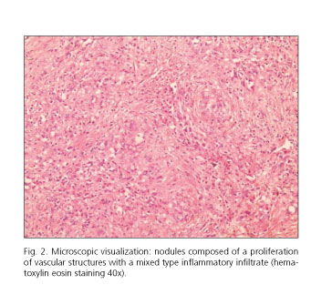
An abdominal ultrasound scan was conducted, where a hypoechogenic lesion was found which deformed the splenic contour without clearly seeing an intralesional vascularization. On abdominal CT, a low attenuation 9 x 6 cm&nbsp;injury was found that deformed the spleen contour with a hypodense behavior in arterial phase, with a little enhancement in portal phase, and which was homogenizing in late phase (<a href="#f1">Fig. 1</a>).</font></p>
    <p>&nbsp;</p>
    <p align="center"><font face="Verdana" size="2"><a name="f1"></a><img src="/img/revistas/diges/v109n3/imagen3_fig1.jpg"></font></p>
    <p>&nbsp;</p>
    <p><font face="Verdana" size="2">
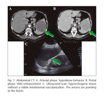
A total splenectomy was performed and the macroscopic study confirmed a welldelimited lesion in the addendum splenic tissue, 9 cm long from the major axis, formed by a fleshy, reddish tissue with whitish and a sort of star shaped foci with an increased consistency. The microscopy examination revealed multiple varying diameter nodules delimited by fibrohyaline tracts, formed by small caliber vascular structures lined by prominent and typical endothelium, with an immunohistochemical profile which refers to the characteristic capillaries one (CD31+, CD34+, CD8) (<a href="#f2">Fig. 2</a>). The SANT diagnosis was reached via a pathological study.</font></p>
    <p>&nbsp;</p>
    <p align="center"><font face="Verdana" size="2"><a name="f2"></a><img src="/img/revistas/diges/v109n3/imagen3_fig2.jpg"></font></p>
    ]]></body>
<body><![CDATA[<p>&nbsp;</p>
    <p><font face="Verdana" size="2"><b>Discussion</b></font></p>
    <p><font face="Verdana" size="2">SANT is a benign vascular condition which has been recently alluded to, the first case being published by Martel et al. in 2004. Therefore, the cases which have been published are very limited. It involves a proliferation of angiomatoid/vascular nodules which predominantly affect women aged 27 to 68. It is usually detected as a coincidental finding in asymptomatic patients.</font></p>
    <p><font face="Verdana" size="2">The etiopathogenesis is not wellknown although it points to several causes: an association with Epstein Barr virus, an abnormal transformation in the red pulp due to a stromal proliferation, or a final stage with a range of benign splenic lesions including harmatomas or inflammatory pseudoneoplasms. In recent studies it has been connected to the typical sclerosing injuries of the disorder related to immunoglobulin G4 (IgG4).</font></p>
    <p><font face="Verdana" size="2">The differential diagnosis must be established with metastases, hemangioma and littoral cell angioma, as well as lymphoproliferative syndrome, harmatoma, inflammatory pseudoneoplasm, and other lesions both benign and malignant.</font></p>
    <p><font face="Verdana" size="2">The presumptive diagnosis is established by imaging tests, especially CT and MR, and the final diagnosis is well established on the basis of pathology. Splenectomy is the treatment of choice since it not only confirms diagnosis but removes the problem as it is a nonrecurrent lesion.</font></p>
    <p>&nbsp;</p>
    <p><font face="Verdana" size="2"><b>References</b></font></p>
    <!-- ref --><p><font face="Verdana" size="2">1. Burneo EM, Franco HR, Castro AY, et al. Transformaci&oacute;n angiomatosa nodular esclerosante del bazo (SANT). Un tumor muy infrecuente. Cirug&iacute;a Esp 2012;9:607-9.    &nbsp;&nbsp;&nbsp;&nbsp;&nbsp;&nbsp;&nbsp;&nbsp;[&#160;<a href="javascript:void(0);" onclick="javascript: window.open('/scielo.php?script=sci_nlinks&ref=5442710&pid=S1130-0108201700030001000001&lng=','','width=640,height=500,resizable=yes,scrollbars=1,menubar=yes,');">Links</a>&#160;]<!-- end-ref --></font></p>    ]]></body>
<body><![CDATA[<!-- ref --><p><font face="Verdana" size="2">2. Raman SP, Singhi A, Horton KM, et al. Sclerosing angiomatoid nodular transformation of the spleen (SANT): Multimodality imaging appearance of five cases with radiology-pathology correlation. Abdom Imaging 2013;38:827-34. DOI: 10.1007/s00261-012-9949-4.    &nbsp;&nbsp;&nbsp;&nbsp;&nbsp;&nbsp;&nbsp;&nbsp;[&#160;<a href="javascript:void(0);" onclick="javascript: window.open('/scielo.php?script=sci_nlinks&ref=5442712&pid=S1130-0108201700030001000002&lng=','','width=640,height=500,resizable=yes,scrollbars=1,menubar=yes,');">Links</a>&#160;]<!-- end-ref --></font></p>    <!-- ref --><p><font face="Verdana" size="2">3. Watanabe M, Shiozawa K, Ikehara T, et al. A case of sclerosing angiomatoid nodular transformation of the spleen: Correlations between contrast-enhanced ultrasonography and histopathologic findings. J Clin Ultrasound 2013;42:103-7. DOI: 10.1002/jcu.22062.    &nbsp;&nbsp;&nbsp;&nbsp;&nbsp;&nbsp;&nbsp;&nbsp;[&#160;<a href="javascript:void(0);" onclick="javascript: window.open('/scielo.php?script=sci_nlinks&ref=5442714&pid=S1130-0108201700030001000003&lng=','','width=640,height=500,resizable=yes,scrollbars=1,menubar=yes,');">Links</a>&#160;]<!-- end-ref --></font></p>
     ]]></body><back>
<ref-list>
<ref id="B1">
<label>1</label><nlm-citation citation-type="journal">
<person-group person-group-type="author">
<name>
<surname><![CDATA[Burneo]]></surname>
<given-names><![CDATA[EM]]></given-names>
</name>
<name>
<surname><![CDATA[Franco]]></surname>
<given-names><![CDATA[HR]]></given-names>
</name>
<name>
<surname><![CDATA[Castro]]></surname>
<given-names><![CDATA[AY]]></given-names>
</name>
</person-group>
<article-title xml:lang="es"><![CDATA[Transformación angiomatosa nodular esclerosante del bazo (SANT): Un tumor muy infrecuente]]></article-title>
<source><![CDATA[Cirugía Esp]]></source>
<year>2012</year>
<volume>9</volume>
<page-range>607-9</page-range></nlm-citation>
</ref>
<ref id="B2">
<label>2</label><nlm-citation citation-type="journal">
<person-group person-group-type="author">
<name>
<surname><![CDATA[Raman]]></surname>
<given-names><![CDATA[SP]]></given-names>
</name>
<name>
<surname><![CDATA[Singhi]]></surname>
<given-names><![CDATA[A]]></given-names>
</name>
<name>
<surname><![CDATA[Horton]]></surname>
<given-names><![CDATA[KM]]></given-names>
</name>
</person-group>
<article-title xml:lang="en"><![CDATA[Sclerosing angiomatoid nodular transformation of the spleen (SANT): Multimodality imaging appearance of five cases with radiology-pathology correlation]]></article-title>
<source><![CDATA[Abdom Imaging]]></source>
<year>2013</year>
<volume>38</volume>
<page-range>827-34</page-range></nlm-citation>
</ref>
<ref id="B3">
<label>3</label><nlm-citation citation-type="journal">
<person-group person-group-type="author">
<name>
<surname><![CDATA[Watanabe]]></surname>
<given-names><![CDATA[M]]></given-names>
</name>
<name>
<surname><![CDATA[Shiozawa]]></surname>
<given-names><![CDATA[K]]></given-names>
</name>
<name>
<surname><![CDATA[Ikehara]]></surname>
<given-names><![CDATA[T]]></given-names>
</name>
</person-group>
<article-title xml:lang="en"><![CDATA[A case of sclerosing angiomatoid nodular transformation of the spleen: Correlations between contrast-enhanced ultrasonography and histopathologic findings]]></article-title>
<source><![CDATA[J Clin Ultrasound]]></source>
<year>2013</year>
<volume>42</volume>
<page-range>103-7</page-range></nlm-citation>
</ref>
</ref-list>
</back>
</article>
