<?xml version="1.0" encoding="ISO-8859-1"?><article xmlns:mml="http://www.w3.org/1998/Math/MathML" xmlns:xlink="http://www.w3.org/1999/xlink" xmlns:xsi="http://www.w3.org/2001/XMLSchema-instance">
<front>
<journal-meta>
<journal-id>1130-0108</journal-id>
<journal-title><![CDATA[Revista Española de Enfermedades Digestivas]]></journal-title>
<abbrev-journal-title><![CDATA[Rev. esp. enferm. dig.]]></abbrev-journal-title>
<issn>1130-0108</issn>
<publisher>
<publisher-name><![CDATA[Sociedad Española de Patología Digestiva]]></publisher-name>
</publisher>
</journal-meta>
<article-meta>
<article-id>S1130-01082017000300013</article-id>
<title-group>
<article-title xml:lang="en"><![CDATA[Portal hydatid with secondary cavernomatosis]]></article-title>
<article-title xml:lang="es"><![CDATA[Hidatidosis portal con cavernomatosis secundaria]]></article-title>
</title-group>
<contrib-group>
<contrib contrib-type="author">
<name>
<surname><![CDATA[Rodríguez-Sanz]]></surname>
<given-names><![CDATA[Mª Belén]]></given-names>
</name>
<xref ref-type="aff" rid="A01"/>
</contrib>
<contrib contrib-type="author">
<name>
<surname><![CDATA[Roldán-Cuena]]></surname>
<given-names><![CDATA[Mª del Mar]]></given-names>
</name>
<xref ref-type="aff" rid="A01"/>
</contrib>
<contrib contrib-type="author">
<name>
<surname><![CDATA[Blanco-Álvarez]]></surname>
<given-names><![CDATA[Carlos Alberto]]></given-names>
</name>
<xref ref-type="aff" rid="A01"/>
</contrib>
<contrib contrib-type="author">
<name>
<surname><![CDATA[Sánchez-Jiménez]]></surname>
<given-names><![CDATA[Raúl]]></given-names>
</name>
<xref ref-type="aff" rid="A01"/>
</contrib>
</contrib-group>
<aff id="A01">
<institution><![CDATA[,Complejo Asistencial de Ávila Department of General Surgery and Digestive Diseases ]]></institution>
<addr-line><![CDATA[Ávila ]]></addr-line>
<country>Spain</country>
</aff>
<pub-date pub-type="pub">
<day>00</day>
<month>03</month>
<year>2017</year>
</pub-date>
<pub-date pub-type="epub">
<day>00</day>
<month>03</month>
<year>2017</year>
</pub-date>
<volume>109</volume>
<numero>3</numero>
<fpage>218</fpage>
<lpage>219</lpage>
<copyright-statement/>
<copyright-year/>
<self-uri xlink:href="http://scielo.isciii.es/scielo.php?script=sci_arttext&amp;pid=S1130-01082017000300013&amp;lng=en&amp;nrm=iso"></self-uri><self-uri xlink:href="http://scielo.isciii.es/scielo.php?script=sci_abstract&amp;pid=S1130-01082017000300013&amp;lng=en&amp;nrm=iso"></self-uri><self-uri xlink:href="http://scielo.isciii.es/scielo.php?script=sci_pdf&amp;pid=S1130-01082017000300013&amp;lng=en&amp;nrm=iso"></self-uri></article-meta>
</front><body><![CDATA[ <p><font face="Verdana" size="2"><b>PICTURES IN DIGESTIVE PATHOLOGY</b></font></p>     <p>&nbsp;</p>     <p><font face="Verdana" size="4"><b>Portal hydatid with secondary cavernomatosis</b></font></p>     <p><font face="Verdana" size="4"><b>Hidatidosis portal con cavernomatosis secundaria</b></font></p>     <p>&nbsp;</p>     <p>&nbsp;</p>     <p><font face="Verdana" size="2"><b>M<sup>a</sup> Bel&eacute;n Rodr&iacute;guez-Sanz, M<sup>a</sup> del Mar Rold&aacute;n-Cuena, Carlos Alberto Blanco-&Aacute;lvarez and Ra&uacute;l S&aacute;nchez-Jim&eacute;nez</b></font></p>     <p><font face="Verdana" size="2">Department of General Surgery and Digestive Diseases. Complejo Asistencial de &Aacute;vila. &Aacute;vila, Spain</font></p>     <p>&nbsp;</p>     <p>&nbsp;</p>     ]]></body>
<body><![CDATA[<p><font face="Verdana" size="2"><b>Case report</b></font></p>     <p><font face="Verdana" size="2">
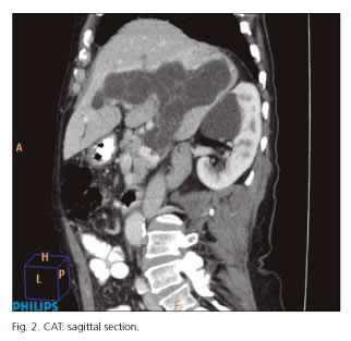
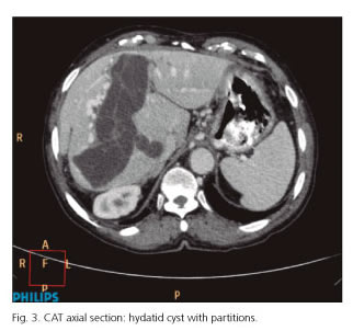
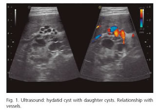
We report the case of an 81-year-old man treated for a hydatid cyst in segments VII-VIII six years before. The patient presented at the clinic due to discomfort in the right hypochondrium over a two-month period. Hydatid serology showed a titer of 1/800. An ultrasound was requested, which showed a cystic lesion with multiple daughter cysts of an elongated morphology and a size of 11 x 5.5 x 8.8 cm in the left, right and common portal vein in relation to portal hydatidosis (<a href="#f1">Fig. 1</a>). The computed tomography (CT) scan revealed a cystic elongated, polylobulated and branched lesion of more than 14 cm of longitudinal diameter and 4 cm of transverse diameter occupying the interior of the right, left and main portal vein. As well as portal cavernomatosis with collateral circulation and esophageal varices (Figs. <a href="#f2">2</a> and <a href="#f3">3</a>). The patient received medical treatment and is currently asymptomatic.</font></p>     <p>&nbsp;</p>     <p align="center"><font face="Verdana" size="2"><a name="f1"></a><img src="/img/revistas/diges/v109n3/imagen6_fig1.jpg"></font></p>     <p>&nbsp;</p>     <p align="center"><font face="Verdana" size="2"><a name="f2"></a><img src="/img/revistas/diges/v109n3/imagen6_fig2.jpg"></font></p>     <p>&nbsp;</p>     <p align="center"><font face="Verdana" size="2"><a name="f3"></a><img src="/img/revistas/diges/v109n3/imagen6_fig3.jpg"></font></p>     <p>&nbsp;</p>     <p><font face="Verdana" size="2"><b>Discussion</b></font></p>     ]]></body>
<body><![CDATA[<p><font face="Verdana" size="2">The complication rate of hydatid cysts in the portal vein with cavernomatosis is very rare. There are few cases reported in the literature (1), the first one being described in 1998 (2).</font></p>     <p><font face="Verdana" size="2">Portal cavernomatosis is a dilatation of the paracholedochal and pericholedochal veins and it is generally secondary to portal vein thrombosis (3). It is an uncommon disease.</font></p>     <p><font face="Verdana" size="2">Portal involvement is caused by several mechanisms:</font></p>     <blockquote>     <p><font face="Verdana" size="2">- Extrinsic compression.</font></p>     <p><font face="Verdana" size="2">- Invasion of vessels which looks like a thrombosis due to an inflammatory reaction in the vessel wall caused by external compression of the cyst, which facilitates the creation of a cyst-portal fistula thus allowing passage of daughter cysts to the vascular lumen.</font></p>     <p><font face="Verdana" size="2">- Cyst rupture, which occurs in 50-90% of cases.</font></p> </blockquote>     <p><font face="Verdana" size="2">The clinical manifestation is usually abdominal pain in the right hypochodrium with or without nausea and vomiting. The diagnosis is based on the detection of specific serum antibodies. Plain standard X-ray is used in imaging tests to detect calcified cysts, which occur in 20-30% of the cases.</font></p>     <p><font face="Verdana" size="2">Abdominal ultrasound is the most widely used technique since it is easy, inexpensive and has a sensitivity of 90-95%. In 1981, Gharbi et al. published an ultrasonographic classification based on structure and morphology. In 2003, the World Health Organization proposed a new classification, the World Health Organization Informal Working Group classification (WHO/IWG), based on the natural evolution of lesions.</font></p>     <p><font face="Verdana" size="2">The treatment of choice for portal hydatid disease is surgery, with the removal of the cyst. In recent years, due to the advances in liver surgery, hepatectomies are being performed with a low morbidity and mortality rate (4), including laparoscopic surgery. The authors have added praziquantel to the treatment schedule (5).</font></p>     ]]></body>
<body><![CDATA[<p>&nbsp;</p>     <p><font face="Verdana" size="2"><b>References</b></font></p>     <!-- ref --><p><font face="Verdana" size="2">1. Gonz&aacute;lez E, Gil Grande L, Del Arbol L, et al. Presinusoidal portal hipertensi&oacute;n secondary to portal invasive echinococosis. J Clin Gastroenterol 2002;34:103-4.    &nbsp;&nbsp;&nbsp;&nbsp;&nbsp;&nbsp;&nbsp;&nbsp;[&#160;<a href="javascript:void(0);" onclick="javascript: window.open('/scielo.php?script=sci_nlinks&ref=5443860&pid=S1130-0108201700030001300001&lng=','','width=640,height=500,resizable=yes,scrollbars=1,menubar=yes,');">Links</a>&#160;]<!-- end-ref --></font></p>    <!-- ref --><p><font face="Verdana" size="2">2. Gil-Egea MJ, Alameda F, Girvent M, et al. Hydatid cyst in the hepatic hileum causing a cavernous transformation in the portal vein. Gastroenterol Hepatol 1998;21:227-9.    &nbsp;&nbsp;&nbsp;&nbsp;&nbsp;&nbsp;&nbsp;&nbsp;[&#160;<a href="javascript:void(0);" onclick="javascript: window.open('/scielo.php?script=sci_nlinks&ref=5443862&pid=S1130-0108201700030001300002&lng=','','width=640,height=500,resizable=yes,scrollbars=1,menubar=yes,');">Links</a>&#160;]<!-- end-ref --></font></p>    <!-- ref --><p><font face="Verdana" size="2">3. Pedrosa I, Saiz A, Ar&aacute;osla J. Hydatid disease: Radiologic and pathologic features and complications. Radiographics 2000;20:795-817. DOI: 10.1148/radiographics.20.3.g00ma06795.    &nbsp;&nbsp;&nbsp;&nbsp;&nbsp;&nbsp;&nbsp;&nbsp;[&#160;<a href="javascript:void(0);" onclick="javascript: window.open('/scielo.php?script=sci_nlinks&ref=5443864&pid=S1130-0108201700030001300003&lng=','','width=640,height=500,resizable=yes,scrollbars=1,menubar=yes,');">Links</a>&#160;]<!-- end-ref --></font></p>    <!-- ref --><p><font face="Verdana" size="2">4. Priego P, Nu&ntilde;o J, L&oacute;pez Herv&aacute;s P, et al. Hidatidosis hep&aacute;tica. Cirug&iacute;a radical vs. no radical: 22 a&ntilde;os de experiencia. Rev Esp Enferm Dig 2008;100(2):82-5. DOI: 10.4321/S1130-01082008000200004.    &nbsp;&nbsp;&nbsp;&nbsp;&nbsp;&nbsp;&nbsp;&nbsp;[&#160;<a href="javascript:void(0);" onclick="javascript: window.open('/scielo.php?script=sci_nlinks&ref=5443866&pid=S1130-0108201700030001300004&lng=','','width=640,height=500,resizable=yes,scrollbars=1,menubar=yes,');">Links</a>&#160;]<!-- end-ref --></font></p>    ]]></body>
<body><![CDATA[<!-- ref --><p><font face="Verdana" size="2">5. Ramia JM, De la Plaza R, Casares M, et al. Profile of patients with hepatic hydatid disease not treated surgically. Rev Esp Enferm Dig 2011;103(9):448-52. DOI: 10.4321/S1130-01082011000900002.    &nbsp;&nbsp;&nbsp;&nbsp;&nbsp;&nbsp;&nbsp;&nbsp;[&#160;<a href="javascript:void(0);" onclick="javascript: window.open('/scielo.php?script=sci_nlinks&ref=5443868&pid=S1130-0108201700030001300005&lng=','','width=640,height=500,resizable=yes,scrollbars=1,menubar=yes,');">Links</a>&#160;]<!-- end-ref --></font></p>      ]]></body><back>
<ref-list>
<ref id="B1">
<label>1</label><nlm-citation citation-type="journal">
<person-group person-group-type="author">
<name>
<surname><![CDATA[González]]></surname>
<given-names><![CDATA[E]]></given-names>
</name>
<name>
<surname><![CDATA[Gil Grande]]></surname>
<given-names><![CDATA[L]]></given-names>
</name>
<name>
<surname><![CDATA[Del Arbol]]></surname>
<given-names><![CDATA[L]]></given-names>
</name>
</person-group>
<article-title xml:lang="es"><![CDATA[Presinusoidal portal hipertensión secondary to portal invasive echinococosis]]></article-title>
<source><![CDATA[J Clin Gastroenterol]]></source>
<year>2002</year>
<volume>34</volume>
<page-range>103-4</page-range></nlm-citation>
</ref>
<ref id="B2">
<label>2</label><nlm-citation citation-type="journal">
<person-group person-group-type="author">
<name>
<surname><![CDATA[Gil-Egea]]></surname>
<given-names><![CDATA[MJ]]></given-names>
</name>
<name>
<surname><![CDATA[Alameda]]></surname>
<given-names><![CDATA[F]]></given-names>
</name>
<name>
<surname><![CDATA[Girvent]]></surname>
<given-names><![CDATA[M]]></given-names>
</name>
</person-group>
<article-title xml:lang="en"><![CDATA[Hydatid cyst in the hepatic hileum causing a cavernous transformation in the portal vein]]></article-title>
<source><![CDATA[Gastroenterol Hepatol]]></source>
<year>1998</year>
<volume>21</volume>
<page-range>227-9</page-range></nlm-citation>
</ref>
<ref id="B3">
<label>3</label><nlm-citation citation-type="journal">
<person-group person-group-type="author">
<name>
<surname><![CDATA[Pedrosa]]></surname>
<given-names><![CDATA[I]]></given-names>
</name>
<name>
<surname><![CDATA[Saiz]]></surname>
<given-names><![CDATA[A]]></given-names>
</name>
<name>
<surname><![CDATA[Aráosla]]></surname>
<given-names><![CDATA[J]]></given-names>
</name>
</person-group>
<article-title xml:lang="en"><![CDATA[Hydatid disease: Radiologic and pathologic features and complications]]></article-title>
<source><![CDATA[Radiographics]]></source>
<year>2000</year>
<volume>20</volume>
<page-range>795-817</page-range></nlm-citation>
</ref>
<ref id="B4">
<label>4</label><nlm-citation citation-type="journal">
<person-group person-group-type="author">
<name>
<surname><![CDATA[Priego]]></surname>
<given-names><![CDATA[P]]></given-names>
</name>
<name>
<surname><![CDATA[Nuño]]></surname>
<given-names><![CDATA[J]]></given-names>
</name>
<name>
<surname><![CDATA[López Hervás]]></surname>
<given-names><![CDATA[P]]></given-names>
</name>
</person-group>
<article-title xml:lang="es"><![CDATA[Hidatidosis hepática. Cirugía radical vs. no radical: 22 años de experiencia]]></article-title>
<source><![CDATA[Rev Esp Enferm Dig]]></source>
<year>2008</year>
<volume>100</volume>
<numero>2</numero>
<issue>2</issue>
<page-range>82-5</page-range></nlm-citation>
</ref>
<ref id="B5">
<label>5</label><nlm-citation citation-type="journal">
<person-group person-group-type="author">
<name>
<surname><![CDATA[Ramia]]></surname>
<given-names><![CDATA[JM]]></given-names>
</name>
<name>
<surname><![CDATA[De la Plaza]]></surname>
<given-names><![CDATA[R]]></given-names>
</name>
<name>
<surname><![CDATA[Casares]]></surname>
<given-names><![CDATA[M]]></given-names>
</name>
</person-group>
<article-title xml:lang="en"><![CDATA[Profile of patients with hepatic hydatid disease not treated surgically]]></article-title>
<source><![CDATA[Rev Esp Enferm Dig]]></source>
<year>2011</year>
<volume>103</volume>
<numero>9</numero>
<issue>9</issue>
<page-range>448-52</page-range></nlm-citation>
</ref>
</ref-list>
</back>
</article>
