<?xml version="1.0" encoding="ISO-8859-1"?><article xmlns:mml="http://www.w3.org/1998/Math/MathML" xmlns:xlink="http://www.w3.org/1999/xlink" xmlns:xsi="http://www.w3.org/2001/XMLSchema-instance">
<front>
<journal-meta>
<journal-id>1130-0108</journal-id>
<journal-title><![CDATA[Revista Española de Enfermedades Digestivas]]></journal-title>
<abbrev-journal-title><![CDATA[Rev. esp. enferm. dig.]]></abbrev-journal-title>
<issn>1130-0108</issn>
<publisher>
<publisher-name><![CDATA[Sociedad Española de Patología Digestiva]]></publisher-name>
</publisher>
</journal-meta>
<article-meta>
<article-id>S1130-01082017000600022</article-id>
<article-id pub-id-type="doi">10.17235/reed.2017.4237/2016</article-id>
<title-group>
<article-title xml:lang="en"><![CDATA[Hepatic hematoma after ERCP: two new case reports]]></article-title>
<article-title xml:lang="es"><![CDATA[Hematoma hepático tras CPRE: presentación de dos nuevos casos]]></article-title>
</title-group>
<contrib-group>
<contrib contrib-type="author">
<name>
<surname><![CDATA[del-Moral-Martínez]]></surname>
<given-names><![CDATA[María]]></given-names>
</name>
<xref ref-type="aff" rid="A01"/>
</contrib>
<contrib contrib-type="author">
<name>
<surname><![CDATA[Delgado-Maroto]]></surname>
<given-names><![CDATA[Ana]]></given-names>
</name>
<xref ref-type="aff" rid="A01"/>
</contrib>
<contrib contrib-type="author">
<name>
<surname><![CDATA[Cervilla-Sáez-de-Tejada]]></surname>
<given-names><![CDATA[María Eloísa]]></given-names>
</name>
<xref ref-type="aff" rid="A01"/>
</contrib>
<contrib contrib-type="author">
<name>
<surname><![CDATA[Casado-Caballero]]></surname>
<given-names><![CDATA[Francisco Javier]]></given-names>
</name>
<xref ref-type="aff" rid="A01"/>
</contrib>
<contrib contrib-type="author">
<name>
<surname><![CDATA[Salmerón-Escobar]]></surname>
<given-names><![CDATA[Francisco Javier]]></given-names>
</name>
<xref ref-type="aff" rid="A01"/>
</contrib>
</contrib-group>
<aff id="A01">
<institution><![CDATA[,Hospital Universitario San Cecilio Clinical Management Unit of Digestive Diseases ]]></institution>
<addr-line><![CDATA[Granada ]]></addr-line>
<country>Spain</country>
</aff>
<pub-date pub-type="pub">
<day>00</day>
<month>06</month>
<year>2017</year>
</pub-date>
<pub-date pub-type="epub">
<day>00</day>
<month>06</month>
<year>2017</year>
</pub-date>
<volume>109</volume>
<numero>6</numero>
<fpage>470</fpage>
<lpage>473</lpage>
<copyright-statement/>
<copyright-year/>
<self-uri xlink:href="http://scielo.isciii.es/scielo.php?script=sci_arttext&amp;pid=S1130-01082017000600022&amp;lng=en&amp;nrm=iso"></self-uri><self-uri xlink:href="http://scielo.isciii.es/scielo.php?script=sci_abstract&amp;pid=S1130-01082017000600022&amp;lng=en&amp;nrm=iso"></self-uri><self-uri xlink:href="http://scielo.isciii.es/scielo.php?script=sci_pdf&amp;pid=S1130-01082017000600022&amp;lng=en&amp;nrm=iso"></self-uri><abstract abstract-type="short" xml:lang="en"><p><![CDATA[ERCP is an endoscopic procedure with a complication risk ranging from 2.5 to 8%. The most frequent complications are pancreatitis, cholangitis, hemorrhage or perforation. Hepatic hematoma after ERCP is a potentially serious, rare complication. Not many cases are reported in the literature. We present here two new cases of hepatic hematoma following ERCP along with a review of the literature and possible therapeutic options.]]></p></abstract>
<abstract abstract-type="short" xml:lang="es"><p><![CDATA[La colangiopancreatografía retrógrada endoscópica (CPRE) es una técnica endoscópica con un riesgo de complicaciones secundarias que oscila del 2,5-8%. Las complicaciones que con más frecuencia se presentan son la pancreatitis, procesos infecciosos, hemorragia o perforación. El hematoma hepático secundario a CPRE es una rara y potencialmente grave complicación con pocos casos descritos en la literatura. A continuación presentamos dos casos de hematomas hepáticos secundarios a la realización de CPRE, realizando una revisión de la bibliografía y de las posibles opciones terapéuticas.]]></p></abstract>
<kwd-group>
<kwd lng="en"><![CDATA[Hepatic hematoma]]></kwd>
<kwd lng="en"><![CDATA[ERCP]]></kwd>
<kwd lng="en"><![CDATA[Complication]]></kwd>
<kwd lng="es"><![CDATA[Hematoma hepático]]></kwd>
<kwd lng="es"><![CDATA[CPRE]]></kwd>
<kwd lng="es"><![CDATA[Complicación]]></kwd>
</kwd-group>
</article-meta>
</front><body><![CDATA[ <p><a name="top"></a>&nbsp;</p>     <p><font face="Verdana" size="2"><b>CASE REPORT</b></font></p>     <p>&nbsp;</p>     <p><font face="Verdana" size="4"><b>Hepatic hematoma after ERCP: two new case reports</b></font></p>     <p><font face="Verdana" size="4"><b>Hematoma hep&aacute;tico tras CPRE: presentaci&oacute;n de dos nuevos casos</b></font></p>     <p>&nbsp;</p>     <p>&nbsp;</p>     <p><font face="Verdana" size="2"><b>Mar&iacute;a-del-Moral-Mart&iacute;nez, Ana Delgado-Maroto, Mar&iacute;a Elo&iacute;sa Cervilla-S&aacute;ez-de-Tejada, Francisco Javier Casado-Caballero and Francisco Javier Salmer&oacute;n-Escobar</b></font></p>     <p><font face="Verdana" size="2">Clinical Management Unit of Digestive Diseases. Hospital Universitario San Cecilio. Granada, Spain</font></p>     <p><font face="Verdana" size="2"><a href="#bajo">Correspondence</a></font></p>     ]]></body>
<body><![CDATA[<p>&nbsp;</p>     <p>&nbsp;</p> <hr size="1">     <p><font face="Verdana" size="2"><b>ABSTRACT</b></font></p>     <p><font face="Verdana" size="2">ERCP is an endoscopic procedure with a complication risk ranging from 2.5 to 8%. The most frequent complications are pancreatitis, cholangitis, hemorrhage or perforation. Hepatic hematoma after ERCP is a potentially serious, rare complication. Not many cases are reported in the literature. We present here two new cases of hepatic hematoma following ERCP along with a review of the literature and possible therapeutic options.</font></p>     <p><font face="Verdana" size="2"><b>Key words:</b> Hepatic hematoma. ERCP. Complication.</font></p> <hr size="1">     <p><font face="Verdana" size="2"><b>RESUMEN</b></font></p>     <p><font face="Verdana" size="2">La colangiopancreatograf&iacute;a retr&oacute;grada endosc&oacute;pica (CPRE) es una t&eacute;cnica endosc&oacute;pica con un riesgo de complicaciones secundarias que oscila del 2,5-8%. Las complicaciones que con m&aacute;s frecuencia se presentan son la pancreatitis, procesos infecciosos, hemorragia o perforaci&oacute;n. El hematoma hep&aacute;tico secundario a CPRE es una rara y potencialmente grave complicaci&oacute;n con pocos casos descritos en la literatura. A continuaci&oacute;n presentamos dos casos de hematomas hep&aacute;ticos secundarios a la realizaci&oacute;n de CPRE, realizando una revisi&oacute;n de la bibliograf&iacute;a y de las posibles opciones terap&eacute;uticas.</font></p>     <p><font face="Verdana" size="2"><b>Palabras clave:</b> Hematoma hep&aacute;tico. CPRE. Complicaci&oacute;n.</font></p> <hr size="1">     <p>&nbsp;</p>     <p><font face="Verdana" size="2"><b>Introduction</b></font></p>     ]]></body>
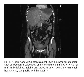
<body><![CDATA[<p><font face="Verdana" size="2">The endoscopic retrograde cholangiopancreatography ERCP is an endoscopic procedure with a complication risk ranging from 2.5 to 8%. The most frequent complications are pancreatitis, cholangitis, hemorrhage or perforation. Hepatic hematoma after ERCP is a potentially serious, rare complication. Not many cases are reported in the literature. We present here two new cases of hepatic hematoma following ERCP along with a review of the literature and possible therapeutic options.</font></p>     <p>&nbsp;</p>     <p><font face="Verdana" size="2"><b>Case report 1</b></font></p>     <p><font face="Verdana" size="2">A 37-year-old woman was admitted to hospital, as programmed, to perform an ERCP due to a diagnosis of a common-bile-duct stone the previous week via a magnetic resonance (MR) cholangiography. On admission, she was asymptomatic. The patient had no significant past medical and family history. During the examination, a marked cutaneous-mucous jaundice was observed. Laboratory tests showed: total bilirubin 7.91 mg/dl, direct bilirubin 7.13 mg/dl, aspartate aminotransferase (AST) 366 U/l, alanine aminotransferase (ALT) 407 U/l, GGT 812 U/l, and alkaline phosphatase (ALP) 332 U/l; the rest of the parameters showed no alteration.</font></p>     <p><font face="Verdana" size="2">An ERCP was performed, revealing a small papilla difficult to cannulate, and gaining access into the pancreatic duct on two occasions. By performing a precut, access was gained into the common bile duct, which was dilated, and an impacted calculus about 2 cm in diameter was found. A wide papillotomy was performed, the calculus was removed with the help of the Fogarty balloon and an outflow of retained bile and pus remains was observed.</font></p>     <p><font face="Verdana" size="2">Six hours after the ERCP was performed, the patient began to complain of intense diffuse abdominal pain associated with diffuse discomfort. She did not present with fever or hyperthermia. An urgent laboratory test showed amylase at 1,702 U/l and no other alterations. The patient was diagnosed with acute pancreatitis post-ERCP and she was prescribed serum therapy, <i>nil per os</i> (NPO), analgesic perfusion and vital signs monitoring. A few hours later, the patient had a presyncopal episode and her diffuse discomfort and intense abdominal pain persisted, observing a blood pressure (BP) of 85/45 mmHg, heart rate (HR) of 120 bpm and SatO<sub>2</sub> of 97% with no O<sub>2</sub> supply, so a new laboratory test was requested in order to evaluate possible complications.</font></p>     <p><font face="Verdana" size="2">A new laboratory test revealed a marked drop in hemoglobin from 12 to 7.6 g/dl, along with the already known hyperamylasemia; there was no rise in acute phase reactants and no other alterations. The patient presented neither tarry stools nor hematemesis, and no blood exteriorization was observed. In view of this findings, an abdominopelvic computed tomography (CT) scan was requested in order to preclude possible complications. The CT scan revealed two large subcapsular/intraparenchymal hypodense collections, one of them (measuring 70 x 107 x 120 mm) in the left hepatic lobe and the other one affecting the entire right hepatic lobe, compatible with hematomas. A small amount of subphrenic intraparenchymal air in contact with the right hepatic lobe collection was observed. Hepatic artery and portal vein permeability was noticed, with no contrast extravasation (<a href="#f1">Fig. 1</a>).</font></p>     <p>&nbsp;</p>     <p align="center"><a name="f1"></a><img src="/img/revistas/diges/v109n6/nota3_fig1.jpg"></p>     <p>&nbsp;</p>     ]]></body>
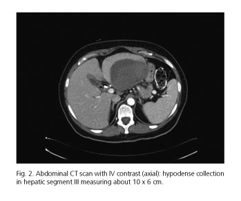
<body><![CDATA[<p><font face="Verdana" size="2">A red blood cell transfusion and intravenous antibiotic therapy were initiated, and the patient was transferred to the Intensive Care Unit for stabilization and monitoring. An urgent arteriography revealed no contrast extravasation. Since the patient was successfully stabilized, afebrile and in good condition, with a stable hemoglobin level, she was managed by close observation. Two weeks later she remained afebrile and there was no evidence of additional bleeding, so she was discharged from the hospital. The patient attended monthly follow-up visits in the clinic, which included laboratory tests and ultrasound monitoring. No clinical or analytical repercussion was observed and the imaging tests showed a progressive reduction in the size of the injuries. Six months after being discharged, a follow-up abdominal CT scan was performed, which revealed that the two hypodense collections in the subphrenic space remained over the right and the left hepatic lobes, though they had decreased significantly, measuring about 11 x 8.5 x 10.5 cm and 2.8 x 2.6 x 2.3 cm (anteroposterior x lateral x transverse diameters, respectively).</font></p>     <p>&nbsp;</p>     <p><font face="Verdana" size="2"><b>Case report 2</b></font></p>     <p><font face="Verdana" size="2">A 60-year-old woman with no significant past medical history was admitted from the Emergency Department with clinical symptoms suggestive of cholangitis. During her stay, an MR cholangiography was performed, which revealed a retained 7.7 mm calculus in the common bile duct, about 4.4 cm from the papilla, with intra- and extrahepatic bile duct dilatation. In view of this finding, we decided to perform an ERCP, but the papilla of Vater could not be cannulated. Two days later, as the jaundice persisted, a new ERCP was performed. A fresh clot covering the papilla was detected. After removing it, access was gained into the bile duct, observing the calculus described during the MR cholangiography, which was removed after performing a biliary sphincterotomy. Finally, diluted epinephrine was injected into the papilla, still with signs of recent bleeding.</font></p>     <p><font face="Verdana" size="2">Seven days after being discharged from the hospital, the patient presented to the Emergency Department with fever, abdominal pain and intense asthenia. Laboratory tests showed hemoglobin (Hb) 8.6 g/dl, hematocrit (Hct) 27.2%, white blood cells (WBC) 10,000 U/ml with 68% polymorphonuclear, platelets 627,000 U/ml, and C-reactive protein (CRP9 187.4 mg/l. An abdominal ultrasound was performed, during which a hepatic space occupying lesion (SOL) in segment III measuring about 10 x 6 cm with ultrasound signs of an abscess was observed. Percutaneous drainage was attempted, but it was not possible due to the lesion density. A fine-needle biopsy was performed and <i>Enterobacter cloacae</i> was isolated in the culture of the obtained material.</font></p>     <p><font face="Verdana" size="2">A follow-up abdominal CT scan after antibiotic therapy revealed the lesion described above, corresponding to a possible hepatic hematoma (<a href="#f2">Fig. 2</a>). Since a high fever persisted, an 8-Fr drainage catheter was placed.</font></p>     <p>&nbsp;</p>     <p align="center"><a name="f2"></a><img src="/img/revistas/diges/v109n6/nota3_fig2.jpg"></p>     <p>&nbsp;</p>     <p><font face="Verdana" size="2">After drainage, the patient remained asymptomatic, afebrile and hemodynamically stable. The patient underwent weekly abdominal ultrasound examinations to monitor the hematoma evolution, observing its progressive reduction. Six weeks later, the drainage was removed with no complications.</font></p>     ]]></body>
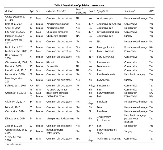
<body><![CDATA[<p>&nbsp;</p>     <p><font face="Verdana" size="2"><b>Discussion</b></font></p>     <p><font face="Verdana" size="2">ERCP is an endoscopic procedure with a complication rate ranging from 2.5 to 8% and a mortality rate ranging from 0.5 to 1%. The most frequently described complications are acute pancreatitis (1-7%), acute cholangitis (1.4%), hemorrhage (1%) and duodenal perforation (1%) (1). Nevertheless, there are some exceptional complications, such as hepatic hematoma.</font></p>     <p><font face="Verdana" size="2">Hepatic hematoma after ERCP is a rare complication, the frequency of which is scarcely reported. We have reviewed the cases reported in the literature, and when entering the terms "hepatic hematoma after ERCP" and "ERCP complications" in PubMed 25 cases reported to date were found (<a href="#t1">Table I</a>).</font></p>     <p>&nbsp;</p>     <p align="center"><a name="t1"></a><img src="/img/revistas/diges/v109n6/nota3_table1.jpg"></p>     <p>&nbsp;</p>     <p><font face="Verdana" size="2">Etiopathogenesis is not completely clear. Two hypothesis have been proposed. One of them states that the lesion would be caused by the guide wire usually used to cannulate the common bile duct, which would perforate the bile duct, thus damaging the hepatic parenchyma. As a result of this, blood vessels would break and air would enter the bile duct and the hematoma, which would explain the presence of subphrenic air in our first patient's CT scan. The other hypothesis states that the hepatic damage is secondary to the traction force exerted with the balloon on the bile duct when trying to remove a retained calculus. This force would cause the rupture of bile ductules and vessels, as well as the consequent bleeding (2).</font></p>     <p><font face="Verdana" size="2">The clinical manifestations are variable, including abdominal pain, anemia, fever and hemodynamic shock. According to the cases reported in the literature, abdominal pain was the first clinical manifestation in 87% of the cases, associated with anemia (29%) and fever (25%). The onset of symptoms can vary as well, ranging from hours to days. The longest period described in a case was fifteen days after ERCP (3).</font></p>     <p><font face="Verdana" size="2">The treatment must be personalized depending on the case. Those patients clinically and hemodynamically stable can be managed conservatively (3,4,5). Prophylactic antibiotic therapy is usually administered, given the high risk of infection. For those cases presenting with hemodynamical instability or associated complications, such as hematoma breakage, urgent surgical treatment should be considered (6,7,8). There are some case reports in which an arteriography with embolization of the bleeding vessel has successfully resolved the clinical situation (9,10).</font></p>     ]]></body>
<body><![CDATA[<p>&nbsp;</p>     <p><font face="Verdana" size="2"><b>References</b></font></p>     <!-- ref --><p><font face="Verdana" size="2">1. Garc&iacute;a-Cano J, Gonz&aacute;lez Mart&iacute;n JA, Morillas Ari&ntilde;o J, et al. Complications of endoscopic retrograde cholangiopancreatography. A study in a small ERCP unit. Rev Esp Enferm Dig 2004;96:163-73.    &nbsp;&nbsp;&nbsp;&nbsp;&nbsp;&nbsp;&nbsp;&nbsp;[&#160;<a href="javascript:void(0);" onclick="javascript: window.open('/scielo.php?script=sci_nlinks&ref=5486113&pid=S1130-0108201700060002200001&lng=','','width=640,height=500,resizable=yes,scrollbars=1,menubar=yes,');">Links</a>&#160;]<!-- end-ref --></font></p>    <!-- ref --><p><font face="Verdana" size="2">2. Baudet JS, Argui&ntilde;arena X, Redondo I, et al. Subcapsular hepatic hematoma: An uncommon complication of endoscopic retrograde cholangiopancreatography. Gastroenterol Hepatol 2011;34(2):79-82. DOI: 10.1016/j.gastrohep.2010.11.002.    &nbsp;&nbsp;&nbsp;&nbsp;&nbsp;&nbsp;&nbsp;&nbsp;[&#160;<a href="javascript:void(0);" onclick="javascript: window.open('/scielo.php?script=sci_nlinks&ref=5486115&pid=S1130-0108201700060002200002&lng=','','width=640,height=500,resizable=yes,scrollbars=1,menubar=yes,');">Links</a>&#160;]<!-- end-ref --></font></p>    <!-- ref --><p><font face="Verdana" size="2">3. Servide MJ, Prieto M, Marquina T. Una rara complicaci&oacute;n tard&iacute;a tras CPRE: Hematoma hep&aacute;tico subcapsular. Rev Esp Enferm Dig 2016;108(4):234-5.    &nbsp;&nbsp;&nbsp;&nbsp;&nbsp;&nbsp;&nbsp;&nbsp;[&#160;<a href="javascript:void(0);" onclick="javascript: window.open('/scielo.php?script=sci_nlinks&ref=5486117&pid=S1130-0108201700060002200003&lng=','','width=640,height=500,resizable=yes,scrollbars=1,menubar=yes,');">Links</a>&#160;]<!-- end-ref --></font></p>    <!-- ref --><p><font face="Verdana" size="2">4. Orellana F, Irarrazaval J, Galindo J, et al. Subcapsular hepatic hematoma post ERCP: A rare or an underdiagnosed complication? Endoscopy 2012;44(Supl 2)UCTN:E108-9. DOI: 10.1055/s-0031-1291493.    &nbsp;&nbsp;&nbsp;&nbsp;&nbsp;&nbsp;&nbsp;&nbsp;[&#160;<a href="javascript:void(0);" onclick="javascript: window.open('/scielo.php?script=sci_nlinks&ref=5486119&pid=S1130-0108201700060002200004&lng=','','width=640,height=500,resizable=yes,scrollbars=1,menubar=yes,');">Links</a>&#160;]<!-- end-ref --></font></p>    ]]></body>
<body><![CDATA[<!-- ref --><p><font face="Verdana" size="2">5. Oliveira-Ferreira A, Tato-Marinho R, Velosa J. Infected hepatic hematoma 10 days after ERCP. Endoscopy 2013;45(Supl 2)UCTN:E402-3.    &nbsp;&nbsp;&nbsp;&nbsp;&nbsp;&nbsp;&nbsp;&nbsp;[&#160;<a href="javascript:void(0);" onclick="javascript: window.open('/scielo.php?script=sci_nlinks&ref=5486121&pid=S1130-0108201700060002200005&lng=','','width=640,height=500,resizable=yes,scrollbars=1,menubar=yes,');">Links</a>&#160;]<!-- end-ref --></font></p>    <!-- ref --><p><font face="Verdana" size="2">6. Gonz&aacute;lez-L&oacute;pez R, Garc&iacute;a-Cano E, Espinosa-Gonz&aacute;lez O, et al. Surgical treatment for liver haematoma following endoscopic retrograde cholangiopancreatography. An unusual case. Cir Cir 2015;83(6):506-9. DOI: 10.1016/j.circir.2015.05.028.    &nbsp;&nbsp;&nbsp;&nbsp;&nbsp;&nbsp;&nbsp;&nbsp;[&#160;<a href="javascript:void(0);" onclick="javascript: window.open('/scielo.php?script=sci_nlinks&ref=5486123&pid=S1130-0108201700060002200006&lng=','','width=640,height=500,resizable=yes,scrollbars=1,menubar=yes,');">Links</a>&#160;]<!-- end-ref --></font></p>    <!-- ref --><p><font face="Verdana" size="2">7. P&eacute;rez-Legaz J, Santos J, Ruiz-Tovar J, et al. Subcapsular hepatic hematoma after ERCP (endoscopic retrograde cholangipancreatography). Rev Esp Enferm Dig 2011;103(10):550-1. DOI: 10.4321/S1130-01082011001000011.    &nbsp;&nbsp;&nbsp;&nbsp;&nbsp;&nbsp;&nbsp;&nbsp;[&#160;<a href="javascript:void(0);" onclick="javascript: window.open('/scielo.php?script=sci_nlinks&ref=5486125&pid=S1130-0108201700060002200007&lng=','','width=640,height=500,resizable=yes,scrollbars=1,menubar=yes,');">Links</a>&#160;]<!-- end-ref --></font></p>    <!-- ref --><p><font face="Verdana" size="2">8. Priego P, Rodr&iacute;guez G, Mena A, et al. Subcapsular liver hematoma after ERCP. Rev Esp Enferm Dig 2007;99:53-4.    &nbsp;&nbsp;&nbsp;&nbsp;&nbsp;&nbsp;&nbsp;&nbsp;[&#160;<a href="javascript:void(0);" onclick="javascript: window.open('/scielo.php?script=sci_nlinks&ref=5486127&pid=S1130-0108201700060002200008&lng=','','width=640,height=500,resizable=yes,scrollbars=1,menubar=yes,');">Links</a>&#160;]<!-- end-ref --></font></p>    <!-- ref --><p><font face="Verdana" size="2">9. Klimov&aacute; K, Padilla-Su&aacute;rez C, Gonz&aacute;lez-Asanza C, et al. Subcapsular hepatic hematoma after ERCP: A case report and revision of literature. Clin Med 2014;03(03):161-6. DOI: 10.4236/crcm.2014.33039.    &nbsp;&nbsp;&nbsp;&nbsp;&nbsp;&nbsp;&nbsp;&nbsp;[&#160;<a href="javascript:void(0);" onclick="javascript: window.open('/scielo.php?script=sci_nlinks&ref=5486129&pid=S1130-0108201700060002200009&lng=','','width=640,height=500,resizable=yes,scrollbars=1,menubar=yes,');">Links</a>&#160;]<!-- end-ref --></font></p>    ]]></body>
<body><![CDATA[<!-- ref --><p><font face="Verdana" size="2">10. Chi KD, Waxman I. Subcapsular hepatic hematoma after guide wire injury during endoscopic retrograde cholangiopancreatography: Management and review. Endoscopy 2004;36(11):1019-21. DOI: 10.1055/s-2004-825861.    &nbsp;&nbsp;&nbsp;&nbsp;&nbsp;&nbsp;&nbsp;&nbsp;[&#160;<a href="javascript:void(0);" onclick="javascript: window.open('/scielo.php?script=sci_nlinks&ref=5486131&pid=S1130-0108201700060002200010&lng=','','width=640,height=500,resizable=yes,scrollbars=1,menubar=yes,');">Links</a>&#160;]<!-- end-ref --></font></p>     <p>&nbsp;</p>     <p>&nbsp;</p>     <p><font face="Verdana" size="2"><a href="#top"><img border="0" src="/img/revistas/diges/v109n6/seta.gif" width="15" height="17"></a><a name="bajo"></a><b>Correspondence:</b>    <br>Mar&iacute;a del Moral Mart&iacute;nez.    <br>UGC Digestive Diseases.    <br>Hospital Universitario San Cecilio.    <br>Calle Dr. Oloriz, 16.    <br>18012 Granada, Spain    ]]></body>
<body><![CDATA[<br>e-mail: <a href="mailto:mdelmoral87@gmail.com">mdelmoral87@gmail.com</a></font></p>     <p><font face="Verdana" size="2">Received: 28-02-2016    <br>Accepted: 27-12-2016</font></p>      ]]></body><back>
<ref-list>
<ref id="B1">
<label>1</label><nlm-citation citation-type="journal">
<person-group person-group-type="author">
<name>
<surname><![CDATA[García-Cano]]></surname>
<given-names><![CDATA[J]]></given-names>
</name>
<name>
<surname><![CDATA[González Martín]]></surname>
<given-names><![CDATA[JA]]></given-names>
</name>
<name>
<surname><![CDATA[Morillas Ariño]]></surname>
<given-names><![CDATA[J]]></given-names>
</name>
</person-group>
<article-title xml:lang="en"><![CDATA[Complications of endoscopic retrograde cholangiopancreatography: A study in a small ERCP unit]]></article-title>
<source><![CDATA[Rev Esp Enferm Dig]]></source>
<year>2004</year>
<volume>96</volume>
<page-range>163-73</page-range></nlm-citation>
</ref>
<ref id="B2">
<label>2</label><nlm-citation citation-type="journal">
<person-group person-group-type="author">
<name>
<surname><![CDATA[Baudet]]></surname>
<given-names><![CDATA[JS]]></given-names>
</name>
<name>
<surname><![CDATA[Arguiñarena]]></surname>
<given-names><![CDATA[X]]></given-names>
</name>
<name>
<surname><![CDATA[Redondo]]></surname>
<given-names><![CDATA[I]]></given-names>
</name>
</person-group>
<article-title xml:lang="en"><![CDATA[Subcapsular hepatic hematoma: An uncommon complication of endoscopic retrograde cholangiopancreatography]]></article-title>
<source><![CDATA[Gastroenterol Hepatol]]></source>
<year>2011</year>
<volume>34</volume>
<numero>2</numero>
<issue>2</issue>
<page-range>79-82</page-range></nlm-citation>
</ref>
<ref id="B3">
<label>3</label><nlm-citation citation-type="journal">
<person-group person-group-type="author">
<name>
<surname><![CDATA[Servide]]></surname>
<given-names><![CDATA[MJ]]></given-names>
</name>
<name>
<surname><![CDATA[Prieto]]></surname>
<given-names><![CDATA[M]]></given-names>
</name>
<name>
<surname><![CDATA[Marquina]]></surname>
<given-names><![CDATA[T]]></given-names>
</name>
</person-group>
<article-title xml:lang="es"><![CDATA[Una rara complicación tardía tras CPRE: Hematoma hepático subcapsular]]></article-title>
<source><![CDATA[Rev Esp Enferm Dig]]></source>
<year>2016</year>
<volume>108</volume>
<numero>4</numero>
<issue>4</issue>
<page-range>234-5</page-range></nlm-citation>
</ref>
<ref id="B4">
<label>4</label><nlm-citation citation-type="journal">
<person-group person-group-type="author">
<name>
<surname><![CDATA[Orellana]]></surname>
<given-names><![CDATA[F]]></given-names>
</name>
<name>
<surname><![CDATA[Irarrazaval]]></surname>
<given-names><![CDATA[J]]></given-names>
</name>
<name>
<surname><![CDATA[Galindo]]></surname>
<given-names><![CDATA[J]]></given-names>
</name>
</person-group>
<article-title xml:lang="en"><![CDATA[Subcapsular hepatic hematoma post ERCP: A rare or an underdiagnosed complication?]]></article-title>
<source><![CDATA[Endoscopy]]></source>
<year>2012</year>
<volume>44</volume>
<numero>^s2</numero>
<issue>^s2</issue>
<supplement>2</supplement>
<page-range>E108-9</page-range><publisher-name><![CDATA[UCTN]]></publisher-name>
</nlm-citation>
</ref>
<ref id="B5">
<label>5</label><nlm-citation citation-type="journal">
<person-group person-group-type="author">
<name>
<surname><![CDATA[Oliveira-Ferreira]]></surname>
<given-names><![CDATA[A]]></given-names>
</name>
<name>
<surname><![CDATA[Tato-Marinho]]></surname>
<given-names><![CDATA[R]]></given-names>
</name>
<name>
<surname><![CDATA[Velosa]]></surname>
<given-names><![CDATA[J]]></given-names>
</name>
</person-group>
<article-title xml:lang="en"><![CDATA[Infected hepatic hematoma 10 days after ERCP]]></article-title>
<source><![CDATA[Endoscopy]]></source>
<year>2013</year>
<volume>45</volume>
<numero>^s2</numero>
<issue>^s2</issue>
<supplement>2</supplement>
<page-range>E402-3</page-range><publisher-name><![CDATA[UCTN]]></publisher-name>
</nlm-citation>
</ref>
<ref id="B6">
<label>6</label><nlm-citation citation-type="journal">
<person-group person-group-type="author">
<name>
<surname><![CDATA[González-López]]></surname>
<given-names><![CDATA[R]]></given-names>
</name>
<name>
<surname><![CDATA[García-Cano]]></surname>
<given-names><![CDATA[E]]></given-names>
</name>
<name>
<surname><![CDATA[Espinosa-González]]></surname>
<given-names><![CDATA[O]]></given-names>
</name>
</person-group>
<article-title xml:lang="en"><![CDATA[Surgical treatment for liver haematoma following endoscopic retrograde cholangiopancreatography: An unusual case]]></article-title>
<source><![CDATA[Cir Cir]]></source>
<year>2015</year>
<volume>83</volume>
<numero>6</numero>
<issue>6</issue>
<page-range>506-9</page-range></nlm-citation>
</ref>
<ref id="B7">
<label>7</label><nlm-citation citation-type="journal">
<person-group person-group-type="author">
<name>
<surname><![CDATA[Pérez-Legaz]]></surname>
<given-names><![CDATA[J]]></given-names>
</name>
<name>
<surname><![CDATA[Santos]]></surname>
<given-names><![CDATA[J]]></given-names>
</name>
<name>
<surname><![CDATA[Ruiz-Tovar]]></surname>
<given-names><![CDATA[J]]></given-names>
</name>
</person-group>
<article-title xml:lang="en"><![CDATA[Subcapsular hepatic hematoma after ERCP (endoscopic retrograde cholangipancreatography)]]></article-title>
<source><![CDATA[Rev Esp Enferm Dig]]></source>
<year>2011</year>
<volume>103</volume>
<numero>10</numero>
<issue>10</issue>
<page-range>550-1</page-range></nlm-citation>
</ref>
<ref id="B8">
<label>8</label><nlm-citation citation-type="journal">
<person-group person-group-type="author">
<name>
<surname><![CDATA[Priego]]></surname>
<given-names><![CDATA[P]]></given-names>
</name>
<name>
<surname><![CDATA[Rodríguez]]></surname>
<given-names><![CDATA[G]]></given-names>
</name>
<name>
<surname><![CDATA[Mena]]></surname>
<given-names><![CDATA[A]]></given-names>
</name>
</person-group>
<article-title xml:lang="en"><![CDATA[Subcapsular liver hematoma after ERCP]]></article-title>
<source><![CDATA[Rev Esp Enferm Dig]]></source>
<year>2007</year>
<volume>99</volume>
<page-range>53-4</page-range></nlm-citation>
</ref>
<ref id="B9">
<label>9</label><nlm-citation citation-type="journal">
<person-group person-group-type="author">
<name>
<surname><![CDATA[Klimová]]></surname>
<given-names><![CDATA[K]]></given-names>
</name>
<name>
<surname><![CDATA[Padilla-Suárez]]></surname>
<given-names><![CDATA[C]]></given-names>
</name>
<name>
<surname><![CDATA[González-Asanza]]></surname>
<given-names><![CDATA[C]]></given-names>
</name>
</person-group>
<article-title xml:lang="en"><![CDATA[Subcapsular hepatic hematoma after ERCP: A case report and revision of literature]]></article-title>
<source><![CDATA[Clin Med]]></source>
<year>2014</year>
<volume>03</volume>
<numero>03</numero>
<issue>03</issue>
<page-range>161-6</page-range></nlm-citation>
</ref>
<ref id="B10">
<label>10</label><nlm-citation citation-type="journal">
<person-group person-group-type="author">
<name>
<surname><![CDATA[Chi]]></surname>
<given-names><![CDATA[KD]]></given-names>
</name>
<name>
<surname><![CDATA[Waxman]]></surname>
<given-names><![CDATA[I]]></given-names>
</name>
</person-group>
<article-title xml:lang="en"><![CDATA[Subcapsular hepatic hematoma after guide wire injury during endoscopic retrograde cholangiopancreatography: Management and review]]></article-title>
<source><![CDATA[Endoscopy]]></source>
<year>2004</year>
<volume>36</volume>
<numero>11</numero>
<issue>11</issue>
<page-range>1019-21</page-range></nlm-citation>
</ref>
</ref-list>
</back>
</article>